| Title |
Resolution of Homonymous Visual Field Loss Documented with Functional Magnetic Resonance and Diffusion Tensor Imaging |
| Creator |
Yoshida, M; Ida, M; Nguyen, TH; Iba-Zizen, MT; Bellinger, L; Stievenart, JL; Nagao, T; Kikuchi, S; Hara, T; Shiba, T; Kitahara, K; Cabanis, EA |
| Affiliation |
Department of Radiology,Tokyo Metropolitan Ebara Hospital, Tokyo, Japan. masakiy@jikei.ac.jp |
| Abstract |
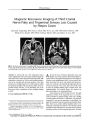
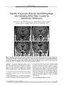
A 68-year-old man developed right homonymous hemianopic paracentral scotomas from acute infarction of the left extrastriate area. He was studied over the ensuing 12 months with visual fields, conventional MRI, functional MRI (fMRI), and diffusion tensor imaging (DTI). As the visual field defect became smaller, fMRI demonstrated progressively larger areas of cortical activation. DTI initially showed that the lesioned posterior optic radiations were completely interrupted. This interruption lessened in time and had disappeared by one year after onset. fMRI and DTI are innovative measures to follow functional and structural recovery in the central nervous system. This is the first reported application of these imaging techniques to acute cerebral visual field disorders. |
| Subject |
Older people; Cerebral Infarction, complications; Cerebral Infarction, diagnosis; Diffusion Magnetic Resonance Imaging, methods; Follow-Up Studies; Hemianopsia, diagnosis; Hemianopsia, etiology; Hemianopsia, physiopathology; Humans; Male; Perimetry; Remission, Spontaneous; Visual Cortex, blood supply; Visual Fields |
| Format |
application/pdf |
| Publication Type |
Journal Article |
| Collection |
Neuro-Ophthalmology Virtual Education Library: Journal of Neuro-Ophthalmology Archives: https://novel.utah.edu/jno/ |
| Publisher |
Lippincott, Williams & Wilkins |
| Holding Institution |
Spencer S. Eccles Health Sciences Library, University of Utah |
| Rights Management |
© North American Neuro-Ophthalmology Society |
| Setname |
ehsl_novel_jno |
| ID |
225542 |
| Reference URL |
https://collections.lib.utah.edu/ark:/87278/s6n90gwz/225542 |