| OCR Text |
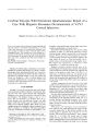
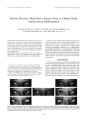
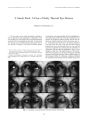
Show Journal of Neuro- Ophthalmology 19( 1): 17- 27, 1999. © 1999 Lippincoll Williams & Wilkins, Inc., Philadelphia Neuro- Ophthalmology of the Pregeniculate Afferent Visual System December, 1997- May, 1998 ( Part I) Laura J. Balcer, M. D., and Steven L. Galetta, M. D. The first half of 1998 was marked by the publication of many timely reports and investigations pertaining to the pregeniculate afferent visual system. This review of the neuro- ophthalmologic literature includes the months of December 1997 through May 1998. The following table of contents has been provided for ease of reference to the topics presented: EVALUATION OF THE AFFERENT VISUAL SYSTEM • Magnetic Resonance Imaging • Visual Field Testing • The Afferent Pupillary Defect • The Pulfrich Phenomenon NEURO- OPHTHALMOLOGY AND THE RETINA • Cancer- Associated Retinopathy • Acute Macular Neuroretinopathy • Terson Syndrome and Intracranial Aneurysms • Vascular Anomalies THE OPTIC NERVE • Anterior Ischemic Optic Neuropathy • Giant Cell Arteritis • Optic Neuritis • Infectious Causes of Optic Neuropathy • Idiopathic Intracranial Hypertension • Leber's Hereditary Optic Neuropathy THE OPTIC CHIASM AND BEYOND • Pituitary Apoplexy • Suprasellar Extramedullar^ Hematopoiesis • Metastatic Tumor to Optic Tract and Lateral Geniculate Nucleus Manuscript received August 11, 1998; accepted December 15, 1998. From the division of Neuro- Ophthalmology, Departments of Neurology and Ophthalmology, Hospital of the University of Pennsylvania, Scheie Eye Institute, University of Pennsylvania School of Medicine, Philadelphia, Pennsylvania, U. S. A. Supported in part by Grant EY00351- 01 ( L. J. B.) from the National Eye Institute, Bethesda, Maryland, U. S. A. Address correspondence and reprint requests to Dr. Steven L. Galetta, Department of Neurology, 3 East Gates, 3400 Spruce Street, Philadelphia, PA 19104, U. S. A. • Infarction of Optic Tract and Nerve • Congenital Optic Tract Syndrome • Homonymous Hemianopsia in Pediatric Patients • Pallidotomy and Visual Field Loss EVALUATION OF THE AFFERENT VISUAL SYSTEM Magnetic Resonance Imaging Within the neuro- ophthalmologist's testing armamentarium, magnetic resonance ( MR) imaging continues to have an important role in the diagnosis of anterior visual pathway lesions. To appreciate the extent of such lesions in three- dimensional space, however, multiple MR images must often be reviewed and synthesized. Groncm-eyer et al. ( 1) describe a " curved reconstruction" technique that enables visualization of the optic chiasm and optic nerves within a single MR image. This method uses a three- dimensional data set acquired using magnetization- prepared rapid- acquisition gradient- echo ( MP-RAGE) sequences. The resultant reconstruction displays the optic nerves and chiasm in a frog's eye view ( Fig. 1). Particularly useful for demonstrating enlargement of the optic nerves and chiasm ( as shown in Fig. 1), curved reconstruction also enables a size- wise comparison of the optic nerves throughout their course from the globe to the chiasm. This technique may provide a useful adjunct to standard MR imaging planes ( axial, sagittal, coronal), especially in the setting of neurofibromatosis type 1 and other disorders of the anterior visual pathway. Visual Field Testing Automated visual field testing is often relied on to provide serial standardized assessments of visual function in patients with anterior visual pathway disorders. In a recent study, Olsson et al. ( 2) confirm that such learning effects may indeed occur, even among asymptomatic people. These investigators devised a perimetric learner's index based on data from 74 normal volunteers, all of whom underwent visual field testing in both eyes during three separate sessions over a 7- month period. They emphasize that the perimetric learner's index may be a useful tool for determining, based on an initial visual field, whether a patient's abnormalities may result largely from lack of experience. Such patients may be 17 18 L. J. BALCER AND S. L. GALETTA FIG. 1. Magnetic resonance image with curved reconstruction technique displaying optic nerves and chiasm within a single image. Reprinted with permission from Gronemeyer SA, Langson JW, Abraham J. Curved reconstruction along the anterior optic pathway. AJNR Am J Neuroradiol 1998; 19: 338- 40. Copyright © by American Society of Neuroradiology. candidates for repeat testing, although the investigators also stress the continued importance of optic disc appearance and other clinical examination findings in the diagnostic process. The Afferent Pupillary Defect The detection of an afferent pupillary defect is critical in the diagnosis of optic nerve, chiasmal, and optic tract lesions. In response to previous debates in the literature regarding the sensitivity of the alternating light test versus the Marcus Gunn test for the identification of an afferent pupillary defect, Enyedi et al. ( 3) compared the two methods directly in a prospective study of 14 patients with unilateral optic neuropathy. Two examiners, masked to the side of the optic neuropathy, performed the tests on each patient in a random order. The starting eye for testing ( right vs. left) was also determined at random in each patient. The Marcus Gunn test was performed by shining a penlight into the first eye for 10 seconds and noting the degree of pupillary redilation at 2 seconds using a grading scale. The process was repeated 3 minutes later in the fellow eye. The alternating light test was accomplished by swinging the light back and forth between the two eyes for six cycles. Among the 14 patients with known unilateral optic neuropathies, the alternating- light test correctly identified the presence and side of the affected eye in 13 patients ( the test was indeterminate in 1 patient). In contrast, the affected eye was correctly identified in 8 of 14 patients, by the Marcus Gunn test. In four patients the Marcus Gunn test did not detect an afferent defect, whereas the unaffected eye was judged to have the afferent pupillary defect in the remaining two patients. The alternating light test did not detect a defect in the unaffected eye in any case. Based on the results of this small study, it can be hypothesized that the alternating- light test may be more sensitive for the detection and lateralization of afferent pupillary defects in patients with known unilateral optic neuropathy. The Pulfrich Phenomenon In addition to afferent pupillary defects, patients with unilateral or asymmetric anterior visual pathway disease may experience the Pulfrich stereo- illusion effect when viewing a swinging pendulum. Whether this same effect could be induced in normal people by the placement of monocular neutral density filters or monocular illumination was examined by Vaphiades and Eggenberger ( 4). They sought to demonstrate that intereye retinal luminance disparity could be the underlying mechanism for the Pulfrich effect. Ten visually asymptomatic volunteers were asked to view a swinging pendulum with a 0.6- log-unit neutral density filter in place over one eye or with monocular illumination of the same eye. The Pulfrich stereo- illusion effect was observed in all 10 visually asymptomatic volunteers when the neutral density filter was placed over either eye. Monocular illumination resulted in perceived elliptical rotation of the pendulum in the direction opposite to that reported for use of the filter over the same eye. The direction of elliptical rotation perceived by the patient was clockwise with the filter over the left eye ( or with illumination of the right eye) and counterclockwise with the filter over the right eye ( or with illumination of the left eye). Intereye retinal luminance disparity is thus implicated by the investigators ( 4) as a potential underlying mechanism for the Pulfrich phenomenon. They emphasize that the swinging pendulum test may be conveniently used to elicit this effect at the bedside in patients with unilateral optic neuropathy, especially in those in whom testing for an afferent pupillary defect is difficult. NEURO- OPHTHALMOLOGY AND THE RETINA Cancer- associated Retinopathy Two recent case reports ( 5,6) emphasize the clinical, electrophysiologic, and immunologic aspects of cancer-associated retinopathy ( CAR). Kiratli et al. ( 5) describe a 66- year- old man who had visual loss and scintillating scotomata in the left eye. The patient had recently undergone a right axillary lymph node biopsy, which was positive for metastatic melanoma. The primary cutaneous site for the melanoma, however, could not be identified. Attenuation of the b- wave by full- field electrore-tinogram was shown in the left eye, with preservation of the a- wave. Autoantibodies to cellular elements in the outer plexiform layer were present in the patient's serum. As the investigators ( 5) point out, this patient was unusual in that he did not have symptoms of night blindness and had metastatic melanoma of unknown primary site. Adamus et al. ( 6) describe a unique patient with CAR in which antirecoverin antibodies were detected in the presence of endometrial cancer. mRNA for recoverin, a protein usually found only in the eye, was expressed in the endometrial cancer cells. This patient was also unusual in ./ Neuro- Ophllwlmol, Vol. 19. No. I. 1999 THE PREGENICULATE AFFERENT VISUAL SYSTEM 19 that the antirecoverin antibodies, thus far reported only in patients with small- cell lung carcinoma, were persistently present in the patient's serum for months after removal of the cancer. These reports emphasize that the clinical spectrum of cancer- associated retinopathies continues to evolve. Acute Macular Neuroretinopathy The visual field, fundus, and fluorescein angiography findings associated with acute macular neuroretinopathy are presented in a photoessay by Amin and Cox ( 7). The patient was a 40- year- old woman with a history of oral contraceptive use who noted the abrupt onset of " swirling" paracentral temporal scotomas. One week before the onset of her symptoms, the patient had experienced a flu- like illness. The paracentral scotomas were confirmed by computed perimetry, and visual acuity was 20/ 20 in both eyes. Retinal photographs, with and without red- free filters, showed dark red areas surrounding the fovea. Findings were normal in fluorescein and in-docyanine green angiography. This case illustrates the salient features of acute macular neuroretinopathy, including the history of viral illness and oral contraceptive use, sudden onset of paracentral scotomas and pho-topsias, normal or mildly reduced visual acuity, and reddish brown deep retinal lesions. The retinal findings may be subtle in this disorder, and occasional patients are mistakenly thought to have optic nerve or chiasmal disease. Terson Syndrome and Intracranial Aneurysms Kuhn et al. ( 8) performed two studies to examine the incidence and significance of Terson syndrome ( vitreous hemorrhage) in patients with subarachnoid hemorrhage. The results of vitrectomy in Terson syndrome were also studied through a retrospective review of records of 23 adult patients ( 26 affected eyes) and 4 children ( 7 affected eyes). All had undergone pars plana vitrectomy for dense vitreous hemorrhages. After a mean follow- up period of 21 months, 81% ( 21/ 26) of adult eyes recovered to a final visual acuity of 20/ 30 or better. A majority of these eyes ( all but five) had shown visual acuity of counting fingers or hand motions before surgery. This indicates, as the investigators point out, that pars plana vitrectomy may be an effective alternative to observation in selected adult patients with dense hemorrhages due to Terson syndrome. Although the visual recovery in children in this study was thought to be less encouraging, Kuhn et al. ( 8) also emphasized that accurate visual acuity testing in this group, especially in infants in the preoperative period, was frequently difficult. Three patients in this study had experienced postoperative retinal detachments and four had development of cataract; therefore, the possibility of postoperative complications must also be weighed when vitrectomy is considered in patients with Terson syndrome. In a related investigation, Kuhn et al. ( 8) performed dilated funduscopic examinations on 100 consecutive patients who underwent surgery for aneurysmal subarachnoid hemorrhage. The presence or absence of intraocular hemorrhages was noted in each case, as were the types of hemorrhages ( subretinal, intraretinal, vitreous cavity). Hemorrhages of various types were noted in 17 of the 200 eyes examined; Terson syndrome ( vitreous hemorrhage) was diagnosed in 8. All of the adult patients with Terson syndrome ( and eight of the nine patients with other types of intraocular hemorrhages) were noted to have a history of transient or prolonged coma during their hospital course. In comparison, coma occurred in only 46% of patients who had no evidence of hemorrhages. These differences were statistically significant ( two- tailed Fisher's exact test, P = 0.0003). Based on the findings of this study, the investigators conclude that the presence of Terson syndrome and other forms of intraocular hemorrhage may be a potential predictor of hospital course for patients with aneurysmal subarachnoid hemorrhage. Vascular Anomalies Intracranial and extracranial vascular disease may be associated with vascular abnormalities of the retina and the optic disc. Massaro et al. ( 9) describe a 5- year- old patient with morning glory disc anomaly and moyamoya disease. Magnetic resonance angiography findings in this patient included narrowing of the ipsilateral intracranial carotid artery and enlargement of the lenticulostriate arteries. The investigators indicate that many associations between retinal vascular abnormalities and intracranial vascular disorders may have been heretofore underrec-ognized. The widespread use of MRI and MR angiography, however, may lead to more frequent identification of such cases in the future. Muci- Mendoza et al. ( 10) reported a 44- ycar- old woman who had circumpapillary cilioretinal collaterals ( also termed Nettleship collaterals) 8 months after a central retinal artery occlusion. In their photoessay, the investigators comment that the formation of ophthalmo-scopically visible collateral vessels requires persistent occlusion of the central retinal artery near the optic disc. The patient in this case had a history of valvular heart disease, and calcific emboli were thought to have caused a persistent central retinal artery occlusion. THE OPTIC NERVE Anterior Ischemic Optic Neuropathy The risk factors and mechanisms for ischemic optic neuropathy ( ION) continue to receive much attention in the neuro- ophthalmologic literature ( 11- 18). In the setting of internal carotid artery dissection, anterior and posterior fON is rare. Biousse et al. ( 11), however, describe four patients in whom ischemic optic neuropathy developed as an early sign of internal carotid artery dissection. In two patients, the initial funduscopic examination revealed optic disc edema, and anterior ischemic optic neuropathy ( AION) was diagnosed. Nonarteritic AION ( NAION) frequently occurs in cardiac surgery and other surgical procedures, as emphasized in two recent reports ( 12,13). Moster et al. ( 12) provide a detailed and instructive discussion of the differential diagnosis and therapeutic considerations for a 70- year- old man who experienced monocular visual loss J Neuw- Opluhulmol, Vol. 19. No. I, 1999 20 L. J. BALCER AND S. L. GALETTA 2 days after coronary artery bypass surgery. The investigators emphasize consideration of NAION and arteritic AION in older patients, even after surgery. Cardiac surgery is not the only procedure associated with the development of AION in patients at risk. Dilger et al. ( 13) describe a 44- year- old patient with bilateral AION after a prolonged lumbar laminectomy and spinal fusion. Whether aspirin use is protective in the prevention of second- eye NATON after a first event was investigated by Kupersmith et al. ( 14) This group reviewed the records of 131 patients in whom a first episode of NAION had been diagnosed. Second- eye NAION developed in 33 of these patients during a follow- up period of at least 2 years. The investigators found that 10 of 57 patients who were taking aspirin had second- eye NAION, whereas 23 of 43 determined not to be taking aspirin had a second event within the follow- up period. The investigators suggest that aspirin use may reduce the incidence of second- eye NAION, but also indicate that further studies involving prospective ascertainment of first and second NAION episodes may be helpful in resolving this question. Ischemic optic nerve injury was the presumed mechanism of bilateral visual loss that occurred after influenza vaccination in two patients reported by Kawasaki et al. ( 15) Two women, aged 47 and 51 years, had visual loss develop sequentially in both eyes within 1 to 4 weeks after an influenza vaccination. Both were noted to have visual field defects and bilateral segmental optic disc edema. Vision did not improve in either patient and worsened from 20/ 25- 20/ 50 to count fingers- 20/ 100 in one patient, despite treatment with intravenous methyl-prednisolone. Results of neuroimaging and spinal fluid studies were normal, and serologic testing revealed no abnormalities that would suggest other autoimmune or infectious causes. Given that there was no visual recovery and noting the presence of segmental optic disc involvement in these patients, the investigators favor an ischemic rather than an inflammatory demyelinating cause. They also suggest that an immune complex-mediated vasculopathy may have caused optic nerve head ischemia in their patients. Giant Cell Arteritis Giant cell arteritis may be associated with AION and posterior ischemic optic neuropathy. Both of these entities were among the many ocular manifestations of biopsy- confirmed giant cell arteritis reported in two studies by Hayreh et al. ( 16,17) These investigators obtained data prospectively from consecutive patients ( 1973 through 1995) who were referred for suspected giant cell arteritis. All patients were asked to provide information about systemic and ocular symptoms of giant cell arteritis and to undergo ophthalmologic examinations and serologic testing ( sedimentation rate, C- reactive protein) at the initial visit. The ocular manifestations among all patients with biopsy- confirmed disease were ascertained. In the first study ( 16), 85 ( 50%) of 170 patients with positive analyses of temporal artery biopsy specimens had ocular signs and symptoms at the time of initial examination. Among those with ocular involvement ( 85 patients), the most common symptom was visual loss ( 97.7%), followed by amaurosis fugax ( 30.6%), eye pain ( 8.2%), and diplopia ( 5.9%). Anterior ION was the most frequent ischemic ocular manifestation ( 81.2%), followed by central retinal artery occlusion ( 14.1%), posterior ION ( 7.1%), and ocular ischemia ( 1.2%). Cilioreti-nal artery occlusion was documented in 12 of 55 patients ( 21.8%) who had satisfactory fluorescein angiography performed. Recognition of the ocular signs and symptoms associated with giant cell arteritis may be critical to prompt diagnosis and treatment. This is especially true in patients who have ocular involvement without systemic signs or symptoms of giant cell arteritis ( 17). In their second report, Hayreh et al. ( 17) found that among the 85 patients in their series who had ocular involvement, 18 ( 2 i .2%) reported no history of systemic symptoms. They suggested maintaining a low threshold for the diagnosis of giant cell arteritis in patients over the age of 55 with isolated ocular signs. The importance of testing the sedimentation rate and C- reactive protein levels is once again emphasized. Giant cell arteritis may cause unusual neurologic manifestations in addition to those affecting the visual system. Galetta et al. ( 18) describe a 75- year- old woman who had a spinal cord infarction and bilateral visual loss despite aggressive corticosteroid treatment. The patient's initial symptoms were headache, jaw claudication, and scalp tenderness. Sedimentation rate was 80 mm/ hour. She initially declined a temporal artery biopsy and was treated with intravenous followed by oral steroid therapy. Two months later, AION developed in the right eye while the patient was on a regimen of 40 mg prednisone per day. She was re- treated with intravenous steroids. However, during the next month, vision in the left eye declined. Left leg numbness and paraparesis also developed. Magnetic resonance imaging of the cervical spine revealed high- signal abnormality on T2- weighted images within the cord at C5- C7. Neuro- ophthalmologic examination revealed visual acuity of no light perception in the right eye and 20/ 100 on the left. The right optic disc was pale and swollen; the left fundus showed creamy chorioretinal lesions. She had moderate spastic paraparesis, with a T10 level sensory to pinprick and decreased vibratory sensation in the left leg. She consented to a temporal artery biopsy, and analysis of the specimen obtained showed active giant cell arteritis with disruption of the internal elastic lamina. A subsequent autopsy confirmed the presence of subacute infarctions in the cervical and thoracic spinal cord. Active granulomatous vasculitis was found within the anterior spinal artery with occlusion of the lumen by thrombus. The investigators emphasize that spinal cord infarction, in this case confirmed by autopsy, may be a rare but devastating manifestation of giant cell arteritis. Optic Neuritis Optic neuritis continues to be an important topic in the neuro- ophthalmologic literature ( 19- 26). In advising pa- J Neiiro- Ophllmlmol, Vol. 19, No. I, 1999 THE PREGENICULATE AFFERENT VISUAL SYSTEM 21 tients with acute monosymptomatic optic neuritis, the prognosis for the development of multiple sclerosis ( MS) is a crucial issue. Data from the Optic Neuritis Treatment Trial ( ONTT) has emphasized the role of MR I in determining prognosis. Soderstrom et al. ( 19) found similarly that both MRI and cerebrospinal fluid ( CSF) findings in patients with monosymptomatic optic neuritis may provide important information regarding a patient's risk for the future development of MS. They observed 147 consecutive patients with monosymptomatic optic neuritis in a population- based study in Sweden. The 5- year study period began on January 1, 1990, and ended on December 31, 1995. Clinically definite MS developed in 53 patients ( 36%) during follow- up. Among the total study group ( 147 patients), 64 of 116 ( 55%) had three or more high- signal lesions on T2- weighted MR imaging, and 103 of 143 ( 72%) had CSF oligoclonal banding at or around the time of the optic neuritis episode. In the ONTT in North America, it was also noted that the presence of three or more MRI- detected lesions ( versus the presence of 0- 2 lesions) was associated with the development of clinically definite MS within the follow- up period ( P < 0.001; Fisher's exact test). In this study, the presence of CSF oligoclonal banding at initial examination was similarly associated with MS development ( P < 0.001; Fisher's exact test). Life table analysis showed that the probability of developing MS at 5 years was approximately 85% in those with three or more MRI-detected lesions compared with a 20% probability in those with none to two lesions. In patients with CSF oligoclonal bands, the 5- year probability of MS by life table analysis was 65% ( vs. < 10% probability in those without bands). Twenty- five percent of patients with either a positive MRI ( three or more lesions) or CSF oligoclonal banding had had MS diagnosed with by 6 months after the optic neuritis episode. Furthermore, the investigators found that the combination of a normal brain MRI and absent CSF oligoclonal bands had a negative predictive value of 100%; that is, MS was diagnosed in none of these patients within the 5- year follow- up period. The ONTT also showed that intravenous methylpred-nisolone hastens visual recovery but does not affect long-term visual outcome. Kapoor et al. ( 20) examined whether MRI characteristics of the optic nerve in acute optic neuritis could be a factor in predicting long- term visual prognosis. They sought to determine whether patients with optic nerve MRI signal abnormalities of greater length and extent ( extension into the optic canal) may have a worse long- term visual prognosis and thus selectively benefit from steroid therapy at the time of diagnosis. They designed a randomized clinical trial of 66 patients with acute optic neuritis to test this hypothesis. Patients with acute optic neuritis without previous episodes in the involved eye were included, with or without a diagnosis of MS. Visual outcomes were assessed 6 months after the administration of intravenous methyl-prednisolone or placebo using visual acuity ( also tested at weeks 1, 2, 4, and 13), contrast sensitivity ( sine- wave gratings), Humphrey 30- 2 perimetry, and the Farn-sworth- Munsell ( FM- 100) test of color vision. The speed and extent of recovery, however, was based on visual acuity, with slow recovery defined as acuity of 6/ 9 ( 20/ 30) or worse at 4 weeks but 6/ 6 ( 20/ 20) or better at 6 months. Poor recovery was defined as visual acuity of 6/ 9 ( 20/ 30) or worse after 6 months of follow- up. At 6 months, there were no statistically significant differences in mean visual acuity, contrast sensitivity, or FM- 100 score between treated and nontreated patients within any of the MRI subgroups ( short vs. long T2- signal abnormality in the optic nerve vs. all patients combined). The investigators concluded that, in this group of patients, intravenous methylprednisolone did not affect long- term visual outcome in patients with acute optic neuritis, even in those with the most extensive MRI signal abnormalities. Magnetic resonance imaging is thus an extremely useful method in patients with acute optic neuritis, not only for prognostic purposes, but also for characterizing the degree of optic nerve involvement. Boorstein et al. ( 21) found that magnetization transfer imaging ( MTI) could reveal optic nerve abnormalities in patients with acute optic neuritis, even in cases in which no abnormality was detected on enhanced or nonenhanced conventional spin-echo MR images. MTI has been previously shown to reveal white matter abnormalities not detectable by spin-echo within the brains of patients with MS, and the method is also potentially helpful in differentiating between edema and demyelination in MS plaques. Obtaining orbital MR images in which the optic nerve may be critically examined is dependent on the use of fat- suppression techniques. The sensitivity for detecting lesions within the optic nerve on T2- weighted images may be improved, according to a study by Jackson et al. ( 22), by combining fat- and water- suppression techniques. Eighteen patients with acute optic neuritis ( 21 nerves, three bilateral cases) and five healthy volunteers were studied using three MR sequences, including a combination of fat- and water- suppression. Abnormalities were detected by imaging on at least one sequence for all 21 of the symptomatic optic nerves studied. However, the number of images depicting the signal abnormality ( extent of the lesion) was significantly higher using the combined fat- and water- suppression sequence ( P < 0.01), as was the contrast ratio between optic nerve and orbital fat, normal nerve, and CSF ( P < 0.001). The investigators emphasize that combined fat- and water-suppression techniques may offer advantages in improving the visibility of optic nerve abnormalities in patients with acute optic neuritis. Differentiating acute demyelinating optic neuritis from other clinical entities, such as optic perineuritis or idiopathic orbital inflammatory syndrome ( orbital pseudotumor), may often be difficult based on clinical history and examination alone. Fay et al. ( 23) showed that MR imaging may be useful in making this distinction. A 47- year- old woman had eye pain and visual loss. The pain was unilateral and worsened with eye movements. Examination revealed visual acuity of 20/ 20 in both eyes with a left afferent pupillary defect. Visual field testing J Neitro- Oplillmlmol, Vol. 19, No. I, 1999 22 L. J. BALCER AND S. L. GALETTA showed marked constriction and a superior altitudinal defect. The left globe was tender to palpation; no other orbital signs were present. Fundus examination findings were normal. Fat- suppressed orbital MR imaging revealed enhancement of the left anterior optic nerve, nerve sheath, and perineural orbital fat. Enhancement of the optic nerve sheath and orbital fat, in addition to enhancement of the optic nerve itself, led the investigators to favor a diagnosis of idiopathic orbital inflammatory syndrome ( rather than demyelinating optic neuritis). The investigators discuss the differential diagnosis of optic nerve sheath enhancement, including glioma, granulomatous diseases, metastatic tumor, lymphoma, and idiopathic orbital inflammatory syndrome. Because the treatment recommendations and prognosis may differ substantially, it is crucial to distinguish typical demyelinating optic neuritis from other causes of inflammatory optic neuropathy. Magnetic resonance imaging may, in selected cases, be helpful in this regard. The Optic Neuritis Study Group ( 24) recently published the 5- year data on visual outcome from the ONTT. Follow- up visual testing, including visual acuity, Pelli- Robson contrast sensitivity, Humphrey 30- 2 perimetry, and FM- 100 color vision testing, was performed on 397 of the 454 ( 87%) enrollees. Of note, none of these assessments of visual outcome differed by treatment group when the 5- ycar results for the intravenous and oral steroid groups were compared with those of the placebo group ( P > 0.10 for all measures; Wilcoxon rank sum test). However, the cumulative probability of recurrent optic neuritis in either eye remained significantly higher in the oral prednisone group ( 41%) than in the intravenous methylprednisolone ( 25%) and placebo ( 25%) groups. Recurrent optic neuritis was also more frequent in patients in whom clinically definite MS developed during the 5- year follow- up ( P = 0.001). Visual acuity at 5 years were 20/ 25 or better in 87% of initially affected eyes, 20/ 25 to 20/ 40 in 7%, 20/ 50 to 20/ 290 in 3%, and 20/ 200 or worse in 3%. Among initially affected eyes, contrast sensitivity was more frequently abnormal at the 5- year follow- up than were the other three visual outcome measures ( P < 0.001; Wilcoxon rank sum test). Further examinations of this cohort of ONTT patients are planned for 2001. Automated perimetry is a widely used, standardized method for evaluating and observing visual function in patients with optic neuritis and other disorders of the afferent visual system. Because reliability is a crucial aspect of such testing, Wall et al. ( 25) examined the long- and short- term variability of automated perimetry results in patients with optic neuritis and healthy volunteers. Seventeen patients with a history of recovered optic neuritis, with or without MS, and 10 paid volunteers were enrolled in the study. To be eligible, patients with optic neuritis had to have a corrected- pattern standard deviation that was worse than the normal 5%, and a mean deviation worse than - 3 dB but better than - 20 dB. All examinees underwent a qualifying test session, followed by testing in one eye for 10 sessions. The first five sessions were performed during the same day at intervals of 1 to 2 hours; the remaining 5 were conducted at varying times of the day at 1- week intervals. Compared with volunteers, the patients with resolved optic neuritis showed greater variability in visual field mean deviation measurements among same- day and different- day test sessions. The potential for such variability in the absence of actual disease progression should be considered in the interpretation of serial visual field examinations in patients with optic neuritis. Emphasizing that the clinical profile of optic neuritis has received little attention in African American patients, Phillips et al. ( 26) sought to determine the demographic, visual function, and imaging characteristics of optic neuritis within this population. The investigators reviewed the medical records of all patients diagnosed with optic neuritis at Emory University Eye Center and Grady Memorial Hospital Eye Clinic in Atlanta, Georgia, U. S. A., from 1989 through 1996. Eligibility criteria for the study included white and African American patients, 15 to 55 years of age, who had been evaluated for a single episode of demyelinating or idiopathic optic neuritis. Thirty- three African American patients and 56 white patients were selected. African American patients were compared with white patients from this study and also with ONTT participants in visual function and other variables. No baseline differences among the three groups were noted in sex, age, percentage of patients with disc edema, percentage of patients with MRI grades 2 to 4 ( ONTT criteria), or percentage of patients with MS at the time of optic neuritis. The African American patients from Emory ( 13 patients) had a significantly higher prevalence of severe visual loss (= s20/ 200) at the time of initial examination ( 13 of 14 patients, 93%) than did white patients from Emory ( 12/ 31 patients, 39%; P = 0.002) or ONTT patients ( 161/ 448 patients, 36%; P < 0.001; chi- square test). This was also true when the same analysis was performed after combining the African American patients from Emory and Grady into a single group. After 1 year of follow- up, there continued to be significantly higher percentages of patients with visual acuity of less than 20/ 40 and 20/ 200 or less among the African American group from Emory compared with white patients from Emory and the ONTT patients. Thus, despite similar baseline characteristics otherwise, the African American patients in this study had worse visual acuity at initial examination and at 1 year than did white patients and ONTT participants. The investigators discuss the possibilities of genetic and environmental factors that may account for these differences, but also address the issues of potential referral and selection bias that may have occurred on the basis of differential patterns of care-seeking. Infectious Causes of Optic Neuropathy Infectious causes of optic nerve involvement highlighted so far this year have included Bartonella henselae ( the bacterial agent in cat scratch disease) ( 27,28), allergic fungal sinusitis with orbital involvement ( 29), and orbital aspergillosis ( 30), among others. Reed et al. J Neum- Ophtlmlmol, Vol. 19, No. I. 1999 THE PREGENICULATE AFFERENT VISUAL SYSTEM 23 ( 27) examined the visual outcomes of seven patients with serologically confirmed cat scratch neuroretinitis who were enrolled in an antibiotic treatment study. All had a known history of cat exposure. Before the beginning of therapy, all patients underwent medical and ophthalmologic examinations. Blood cultures and serologies were obtained to exclude alternative diagnoses such as tuberculosis, Lyme disease, toxoplasmosis, syphilis, and sarcoidosis. Patients ranged in age from 19 to 43 years. Fever, malaise, and blurred vision were prominent symptoms. Visual acuity in affected eyes ranged from 20/ 25 to counting fingers. All patients had papillitis at the time of initial examination, and five of the seven had a macular star. White dots, hemorrhages, and cotton wool spots were variably present on retinal examinations. Treatment consisted of 100 mg oral doxycycline twice daily and 300 mg rifampin two to three times daily for 4 to 6 weeks. Four to 59 days after the end of treatment, visual acuity had returned to 20/ 20 in six patients and 20/ 30 in one. Late persistent findings in some patients included mild optic disc pallor and macular pigmentary changes. No significant adverse reactions to the medications were reported. Based on comparison with historical data, the investigators conclude that early antibiotic therapy appears to shorten the time to visual recovery. Further studies and experience regarding the use of the doxycycline/ rifampin combination are needed, especially to assess the incidence of potential short- and long- term side effects. As shown in the series by Reed et al. ( 27), the fundu-scopic manifestations in the acute phase of cat scratch disease may vary. Cunningham et al. ( 28) described two patients with histories of cat exposure who were noted to have inflammatory masses on the optic nerve head. Both patients experienced acute and profound loss of vision in the affected eye and recovered vision only to the level of 20/ 50- 20/ 60 after oral antibiotic therapy ( one patient received doxycycline and the other trimethoprim and sulfamethoxazole). Both had positive titers for Bartonella henselae, which were documented using a recently developed indirect fluorescence antibody test. Allergic fungal sinusitis has been reported rarely in the neuro- ophthalmologic literature but may be readily recognized based on its characteristic clinical, radiologic, and pathologic features. Klapper et al. ( 29) described two patients, both of whom were evaluated for proptosis and other orbital signs. The first patient, a 51- year- old woman, also had a constricted visual field in the right eye and a trace right afferent pupillary defect, indicating optic nerve involvement. Computed tomographic scanning of the orbits in both patients showed diffuse opacification of the paranasal sinuses; patient 1 also had abnormal soft tissue enhancement of the right orbital apex and cavernous sinus. Surgical sinus debridement was performed in both patients. Microscopic pathologic examination of the sinus material revealed chronic inflammation, numerous eosinophils, and septate fungal hyphae- characteristic of allergic fungal sinusitis. Aspergillus flavus and Bipolaris spicifera grew in cultures from patients 1 and 2, respectively. In addition to initial debridement, one patient received antifungal therapy ( 200 mg oral itraconazole every 12 hours) and corticosteroids, whereas the other underwent a second sinus surgery 2 months later. The investigators emphasize the chronic, localized, and noninvasive nature of allergic fungal sinusitis and its occurrence in immunocompetent, atopic hosts. In contrast, acute and invasive forms of aspergillosis may prove fatal, as is illustrated by a patient reported by Hutnik et al. ( 30). They emphasize the increasingly atypical orbital and neuro- ophthalmologic manifestations of aspergillosis, including its occurrence in apparently immunocompetent people. This clinical scenario is undoubtedly one of the most feared among neuro-ophthalmologists and is worth reviewing in detail. The patient was a 75- year- old man who described a 1 - week history of progressive loss of vision in the right eye. He had been admitted to a hospital for pneumonia, and intravenous antibiotics had been administered. Examination revealed no light perception vision in the right eye with pallid optic disc edema. Right temporal artery prominence and tenderness were also present, but examination of a temporal artery biopsy specimen revealed no evidence of inflammation. Oral steroids, which had been initiated before biopsy, were slowly tapered. Six weeks later, a CT scan, performed to evaluate increasing right-side headaches, revealed a mass in the right orbital apex with extension into the posterior ethmoid sinus. A biopsy was not thought feasible. Two weeks later, proptosis developed, along with an ocular motility deficit. Right ptosis, complete ophthalmoplegia, left hemiparesis, and visual loss in the left eye rapidly ensued. Magnetic resonance imaging showed a right temporal hematoma and diffuse meningeal enhancement. The patient died 1 week later, despite the initiation of broad antifungal ( amphotericin B and fluconazole) and antibacterial coverage. The immediate cause of death was determined to be a rupture of the right internal carotid artery secondary to invasion by Aspergillus fumigatus. Septate branching hyphae were also found at autopsy within structures of the right orbit, but not in any other extracranial location. The inclusion of invasive aspergillosis in the differential diagnosis of orbital syndromes and atypical headache, even in apparently immunocompetent hosts, is strongly emphasized by the investigators. Idiopathic Intracranial Hypertension Another potential cause of optic nerve- related visual loss is idiopathic intracranial hypertension ( pseudotumor cerebri) ( 31- 37). Obesity and female sex are known risk factors. However, the effects of weight loss on the outcome of idiopathic intracranial hypertension ( IIH) have been difficult to demonstrate for two reasons, among others: Initiation of weight- loss programs should be recommended to all patients who are above their ideal body weight, making randomized trials difficult, and the natural history of IIH in most cases is one of gradual improvement of signs and symptoms. A retrospective study by Kupersmith et al. ( 31) was designed to address the question of whether patients who had documented his- J Neum- Ophlluiliiiol, Vol. 19, No. I. 1999 24 L. J. BALCER AND S. L. GALETTA tories of weight loss versus no weight loss during their clinical course showed a more rapid or complete visual recovery. Adult patients with documented weight loss status who met clinical criteria for the diagnosis of I1H were chosen for inclusion in the study through the review of records from a large database. Among the 250 patients in the database with a diagnosis of IIH, 58 met inclusion criteria. These patients were then categorized into two groups based on whether they had lost 2.5 kg or more within any 3- month interval during follow- up; these patients constituted the weight loss ( 38 patients) and no weight loss ( 20 patients) groups. Visual function at the initial visit and at 6 months or longer of follow- up was recorded based on visual acuity, visual field grade, and papilledema grade ( Frisen grading scale). The weight loss group had a mean weight loss of 13.3 kg versus 0.2 kg in the no weight loss group. At 6 months or longer after the baseline examination, no significant differences in mean visual acuity or mean visual field grade between the two groups were shown. However, the weight loss group had a slightly lower mean papilledema grade for the worse eyes. The investigators emphasize, further studies with standardized follow- up intervals and complete ascertainment of serial weight and height measurements are needed. Defining weight loss status in terms of a predetermined percentage of body weight or change in body mass index may also be helpful. Friedman and Streeten ( 32) performed a recent investigation of 30 women with IIH, the results of which have indicated that this disorder may share a common pathogenesis with idiopathic orthostatic edema ( a disorder of salt or water excretion in the upright posture). During the study, the 30 women with IIH, 22 weight- matched obese volunteers, 30 women with orthostatic edema, and 20 lean volunteers were observed to determine their magnitudes of weight gain from morning to evening ( as a measure of orthostatic fluid retention). Orthostatic changes in urinary sodium, creatinine, and water, and the clinical response of overall body weight, headache, and disc swelling to therapy with diuretics alone versus diuretics plus sympathomimetic agents were also assessed. Eighty percent of the patients with IIH were found to have evidence of orthostatic retention of sodium or water. Both the IIH patients and those with orthostatic edema also had evidence of impaired sodium- and water- load excretion in upright posture compared with that of the obese and lean volunteers. Greater amounts of weight loss between morning and evening were achieved in IIH patients with the use of diuretics plus sympathomimetic ( 10- 40 mg dextroamphetamine daily or 15- 30 mg phen-termine daily) than with diuretics alone. Given the similar patterns of sodium and water retention observed in this study for both the IIH and orthostatic edema patients, the investigators recommend future evaluation of therapies to achieve dietary salt and water restriction and diuresis. Cases of idiopathic intracranial hypertension ( IIH) without papilledema are uncommon but may occur as shown in a report by Krisha et al. ( 33) A 17- year- old woman was evaluated for a 1 - week history of holocranial headache, nausea, and binocular horizontal diplopia. Examination revealed that her weight was 114 kg. Visual acuity was 20/ 20 in both eyes. A left abduction deficit was present; forced ductions were free. There was no optic disc swelling or pallor. Magnetic resonance images and MR venograms of the head were normal. Lumbar puncture, however, revealed an opening pressure of 440 mm H2 0 with normal constituents ( 1 white blood cell, protein 11 mg/ dl, glucose 60 mg/ dl). The headache was partially relieved by the lumbar puncture, and the patient began therapy with 500 mg acetazolamide twice daily. By 2 months after her initial visit, the abduction deficit and headache had resolved; optic disc swelling continued to be absent. This patient thus met clinical criteria for IIH, yet she did not have papilledema at any point during her course. Possible mechanisms for IIH without papilledema are discussed by the investigators, including a block of spinal fluid pressure at the level of the optic canal, preventing swelling of the distally located optic discs. Consideration of IIH in young patients with headache and unilateral or bilateral sixth nerve palsy, even in the rare absence of apparent papilledema, is emphasized. Patients with IIH may have a varied symptom profile, including headache, transient visual obscurations, diplopia, and pulsatile tinnitus. Often perceived only by the patient, pulsatile tinnitus may occasionally be audible to the examiner with or even without a stethoscope. Biousse et al. ( 34) described three patients with IIH, all of whom reported pulsatile " whooshing" sounds that were intermittently audible to their spouses and physicians. The patients met clinical criteria for IIH, and the tinnitus resolved after a lumbar puncture in each case. Since du-ral arteriovenous malformations may also be associated with increased intracranial pressure and papilledema, as recently emphasized by Martin et al. ( 35), a reasonably careful search for such entities is warranted in patients with audible pulsatile tinnitus. Gaze- evoked amaurosis, most commonly associated with orbital mass lesions or optic nerve sheath meningiomas, was reported for the first time as a rare initial symptom of IIH. O'Duffy et al. ( 36) described a 46- year-old man who described a 1- month history of recurrent visual loss in the left eye. The episodes of visual loss occurred only during sustained left gaze. Headaches, nausea, and transient visual obscurations had also been present for 3 months. Visual acuity was 20/ 20 and 20/ 30; chronic bilateral disc swelling was present. Goldmann perimetry was remarkable for enlarged blind spots and concentric constriction. Magnetic resonance images and MR venograms were normal with the exception of dilated optic nerve sheaths. Spinal fluid opening pressure was 370 mm H2 0 with normal constituents. The gaze-evoked amaurosis in the left eye did not respond to acetazolamide therapy but resolved completely after optic nerve sheath fenestration. The investigators propose that pressure elevation within the sheath may have led to temporary optic nerve ischemia in the setting of extreme gaze in this patient. Gaze- evoked amaurosis has thus J Neiim- Oplilluilmol. Vol. 19, No. I, 1999 THE PREGENICULATE AFFERENT VISUAL SYSTEM 25 been added to the long list of symptoms that may occur as presenting features of IIH. Visual loss is the most serious potential complication of idiopathic intracranial hypertension ( IIH). Documentation of visual function at diagnosis and at intervals during follow- up is critical in the management of such patients. Rowe and Sarkies ( 37) prospectively observed the visual function in 35 patients with IIH over a 6- month to 3- year period. They sought to compare three commonly used examination techniques with respect to their ability to detect visual loss in asymptomatic patients: Snellen visual acuity, Goldmann and Humphrey perimetry, and Pelli- Robson contrast sensitivity. During the study, each patient had both eyes tested on at least two occasions, resulting in 150 eye tests using all three parameters. Of these 150 test sessions, 110 showed abnormalities on one or more tests, whereas 40 were normal on all three tests. Visual field testing ( either Humphrey or Goldmann) was abnormal for 103/ 110 test sessions ( 94%), whereas abnormalities of Snellen acuity and contrast sensitivity were noted in only 36 ( 33%) and 28 ( 25%) eye tests, respectively. The investigators also enumerate the types of visual field defects and the patterns of visual recovery in the 35 patients studied, providing the first prospective study of visual function for patients with IIH in the United Kingdom. Leber's Hereditary Optic Neuropathy The availability of DNA testing for Leber's hereditary optic neuropathy ( LHON) has greatly enhanced our knowledge of this disorder. Mitani et al. ( 38) showed that postmortem diagnosis of the 11778 LHON point mutation may be possible using archival histopathologic specimens. Using polymerase chain reaction ( PCR) techniques, this group detected the 11778 mutation within various archival tissues from the autopsy of a patient who carried a clinical diagnosis of LHON and within optochiasmal arachnoidea samples ( from surgeries) from five of six patients who had bilateral optic neuropathies consistent with HON. Although the investigators emphasize the importance of demonstrating the ability to identify mutations in fixed, embedded tissues, they also point out the limitations of PCR technologies, including the possibility of contamination of normal with mutant DNA within the laboratory. Most patients with LHON show symptoms and signs during the second or third decade of life. However, initial manifestations of this phenotype may emerge much later, as illustrated by a 73- year- old man described by Ajax and Kardon ( 39). Bilateral sequential visual loss developed in this patient and progressed gradually over a 3- month period. Visual acuity declined rapidly from 20/ 40 and 20/ 200 to count fingers at 6 feet in both eyes. Bilateral cecocentral scotomas and dyschromatopsia were present. Funduscopic examination showed temporal pallor of both optic discs. The patient admitted to a history of pipe smoking but reported no other toxic exposures. B | 2 levels and results of syphilis serology were normal. The family history, however, revealed unexplained central visual loss in the patient's sister at age 36 and in her son ( the patient's nephew) at age 27. Screening for LHON mutations showed the 11778 mitochondrial DNA point mutation. The patient's visual loss remained otherwise unexplained. This patient is the oldest reported to date ( a 72- year- old was described previously) to experience new- onset LHON signs and symptoms. The importance of considering this diagnosis in patients of any age with otherwise unexplained visual loss is emphasized by the investigators. THE OPTIC CHIASM AND BEYOND Pituitary Apoplexy Pituitary apoplexy, characterized by sudden hemorrhage or infarction of the pituitary gland, requires urgent medical and surgical management. The clinical signs and symptoms of this syndrome, however, may be varied, and MRI may often be required to establish the diagnosis. Dodick and Wijdicks ( 40) reported a 50- year- old woman who had thunderclap headache as the initial manifestation of pituitary apoplexy. She had a sudden, intense headache accompanied by nausea and vomiting. A CT scan of the brain 3 days later was interpreted as normal. The next day, however, she had third nerve palsy involving the right pupil with visual loss and was admitted for further evaluation. Magnetic resonance imaging revealed a sellar and suprasellar mass of mixed signal intensity. Hyperintensities consistent with subacute hemorrhage were noted at the borders of the mass. A second CT scan also showed a mass within the suprasellar cistern. Corticosteroids were administered, and the pituitary mass was resected through the transsphenoidal route. The investigators emphasize the importance of suspecting pituitary apoplexy in patients with new- onset acute headache, even in the presence of normal CT scans and neurologic examination results. As illustrated by this patient, pituitary apoplexy may also cause delayed visual loss, ophthalmoplegia, and altered mental status. Emergent MRI, corticosteroid therapy, and neurosurgical evaluation are all crucial to the diagnosis and management of pituitary apoplexy. Suprasellar Extramedullary Hematopoiesis Suprasellar extramedullary hematopoiesis was the cause of visual loss in a patient reported by Aarabi et al. ( 41). The patient was a 21- year- old man with a history of homozygous beta thalassemia. He described a gradual dimming of his vision over several months. Examination revealed markedly reduced visual acuity ( 3/ 400 on the right and 2/ 400 on the left), bitemporal visual field loss, and optic atrophy. A CT scan of the head showed an enhancing suprasellar and parasellar mass. On resection, pathologic analysis of the mass revealed hematopoietic elements, consistent with a focus of extramedullary hematopoiesis. Monthly blood transfusions and low- dose radiation therapy did not result in an improvement of vision. This unique patient serves to illustrate the variety of skull base and sellar masses that may lead to visual loss and optic atrophy. Cranial extramedullary hemato- ./ Neuro- Ophlliaimol. Vol. 19, No. I. 1999 26 L. J. BALCER AND S. L. GALETTA poicsis should be considered in patients with thalassemia and neuro- ophthalmologic signs. Tumor Metastasis to the Optic Tract and Lateral Geniculate Nucleus Among mass lesions that may affect the anterior visual pathway, metastatic tumors are always a consideration. Metastatic disease, however, represents an uncommon cause of optic tract and lateral geniculate- related visual field defects. Groom ct al. ( 42) described a 44- year- old woman with an optic tract syndrome secondary to metastatic breast cancer. Several weeks before examination, the patient noted difficulty reading and stated she was " losing the right sides of words." Visual acuity was 20/ 2 0 - 1 in both eyes. There was a minimal right afferent pupillary defect. Computed perimetry confirmed the presence of a slightly incongruous incomplete right homonymous hemianopsia. Horizontal " band pallor" of the right optic nerve was noted. An MRI of the brain with gadolinium showed an isolated area of abnormal enhancement within the right lateral geniculate body with extension into the optic tract. Radiation therapy was initiated for presumed metastatic breast carcinoma. Bilateral disc pallor developed later along with a complete right homonymous hemianopsia. The importance of the afferent pupillary defect band pallor in the localization of homonymous visual field defects to the optic tract is emphasized. Infarction of Optic Tract and Nerve Infarction of the optic tract and ipsilateral optic nerve was reported by Archer et al. ( 43) in a patient with unilateral internal carotid artery occlusion. A 52- year- old man experienced the sudden onset of complete visual loss in the left eye, temporal visual loss in the right eye, global aphasia, right- side weakness, and right- side sensory loss to pain and touch. Areas of hypodensity were noted on a CT scan within the left internal capsule, thalamus, and medial temporal lobe. A CT angiogram showed narrowing and occlusion of the left internal carotid artery. Ten months later, neuro- ophthalmologic examination showed visual acuity of 20/ 40 ( 6/ 12) on the right and no light perception on the left. There was a macula-splitting temporal visual field defect in the right eye, bow- tie atrophy of the right disc, and diffuse pallor of the left disc. Transcortical motor aphasia was present. An MRI scan confirmed the location of the infarction within the left lateral geniculate body, optic tract, medial temporal lobe, thalamus, posterior limb of the internal capsule, intermediate zone of the occipital lobe, and cerebral peduncle. This patient thus showed an infarction of regions within the left anterior choroidal artery distribution, as well as a probable posterior infarction of the left optic nerve caused by a low- flow state. This manifestation highlighted the salient and unique clinical features of the optic tract syndrome. Congenital Optic Tract Syndrome Just as acquired processes, such as tumor, infarct, de-myelinating disease, aneurysms, and trauma have been reported to cause optic tract syndromes, congenital lesions may be associated with the same clinical features. Murphy et al. ( 44) described the neuro- ophthalmologic, MRI, and scanning laser ophthalmoscopic findings in a 30- year- old asymptomatic woman with congenital optic tract syndrome. A screening visual field test performed by her optometrist, however, revealed an incongruous left homonymous hemianopsia. Visual acuity was reduced in the left eye ( 20/ 60) compared with that in the right ( 20/ 20), and color vision was poor in both eyes ( 1- 2/ 16 Ishihara plates correct). There was temporal pallor of the right disc and bow- tie atrophy on the left. A double- ring sign was present in both eyes. An arachnoid cyst as well as abnormal increased signal intensity in the right cerebral peduncle was noted on MRI. Mass effect on the adjacent right optic tract was also present, and the ipsilateral occipital lobe was significantly smaller than that on the left. Examination of the nerve fiber layer by confocal scanning laser ophthalmoscopy showed loss of normal striations in the right eye temporally and in the left eye nasally. Scanning laser ophthalmoscopy may continue to be helpful in the future for defining and characterizing defects within the nerve fiber layer of patients with anterior visual pathway lesions. Homonymous Hemianopsia in Pediatric Patients In a large series of pediatric patients with acquired homonymous field loss reported by Liu and Galetta ( 45), nearly 25% had structural lesions involving the optic tract. Among the eight children with optic tract lesions, seven had neoplasms ( the visual field loss developed in two after surgery), and one had an intracranial hemorrhage. The tumors responsible for the optic tract- related visual field loss in these patients included craniopharyngioma, hypothalamic/ chiasmal glioma, glioblastoma multiforme, and brain stem/ thalamic glioma. The investigators conclude that, in contrast to results in adult series, neoplasm has been found to be the most common cause of acquired homonymous hemifield defects in children. Pallidotomy and Visual Field Loss Damage to the optic tract resulting in homonymous visual field loss may occur in posterior globus pallidus internus ( GPi) pallidotomy ( 46). Biousse et al. ( 46) described the incidence and types of visual field defects occurring in a large series of patients who underwent GPi pallidotomy for medically intractable Parkinson's disease. Forty consecutive patients were examined using Goldmann perimetry 3 months after surgery; 33 of these patients had also undergone visual field testing before surgery. In 35 of the 40 patients, the optic tract had been identified by microelectrode stimulation as part of the localization process during surgery. In three patients ( all of whom had received microelectrode stimulation of the optic tract) contralateral homonymous superior quadran-tanopia developed after pallidotomy. Two of these patients were symptomatic, with one reporting reading difficulties and one experiencing defects in central vision. A common feature among the three patients with postpal-lidotomy homonymous defects was the relatively ventral ./ Neum- Oplilluiliiial, Vol. 19. No. I. 1999 THE PREGENICULATE AFFERENT VISUAL SYSTEM 27 placement of the GPi lesion. The investigators attribute the visual field loss to possible penetration and stimulation of the optic tract during the localization procedure. The relative safety of pallidotomy when performed by experienced electrophysiologists, however, is also stressed. As surgical techniques are advanced further, the occurrence of visual field defects may become even more uncommon. Careful visual field testing before and after GPi pallidotomy is recommended for all patients. REFERENCES 1. Gronemeyer SA, Langston JW, Abraham J. Curved reconstruction along the anterior optic pathway. AJNR Am J Neuroradiol 1998; 19: 338- 40. 2. Olsson J, Asman P, Heijl A. A perimetric learner's index. Acta Ophthalmol Scand 1997; 75: 665- 8. 3. Enyedi LB, Sundeep D, Cox TA. A comparison of the Marcus Gunn and alternating light tests for afferent pupillary defects. Ophthalmology 1998; 105: 871- 3. 4. Vaphiades MS, Eggenberger E. The Pulfrich effect and its relationship to retinal illumination. J Neitro- Ophthalmol 1997; 17: 240- 2. 5. Kiratli H, Thirkill CE, Bilgie S, Eldem B, Kececi A. Paraneoplastic retinopathy associated with metastatic cutaneous melanoma of unknown primary site. Eye 1997; l 1: 889- 92. 6. Adamus G, Amuiidson D, MacKay C, Gouras P. Long- term persistence of antirccovcrin antibodies in endometrial cancer-associated retinopathy. Arch Ophthalmol 1998; I 16: 251- 3. 7. Amin P, Cox TA. Acute macular neurorelinopathy. Arch Ophthalmol 1998; 116: 112- 3. 8. Kuhn E, Morris R, Witherspoon CD, Mester V. Terson syndrome: results of vitrectomy and the significance of vitreous hemorrhage in patients with subarachnoid hemorrhage. Ophthalmology 1998; 105: 472- 7. 9. Massaro M, Thorarensen O, Liu GT, Maguire AM, Zimmerman RA, Brodsky MC. Morning glory disc anomaly and moyamoya vessels. Arch Ophthalmol 1998; 116: 253- 4. 10. Muci- Mendoza R, Parsa CF, Hoyt WF. Development of cilioreti-nal collaterals in a patient with calcific valvular heart disease. Arch Ophthalmol 1998;! 16: 255. 11. Biousse V, Schaison M, Touboul PJ, D'Anglejan- Chatillon J, Bousser MG. Ischemic optic neuropathy associated with internal carotid artery dissection. Arch Neurol 1998; 55: 715- 9. 12. Moster ML, Katz BJ, Sedwick LA. Visual loss after coronary artery bypass surgery. Sitrv Ophthalmol 1998; 42: 453- 7. 13. Dilger JA, Tetzlalf JE, Bell GR, Kosmorsky GS, Agnor RC, O'Hara JF. Ischaemic optic neuropathy after spinal fusion. Can J Anesth 1998; 35: 63- 6. 14. Kupersmith MJ, Frohman L, Sanderson M, et al. Aspirin reduces the incidence of second eye NAION: a retrospective study. J Neuro- Ophlhalmol 1997; 17: 250- 3. 15. Kawasaki A, Purvin VA, Tang R. Bilateral anterior ischemic optic neuropathy following influenza vaccination. J Neuro- Ophthalmol 1998; 18: 56- 9. 16. Hayreh SS, Podhajsky PA, Zimmerman B. Ocular manifestations of giant cell arteritis. Am J Ophthalmol 1998; 125: 509- 20. 17. Hayreh SS, Podhajsky PA, Zimmerman B. Occult giant cell arteritis: ocular manifestations. Am J Ophthalmol 1998; 125: 521- 6. 18. Galelta SL, Balcer LJ, Lieberman AP, Sycd NA, Lee JM, Ober-holtzer JC. Refractory giant cell arteritis with spinal cord infarction. Neurology 1997; 49: 1720- 3. 19. Soderstrom M, Ya- Ping J, Hillert J, Link H. Optic neuritis: prognosis for multiple sclerosis from MR1, CSF, and HLA findings. Neurology 1998; 50: 708- 14. 20. Kapoor R, Miller DH, Jones SJ, et al. Effects of intravenous meth-ylprednisolone on outcome in MRI- based prognostic subgroups in acute optic neuritis. Neurology 1998; 50: 230- 7. 21. Boorstcin JM, Moonis G, Boorstcin SM, Patel YP, Culler AS. Optic neuritis: imaging with magnetization transfer. AJR Am .1 Roentgenol 1997; 169: 1709- 12. 22. Jackson A, Sheppard S, Laitt RD, Kassner A, Moriarty D. Optic neuritis: MR imaging with combined fat- and water- suppression techniques. Radiology 1998; 206: 57- 63. 23. Fay AM, Kane SA, Kazim M, Millar WS, Odd JG. Magnetic resonance imaging of optic perineuritis. ./ Neuro- Ophlhalmol 1997; 17: 247- 9. 24. The Optic Neuritis Study Group. Visual function 5 years after optic neuritis: experience of the Optic Neuritis Treatment Trial. Arch Ophthalmol 1997; 1 15: 1545- 52. 25. Wall M, Johnson CA, Kutzko KE, Nguyen R, Brito C, Kcltncr JL. Long- and short- term variability of automated perimetry results in patients with optic neuritis and healthy subjects. Arch Ophthalmol 1998; 116: 53- 61. 26. Phillips PH, Newman NJ, Lynn MJ. Optic neuritis in African Americans. Arch Neurol 1998; 55: 186- 92. 27. Reed . IB, Scales DK, Wong MT, Lattuada CP, Dolan MJ, Schwab 1R. Bartonella henselae neuroretinitis in cat scratch disease: diagnosis, management, and sequelae. Ophthalmology 1998: 105: 459- 66. 28. Cunningham ET, McDonald HR. Schalz H, Johnson RN, Ai E, Grand MG. Inflammatory mass of the optic nerve head associated with systemic Bartonella henselae infection. Arch Ophthalmol 1997; 115: 1596- 7. 29. Klapper SR, Lee AG, Patrinely JR, Stewart M, All'ord EL. Orbital involvement in allergic fungal sinusitis. Ophthalmology 1997; 104: 2094- 100. 30. Hutnik CML, Nicolle DA, Munoz DG. Orbital aspergillosis: a fatal masqticradcr. ./ Neuro- Ophlhalmol 1997; 17: 257- 61. 31. Kupersmith MJ, Gamell L, Turbin R, Peck V, Spiegel P, Wall M. Effects of weight loss on the course of idiopathic intracranial hypertension in women. Neurology 1998; 50: 1094- 8. 32. Friedman DI, Slreeten DHP. Idiopathic intracranial hypertension and orthostatic edema may share a common pathogenesis. Neurology 1998; 50: 1099- 104. 33. Krishna R, Kosmorsky GS, Wright KW. Pseudotumor cerebri sine papilledema with unilateral sixth nerve palsy../ Neuro- Ophlhalmol 1998; 18: 53- 5. 34. Biousse V, Newman NJ, Lcsscll S. Audible pulsatile tinnitus in idiopathic intracranial hypertension. Neurology 1998; 50: 1185- 6. 35. Martin TJ, Bell DA, Wilson JA. Papilledema in a man with an " occult" dural arteriovenous malformation. ./ Neuro- Ophlhalmol 1998; 18: 49- 52. 36. O'Duffy D, James B, Elston J. Idiopathic intracranial hypertension presenting with gaze- evoked amaurosis. Ada Ophthalmol Scant! 1998; 76: 119- 20. 37. Rowe FJ, Sarkics NJ. Assessment of visual function in idiopathic intracranial hypertension: A prospective study. Eye 1998; 12: 111- 8. 38. Mitani 1, Miyazaki S, Hayashi T, Fukidomc Y, Shimo- oko M. Detection of mitochondrial DNA nucleotide 1 1778 point mutation of Leber hereditary optic neuropathy from archival stained histo-palhological preparations. Acta Ophthalmol Scaucl 1998; 76: 14- 9. 39. Ajax ET, Kardon R. Late- onset Leber's hereditary optic neuropathy. ./ Neuro- Ophlhalmol 1998; 18: 30- 1. 40. Dodick DW, Wijdieks EFM. Pituitary apoplexy presenting as a thunderclap headache. Neurology 1998; 50: 1510- 1. 41. Aarabi B, Haghshenas M, Rakeii V. Visual failure caused by suprasellar extramedullar^ hematopoiesis in beta thalassemia: case report. Neurosurgery 1998; 42: 922- 6. 42. Groom M, Kay MD, Vicinanza- Adami C, Sanlini R. Optic tract syndrome secondary to metastatic breast cancer. Am .1 Ophthalmol 1998; 125: 115- 8. 43. Archer . IS, Gracies JM, Tohver E, Crimmins DS, O'Lcary BJ, Milder DG. Bilateral optic disk pallor after unilateral internal carotid artery occlusion. Neurology 1998; 50: 809- 11. 44. Murphy MA, Grosof DH, Hart WM. Congenital optic tract syndrome: magnetic resonance imaging and scanning laser ophthalmoscopy findings. J Neuro- Ophthalmol 1997; 17: 226- 30. 45. Liu GT, Galetta SL. Homonymous hemificld loss in childhood. Neurology 1997; 49: 1748- 9. 46. Biousse V, Newman NJ, Carroll C, et al. Visual fields in patients with posterior GPi pallidotomy. Neurology 1998; 50: 258- 65. J Nnim- Ophtlwlmol, Vol. 19, No. I, 1999 |