| OCR Text |
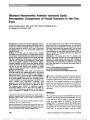
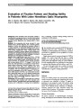
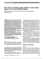
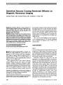
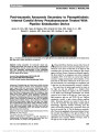
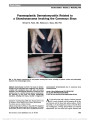
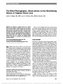
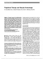
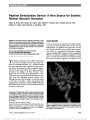
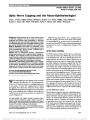
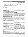
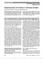
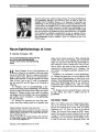
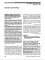
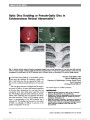
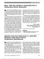
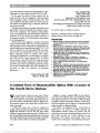
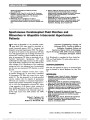
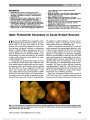
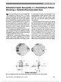
Show Acute Monocular Visual Loss in an Elderly Woman: A Neuro-Ophthalmologic Emergency Michael S. Vaphiades, DO, Jennifer Doyle, MD, Patricia A. Hudgins, MD, Daniel J. Brat, MD Drs. Vaphiades and Doyle: An 84-year-old woman complained of a 2-week history of frontal headache and a 1-day history of sudden visual loss in the left eye. Her medical history included hypertension and hyperlipidemia. She denied jaw claudication, scalp tender-ness, weight loss, fever, and chills. Her examination revealed normal blood pressure and heart rate. Visual acuity was 20/30, right eye, and no light perception, left eye. Color vision was reduced in the right eye. Pupils were isocoric, and the left pupil was amaurotic. The visual field of the right eye was full to confrontation and funduscopy was normal bilaterally. Suspected of having giant cell arteritis, the patient was treated with 80 mg/d of prednisone and 100 mg of doxycycline twice a day. Laboratory studies were obtained that showed a white blood cell count of 15,500/mL (normal, 4000- 10,500/mL), platelets of 445,000/mL (normal, 140,000- 415,000/mL), C-reactive protein of 12.1 mg/dL (normal, 0-4.9 mg/dL), and erythrocyte sedimentation rate of 62 mm/h. A left temporal artery biopsy was performed. Dr. Brat: Numerous cross sections of the temporal artery show slightly thickened smooth muscular walls and focal calcifi-cation but no acute or chronic inflammatory infiltrates (Fig. 1A). An elastic Van Gieson stain reveals an intact inter-nal elastic lamina (Fig. 1B). There is no evidence of arteritis. Drs. Vaphiades and Doyle: In view of the negative temporal artery biopsy, plans were made to perform a contralateral biopsy, and urgent brain magnetic resonance imaging (MRI) was performed. Dr. Hudgins: MRI demonstrates opacification of the left sphenoid sinus (Fig. 2). In addition, the left anterior clinoid process is pneumatized, and the pneumatized area also is opacified. Because of the pneumatization, the sinus essentially sur-rounds the canalicular portion of the left optic nerve. Drs. Vaphiades and Doyle: In view of the MRI findings, the patient was admitted to hospital directly from the MRI facility. To clarify the relationship of the lesion with the surrounding bone and soft tissue, we elected to obtain an emergent computed tomography (CT) of the paranasal sinuses. Dr. Hudgins: The noncontrast axial CT of the brain demonstrates left sphenoid sinus opacification and focal dehiscence of the optic canal (Fig. 3). Drs. Vaphiades and Doyle: The patient was taken to the operating room where she underwent endoscopic sinus surgery that revealed pus surrounding the left optic nerve and destruction of the optic canal. Marsupialization of the left sphenoid sinus was performed, and tissue was removed from around the optic nerve and from the optico-carotid recess and sent for pathological examination. At the conclusion of the opera-tion, gentamycin-soaked sinus foam was placed into the operative bed. Dr. Brat: Histopathologic evaluation of the sphenoid sinus contents shows respiratory mucosa and submucosa with marked acute and chronic inflammation. The chronic inflammatory infiltrate is mostly lymphoplasmacytic and associated with Departments of Ophthalmology (MSV, JD), Neurology (MSV), and Neurosurgery (MSV), University of Alabama at Birmingham, Birmingham, Alabama; and Departments of Radiology (PAH) and Pathology and Laboratory Medicine (DJB), Emory University, Atlanta, Georgia. Supported in part by an unrestricted grant from the Research to Prevent Blindness, Inc., New York, NY. The authors report no conflicts of interest. Address correspondence to Michael S. Vaphiades, DO, Department of Ophthalmology, University of Alabama at Birmingham, Suite 601, 700 South 18th Street, Birmingham, AL 35233; E-mail: vaph@uab.edu 390 Vaphiades et al: J Neuro-Ophthalmol 2013; 33: 390-393 Clinical-Pathological Case Study Section Editor: Neil R. Miller, MD Copyright © North American Neuro-Ophthalmology Society. Unauthorized reproduction of this article is prohibited. prominent myxoid change and edema in the submucosa (Fig. 4A, B). In other regions, the inflammatory process was more acute, with necrobiotic material associated with tissue destruction and bone erosion (Fig. 4C). Drs. Vaphiades and Doyle: Cultures of material removed at the time of surgery grew methicillin-resistant Staphylococcus aureus (MRSA). The patient was treated with intravenous clindamycin and oral sulfamethoxazole/trimethoprim as well as high-dose intra-venous corticosteroids. On postoperative day 2, visual acu-ity in the left eye had improved from no light perception to 20/20. Twenty days later, the patient was off all antibiotics and steroids, and her vision remained stable. Final Diagnosis Acute left optic neuropathy from a sphenoid sinus mucopyocele. Drs. Vaphiades and Doyle: Mucoceles are cyst-like lesions lined with respiratory epithe-lium that contain mucoid secretions that lead to thinning and erosion of the bony walls of the affected paranasal sinus (1,2). When the lesion contains pus, it is termed a mucopyocele. More than half of these lesions are located in the frontal sinuses, with most of the remainder being in the ethmoid sinuses. Sphenoid sinus mucoceles are relatively rare, repre-senting 1% of all paranasal sinus mucoceles (3,4). They usu-ally start unilaterally, but by the time of presentation, the entire sphenoid sinus complex may be opacified and expanded with deossification and thinning of the bony walls. Mucoceles and mucopyoceles most often develop after long-standing sinus ostial obstruction in the setting of sinusitis, allergy, or trauma. On MRI, these lesions show variable intensity depending on protein concentration and viscosity but commonly demonstrate high signal on both T1 and T2 images. There is peripheral enhancement in the FIG. 1. Cross sections of temporal artery biopsy specimen show no evidence of inflammation and the internal elastic lamina is intact (A, hematoxylin and eosin, ·100; B, elastic Van Gieson, ·100). FIG. 2. Contrast-enhanced T1 axial (A) and coronal (B) magnetic resonance imaging with fat suppression demonstrates opacification of the left sphenoid sinus with a pneumatized but opacified anterior clinoid process (arrows). Vaphiades et al: J Neuro-Ophthalmol 2013; 33: 390-393 391 Clinical-Pathological Case Study Copyright © North American Neuro-Ophthalmology Society. Unauthorized reproduction of this article is prohibited. edematous and inflamed mucoperiosteum, but no central enhancement, a useful pattern to help distinguish a muco-cele from sinus neoplasm. CT is helpful in demonstrating the relationship of the lesion with surrounding bone and soft tissue, showing an isodense smooth mass with an enhancing rim, usually associated with bowing and thinning of the bony margins of the affected sinus (5). Cranial neuropathies are a feature in as many as 50% of mucoceles and mucopyoceles (4). Compression of the cav-ernous sinus may cause proptosis and periocular pain (2,3,6-8). The cavernous sinus expands anteriorly at the level of the anterior clinoid process in close proximity to the third nerve (9). The third nerve is most commonly affected (9), with sparing of pupillary function mimicking a vasculopathic process in 46.6% of cases (7). Patients may present with periocular pain and a third nerve palsy with pupillary involvement, suggesting aneurysmal compression (9). In some cases, the third nerve palsy may wax and wane (2). Frontal sinus mucoceles can cause downward displace-ment of the globe and limitation of elevation by erosion into the orbit from above (10,11). Optic neuropathy from sphenoid sinus mucocele, as occurred in our patient, is rare and may be caused by direct compression of the nerve or its blood supply by the expanding mucocele or by spread of inflammation or infection (12). Mild elevation of the optic chiasm usually does not cause visual loss. However, involvement of the optic canal often is associated with an optic neuropathy, where the optic nerve is particularly susceptible to vascular compromise (12-14). The rapid improvement in visual function from no light percep-tion to 20/20 in the left eye in our patient suggests that in our case, visual loss was related to direct compression of the can-alicular portion of the optic nerve by the mucocele. With respect to the causative organism of mucopyocele, MRSA has been isolated in up to 20.7% of patients with both acute and chronic rhinosinusitis (15). Indeed, S. aureus was the most common etiologic agent associated with acute and chronic sinusitis in a review of bacterial causes of sphenoid sinus inflammation (16). However, no specific organism has been found to be associated with mucopyo-celes (17-20). One review of maxillary sinus mucopyoceles found coagulase-negative S. aureus to be the most frequently encountered organism (17), whereas another found alpha-hemolytic Streptococcus to be the most common pathogen (18). A chart review of patients with cystic fibrosis presenting for lung transplantation found asymptomatic frontal sinus FIG. 3. Noncontrast axial computed tomography of the orbits demonstrates left sphenoid sinus opacification and a focal dehiscence in the medial and superior aspect of the left optic canal (arrow). FIG. 4. Histopathology of the sphenoid sinus contents. The sphenoid sinus mucosa and submucosa show marked chronic inflammation with myxoid change and edema (A) and a dense lymphoplasmacytic infiltrate (B). Other regions showed acute inflammation and necrobiotic material along with fragments of eroded bone (C) (hematoxylin and eosin; A, ·40; B, ·400; C, ·40). 392 Vaphiades et al: J Neuro-Ophthalmol 2013; 33: 390-393 Clinical-Pathological Case Study Copyright © North American Neuro-Ophthalmology Society. Unauthorized reproduction of this article is prohibited. mucoceles in 3 patients; endoscopic surgery was performed and intraoperative culture results showed Pseudomonas aeruginosa from all 3 patients (20). This was not surpris-ing as patients with cystic fibrosis frequently are colonized with P. aeruginosa; however, 3 other isolates were found in these cases, 1 of which was MRSA (20). Another study reviewed isolated sphenoid sinus disease in 29 patients with 4 different diagnoses: sinusitis, noninvasive fungus balls, neo-plasms, and mucoceles (19). Mucoceles were the least com-mon source for sphenoid disease with a prevalence of 12%. Although MRSA was isolated from 8 of 11 cultures in this study, the authors did not indicate the specific source of the organism (i.e., mucocele vs sinusitis). A case report of bilat-eral sphenoid sinus disease with visual impairment due to S. aureus has been reported, although the causative organism was not specifically identified as MRSA (21). Treatment of both mucoceles and mucopyoceles consists of relieving the sinus ostial obstruction, marsupialization of the lesion followed by antibiotics. This usually results in rapid regression of ophthalmic and other manifestations (19,22). In our case, recognition of the lesion with MRI and CT as a sphenoid sinus mucocele or mucopyocele lead to urgent surgery and resultant visual recovery. However, delay in treatment may result in permanent blindness (22). REFERENCES 1. Akan H, Cihan B, Celenk C. Sphenoid sinus mucocele causing causing third nerve paralysis: CT and MR findings. Dentomaxillofac Radiol. 2004;33:342-344. 2. Friedmann G, Harrison S. Mucocoele of the sphenoidal sinus as a cause of recurrent oculomotor nerve palsy. J Neurol Neurosurg Psychiatry. 1970;33:172-179. 3. Prepageran N, Subramaniam KN, Krishnan GG, Raman R. Ocular presentation of sphenoid mucocele. Orbit. 2004;23:45-47. 4. Sethi DS, Lau DPC, Chan C. Sphenoid sinus mucocoele presenting with isolated oculomotor nerve palsy. J Laryngol Otol. 1997;111:471-473. 5. Yousem DM, Grossman RI. Neuroradiology: The Requisites, 3rd edition. Philadelphia, PA: Mosby, 2010. 6. Chen HJ, Kao LY, Lui CC. Mucocele of the sphenoid sinus with the apex orbitae syndrome. Surg Neurol. 1986;25:101-104. 7. Johnson LN, Hepler RS, Yee RD, Batzdorf U. Sphenoid sinus mucocele (anterior clinoid variant) mimicking diabetic ophthalmoplegia and retrobulbar neuritis. Am J Ophthalmol. 1986;102:111-115. 8. Roberson GH, Patterson AK, Deeb ME, Maisel RH, Bone RC. Sphenoethmoidal mucocele: radiographic diagnosis. AJR Am J Roentgenol. 1976;127:595-599. 9. Ashwin PT, Mahmood S, Pollock WST. Sphenoid sinus mucocoele mimicking aneurysmal oculomotor nerve palsy. Eye (Lond). 2001;15:108-110. 10. Lin CJ, Kao CH, Kang BH, Wang HW. Frontal sinus mucocele presenting as oculomotor nerve palsy. Otolaryngol Head Neck Surg. 2002;126:588-590. 11. Ehrenpreis SJ, Biedlingmaier JF. Isolated third-nerve palsy associated with frontal sinus mucocele. J Neuroophthalmol. 1995;15:105-108. 12. Levy J, Monos T, Puterman M. Bilateral consecutive blindness due to sphenoid sinus mucocele with unilateral partial recovery. Can J Ophthalmol. 2005;40:506-508. 13. Slavin ML, Liebergall DA. Acute visual loss in the elderly due to retrobulbar optic neuropathy. Surv Ophthalmol. 1996;41:261-267. 14. Yamaguchi K, Ohnuma I, Takahashi S, Fuse T, Aoyagi M, Nagahata M, Hosoya T, Yamaguchi K. Magnetic resonance imaging in acute optic neuropathy by sphenoidal mucocele. Int Ophthalmol. 1997;21:9-11. 15. McCoul ED, Jourdy DN, Schaberg MR, Anand VK. Methicillin-resistant Staphylococcus aureus sinusitis in nonhospitalized patients: a systematic review of prevalence and treatment outcomes. Laryngoscope. 2012;122: 2125-2131. 16. Brook I. Bacteriology of acute and chronic sphenoid sinusitis. Ann Otol Rhinol Laryngol. 2002;111:1002-1004. 17. Busaba NY, Salman SD. Maxillary sinus mucoceles: clinical presentation and long-term results of endoscopic surgical treatment. Laryngoscope. 1999;109:1446-1449. 18. Busaba NY, Siegel N, Salman SD. Bacteriology of nontraumatic maxillary sinus mucoceles versus chronic sinusitis. Laryngoscope. 2000;110:969-971. 19. Friedman A, Batra PS, Fakhri S, Citardi MJ, Lanza DC. Isolated sphenoid sinus disease: etiology and management. Otolaryngol Head Neck Surg. 2005;133:544-550. 20. Solares CA, Citardi MJ. Management of frontal sinus mucoceles with posterior table erosion in the pretransplant cystic fibrosis population. Am J Otolaryngol. 2007;28:110-114. 21. Heylbroeck P, Watelet JB, Delbeke P, Van Cauwenberge P, Bachert C. Vision impairment as a presenting symptom of a sphenoidal mucocele. Rhinology. 2003;41:187-191. 22. Vaphiades MS, Yunker JJ, Roberson GH, Meyer DR, Mills DM. Optic neuritis is nothing to sneeze at. Surv Ophthalmol. 2007;52:106-110. Vaphiades et al: J Neuro-Ophthalmol 2013; 33: 390-393 393 Clinical-Pathological Case Study Copyright © North American Neuro-Ophthalmology Society. Unauthorized reproduction of this article is prohibited. |