| OCR Text |
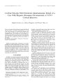
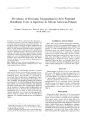
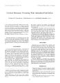
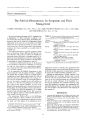
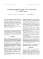
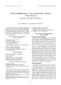
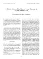
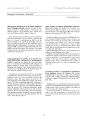
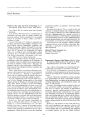
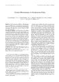
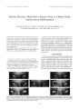
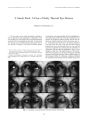
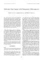
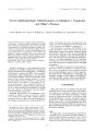
Show Journal of Nairn- Ophthalmology 19( 1): 28- 33, 1999. © 1999 Lippincotl Williams & Wilkins, Inc., Philadelphia A Multiple Sclerosis- Like Illness in a Man Harboring the mtDNA 14484 Mutation M. Tariq Bhatti, M. D., and Nancy J. Newman, M. D. In most cases of Leber's hereditary optic neuropathy ( LHON) the only clinical manifestation is visual loss. A multiple sclerosis- like illness has been infrequently reported in association with LHON. Most patients are women harboring the mtDNA 11778 mutation. We present a young man with clinical and paraclinical evidence of a dcmyelinating process with profound bilateral visual loss who harbored the mtDNA 14484 mutation associated with LHON. Key Words: Leber's optic neuropathy- Multiple sclerosis- Visual loss. Leber's hereditary optic neuropathy ( LHON) is associated with three so- called primary mitochondrial DNA ( mtDNA) mutations at nucleotide positions 11778, 3460, and 14484. The 14484 mutation accounts for 10% to 25% of cases of LHON ( 1). The clinical characteristics of LHON in patients with the 14484 mutation are similar to those of patients with the other primary mutations, except for a better visual prognosis ( 2) and the paucity of associated neurologic symptoms and signs ( 3). A multiple sclerosis ( MS)- like illness has been reported in several patients with the 11778 mutation and, less frequently, in patients with the 3460 mutation ( 4- 13). We describe a 28- year- old man with bilateral, profound, and permanent visual loss and clinical and paraclinical evidence of demyelinating disease, who harbored the 14484 mutation. CASE REPORT An otherwise healthy 28- year- old man with no family history of visual loss or neurologic disease noted left hand, chest, and back numbness in January 1992. He also saw " tiny spots" associated with blurred vision in both Manuscript received March 12, 1998; accepted June 12, 1998. From the Departments of Ophthalmology ( M. T. B., NJ. N), Neurology ( N. J. N), and Neurological Surgery ( N. J. N); Emory University School of Medicine, Atlanta, Georgia, U. S. A. Supported in part by a departmental grant ( ophthalmology) from Research to Prevent Blindness, New York, New York; and by Core Grant P30- EYO 6360 from the National Institutes of Health, Bethesda, Maryland, U. S. A. Address correspondence and reprint requests to Nancy J. Newman, MD, Ncuro- ophtlialmology Unit, Emory Eye Center, I365B Clifton Rd., NE, Atlanta, GA 30322, U. S. A. eyes and had difficulty with depth perception. Neurologic examination revealed decreased sensation of all left fingers except the thumb. An electromyogram of the left upper extremity was normal. Magnetic resonance imaging ( MRI) revealed T2- weighted hyperintense lesions of the left forceps major ( 3 mm) and the upper cervical cord at the odontoid level ( 6 mm) ( Fig. 1). Cerebrospinal fluid ( CSF) had minimally elevated protein ( 47 mg/ dl), 20 lymphocytes per cu mm, and no oligoclonal bands. Lyme antibody and antinuclear antibody titers were negative as were syphilis test results. Normal P100 latencies were recorded on visual evoked responses. In November 1992 he noted painless decreased vision in the left eye, and optic neuritis was diagnosed. Vision did not improve after treatment with 320 mg intravenous methylprednis-olone every 8 hours for 3 days followed by a 1 week tapering dose of 60 mg oral prednisone. Brain MRI showed possible enhancement of the left optic nerve ( Fig. 2A) and a few T2- weighted bright cerebral white-matter lesions ( Fig. 2B). Visual deterioration of the left eye continued , and in March 1993 mtDNA analysis was negative for the point mutations at positions 11778 and 3460 associated with LHON. In May 1996 neurologic examination was remarkable for slightly ataxic gait, left optic nerve pallor, and horizontal nystagmus of the left eye. Magnetic resonance imaging revealed abnormal, T2- weighted hyperintense punctate signals in the medulla and pons and periventricular white- matter lesions ( Fig. 3). In November 1996 visual acuity was 20/ 50 right eye and 20/ 400 left eye with an intermittent nystagmoid movement of the left eye and mild left peripheral facial weakness. Vision continued to worsen progressively, exacerbated by exertion or anger. Visual acuity in March 1997 was 20/ 200 right eye and 20/ 400 left eye. Analysis of CSF showed an elevated protein level of 91 mg/ dl, an immunoglobulin ( Ig) G concentration of 13.4 mg/ dl and no cells. By July 1997, visual acuity was 20/ 400 right eye and was restricted to counting fingers in the left eye. There was temporal pallor of the right optic disc and marked pallor of the left optic disc. Levels of vitamin B12, serum and erythrocyte folate, long chain fatty acids, and angio-tensin- converting enzyme were all normal and results of syphilis serology was negative. Mitochondrial DNA 28 MULTIPLE SCLEROSIS- LIKE ILLNESS 29 FIG. 1. Axial ( A) and sagittal ( B) T2- weighted MRI showing a hyper-intense lesion of the upper cervical spinal cord ( arrows). analysis was positive for the point mutation at position 14484 associated with LHON. DISCUSSION Neurologic abnormalities have been reported in as many as 70% of patients with LHON, but the findings are usually minor, such as sleep- pattern disturbances, mild movement disorders or tremor, peripheral neuropathies, migraine, and cerebellar abnormalities ( 4). The term " Leber's plus" has been used to describe families with members who have LHON and more severe neurologic abnormalities. These pedigrees are probably genetically distinct from those with the primary LHON mutations ( 1). One exception to this genetic distinction appears to be the occasional occurrence of an MS- like illness in patients harboring one of the primary LHON mutations. Before molecular genetic confirmation of LHON, several LHON pedigrees were reported in which patients, usually women, exhibited optic atrophy and an MS- like illness ( 14- 17) ( Table 1). Ferguson et al. ( 14) first suggested an association in a young woman with bilateral visual loss who showed pyramidal signs and had ataxia. Lees et al. ( 15) described two children of a mother affected with LHON who had permanent visual loss fol- FIG. 2. Axial T1- weighted MRI showing possible subtle enhancement of the left optic nerve [ arrow) ( A) and T2- weighted image demonstrating cerebral white matter lesions ( B). J Nettro- Ophtlwlnmt, Vol. 19, No. I, 1999 30 M. T. BHATTI AND N. J. NEWMAN FIG. 3. T2- weighted and proton density MRI showing hyperintense lesions of dorsal- lateral medulla ( A) and ventral- lateral pons ( B) ( arrows). lowed by neurologic disease resembling MS. De Weerdt et al. ( 16) reviewed nine patients in eight pedigrees ( eight women) with an MS- like illness and optic atrophy. Since the availability of molecular genetic confirmation of LHON, several patients have been described with the primary LHON mutations and clinical or paraclinical evidence of MS ( 4- 13,18,19) ( Table 2). Harding et al. ( 5) reported eight women with bilateral visual loss associated with the 11778 mutation and clinical or radiologic evidence of demyelinating disease. An MRI revealed abnormal signal in the periventricular, subcortical white matter or brainstem in all 7 patients imaged, and CSF analysis yielded abnormal findings in three of five patients tested. Subsequently, there have been 15 other well-described cases, 8 women and 7 men, with visual loss associated with the 1 1778 or 3460 mutation and an MS- like illness ( 4,6- 13). In all patients abnormal signals on MRI and the presence of oligoclonal bands or increased synthesis of IgG in the CSF ( in 11 of 13 patients) were highly suggestive of a demyelinating process. Findings in magnetic resonance spectroscopy performed on two patients were compatible with a chronic demyelinating process ( 8). Two further cases of neurologic dysfunction in LHON patients are less suggestive of demyelinating disease ( 18,19). In both cases, there was isolated brainstem involvement with a single lesion on MRI and normal CSF analysis. In one patient, it was associated with the 3460 mutation ( 18) and the other with the 14484 mutation ( 19). Several investigators have examined the frequency of mtDNA mutations in a population of patients with clinically definite MS. Kellar- Wood et al. ( 9) screened 307 random MS patients and did not detect the 11778 or 3460 mutations. However, when they analyzed 20 MS patients with early and severe visual loss, 1 harbored the 11778 mutation and 2 the 3460 mutation. Similarly, Leuzzi et al. ( 10) detected 1 out of 74 MS patients with early and prominent optic nerve involvement who harbored the 11778 mutation. Carrara et al. ( 11) screened 53 MS patients with severe optic nerve involvement and verified one man harboring the 11778 mutation. However, in several other studies the 3460 or 11778 mutations were not detected in the study populations of MS patients, with or without prominent optic neuritis ( 20- 24). In Japan, where most cases of LHON are associated with the TABLE 1. Before molecular genetic confirmation: MS and LHON Author Age/ sex Neurologic symptoms Paraclinical studies Family history 1. Ferguson ct al, 1928 2. Bruyn et al, 1964 3. Leeset al, 1964 4. Lees et al, 1964 5. de Weerrdt et al, 1969 6. de Weerdt et al, 1969 7. de Weerdt et al, 1969 8. de Weerdt et al, 1969 9. de Weerdt et al, 1969 17/ female Female 15/ female Male Female Male Female Female Female Parathesias, sensory loss Ataxia, micturition difficulties, spasticity Gait disturbance, urinary retention, slurred speech Transient left- sided weakness MS MS Spastic paralysis MS MS CSF: mononuclear cell pleocytosis, increased globulin CSF: pleocytosis CSF: increased globulin CSF: normal EEG: normal Air encephalogram: normal Brother- optic atrophy Positive Mother- LHON Mother- LHON Positive Positive Positive Positive Positive CSF, cerebral spinal fluid; MS, multiple sclerosis; EEG, electroencephalogram; LHON, Leber's hereditary optic neuropathy. ,/ Neum- Oplitluilmol, Vol. 19. No. I, 1999 MULTIPLE SCLEROSIS- LIKE ILLNESS 31 TABLE 2. Molecular genetic confirmation: MS and LHON Author Age Sex/ race Neurologic symptoms Visual acuity Imaging Paraclinical studies Family history LHON mutation i. Harding ct al, 1992 2. Harding cl al, 1992 3. Harding ct al, 1992 4. Harding cl al, 1992 5. Harding cl al, 1992 6. Harding cl al, 1992 7. Harding cl al, 1992 8. Harding et al, 1992 9. Flanigan ct al, 1993 10. Flanigan ct al, 1993 11. Flanigan cl al, 1993 12. Flanigan cl al, 1993 13. Paulus et al, 1993 14. Kcllar- Wood cl al, 1994 15. Kcllar- Wood c l a l , 1994 16. Kcllar- Wood el al, 1994 17. Carrara cl al, 1995 18. Vergani ct al, 1995 19. Olscn el al, 1995 20. Nikoskclaincn ct al, 1995 21. Mcire et al, 1995 40 Female/ while Transient parasthesia 00: 1/ 60 Lhermitlc's OS: 6/ 60 46 Female/ while 39 Female/ while 62 Fcmalc/ whitc 56 Female/ white Fcmalc/ whitc 25 Fcmalc/ whitc Seizures, clumsiness, weakness and ataxia Transient leg numbness, weakness, spasticity, ataxia, left INO Oiplopia, vertigo, leg weakness, right INO Transient vertigo, OD: LP OS: HM OD: NLP OS: 1/ 36 OD: CF OS: 6/ 60 OD: 6/ 9 hand clumsiness OS: 6/ 6 Transient numbness, OD: 2/ 60 left arm weakness OS: 1/ 60 Normal OU: CF 59 Female/ while Normal 42 Female/ white Transient left hemiparesis and dizziness 50 Female/ while Hand tremor, paraparesis, clonus and rotary nystagmus 43 Female/ black Qiiadriparcsis 33 Male/ black 23 Male/ white Tinnitus, palatal myoclonus, global ocular paresis 35 Female OO: CF OS: 20/ 400 OD: LP OS: 20/ 20 OD: LP OS: 20/ 400 OU: CF 33 Female Male 30 Male Transient paresthesias Dysarthria, parasthesia, numbness MS Pyramidal signs, spastic paresis, ankle clonus 44 Male/ white Raynaud's phenomenon, clumsiness, limb ataxia 57 Female Spastic paraparesis, transient diplopia 47 Male Normal OD: 6/ 60 OS: 6/ 24 OD: 6/ 12 OS: 6/ 36 OD: 6/ 60 OS: CF OU: 4/ 50 OU: 1/ 60 OD: CF OS: HM OU: < 0. l CT: normal MR1: multiple periventricular, subcortical, brainstem and cerebellar white mailer lesions CT: generalized atrophy MRI: periventricular and cerebral white matter lesions and increased signal in medulla MRI: cerebral atrophy, periventricular white mailer lesions MRI: multiple cerebral, cerebellar and brainstem white mailer lesions MRI: periventricular while matter lesions MRI: multiple periventricular and subcortical while matter lesions MRI: multiple periventricular and subcortical while matter lesions MRI: multiple periventricular and subcortical while matter lesions MRI: periventricular white mailer lesions MRI: increased signal in cerebral hemisphere, pons, and CI- C7 MRI: increased signal in cerebral hemispheres and medulla CT: hypodensc midbrain lesion MRI: hyperintense periaqueductal lesion MRI: MS lesions MRI: MS lesions MRI: MS lesions MRI: demyelinating lesions MRI: multiple periventricular and subcortical lesions CT: widened sulci MRI: periventricular and proximal brainstem lesions MRI: cerebral white matter and putaminal lesions MRI: periventricular white matter lesions VCP: unrceordable CSF: increased IgG PVEP: absent BAEP: normal CSF: oligoclonal bands EEG: abnormal spikes Brolher- LHON Brother- visual loss Air encephalogram: normal Right carotid angio: normal Pattern VFP: normal CSF: normal CSF: oligoclonal bands CSF: oligoclonal bands, increased IgG CSF: oligoclonal bands BAEP: increased latency CSF: oligoclonal bands CSF: normal Pattern VFP: absent Muscle biopsy: negative BAFP: abnormal Vitamin B12: 142 Pattern VFP: absent CSF: oligoclonal bands Pattern VFP: delayed latency CSF: normal CSF: oligoclonal bands Muscle biopsy: myopathic changes CSF: oligoclonal bands BAEP: abnormal CSF: oligoclonal bands CSF: normal BAEP: abnormal Son- ou sequential visual loss 11778 CSF: lymphoe pleocytosis, IgG VEP: delayed CSF: normal ytic increase Brother- visual loss Maternal eousins- visnal loss Son- LHON Brother- blind ( 20' s) Maternal brother- I. HON Maternal male cousin- I. HON Maternal male cousin- I. HON Brother- visual loss 11778 11778 11778 11778 11778 11778 1778 Maternal 1778 ncphew- LHON Maternal male cousin- LHON Mother and 3460 !> randmother- LHON None None 3460 11778 11778 Maternal 11778 hall'- brolhcr- LllON 1778 Grandmothcr- LHON 3460 { continues) J Naim- Ophlhalnml. Vol. 19, No. I. 1999 32 M. T. BHATTI AND N. J. NEWMAN TABLE 2. Continued Author 22. Jansen el al, 19% 23. Jansen el al, 1996 24. Funalot et al, 1996 25. Leu/ y. i e( al. 1997 Age 65 44 30 30 Scx/ r ace Female/ white Malc/ w' Male Male hile Neurological symptoms Paraparesis Spastic ataxia, scissoring gait Vertigo, global gaze palsy, spastic ataxia, ankle clonus Pyramidal, cerebellar, and sensory deficits Visual acuity OD: 2/ 60 OS: 3/ 60 OD: 1/ 36 OS: 0.2 OD: 1/ 10 OS: 4/ 10 OU: LP Imaging CT: normal MRI: increase signal cerebral hemispheres CT: normal MRI: increase signal cerebral hemispheres CT: normal MRI: brainstem and cervical cord lesions MRI: multiple cerebral and cerebellar white matter lesions Paraclinical studies CSF: increased IgG ERG/ EOG: normal VEP: absent Cerebral angio: normal BAEP: delayed CSF: increased IgG EOG/ ERG/ EEG: normal CSF: normal BAEP: prolonged Hep C: positive VEP: abnormal CSF: lymphocytic pleoeytosis, increased IgG Family history Sister, son, male cousins- LHON Molher- LHON Maternal cousins- LHON Maternal uncle- MS LHON mutation 11778 11778 14484 11778 CT, brain computed loniography; BAEP, brainstem auditory evoked potentials; MRI, brain magnetic resonance imaging; ERG, clcctrorctinogram; CSF, cerebral spinal fluid; EOG, electrooculogram; VEP, visual evoked potentials; MS, multiple sclerosis; EEG, electroencephalogram; LHON, Leber's hereditary optic neuropathy; INO, internuclear ophthalmoplegia. 11778 mutation and optic neuritis is particularly severe, the 11778 mutation was not detected in 80 clinically definite MS patients or in 18 patients with severe visual loss ( 25). However, in several of these studies secondary mtDNA mutations were detected in a larger proportion of MS patients than in control subjects, leading to speculation that there may be a pathogenic role of mitochondrial mutations in the susceptibility to MS ( 20,23). In four published reports, MS patients were screened for the 14484 mutation, and it was found in none of them ( 10, 20,22,23). Few investigators have screened populations of LHON patients for the coexistence of a MS- like illness. In a series of 107 patients with LHON, 5 of 11 ( 45%) women harboring the 11778 mutation had an MS- like illness ( 2). Newman et al. ( 26) reported only 1 man with an MS- like illness among 72 patients with the 11778 mutation. Nikoskelainen et al. ( 4) reviewed 46 LHON cases and found 2 patients with a process similar to MS. Clinically and paraclinically, our patient manifested a process consistent with clinically definite MS and harbored the 14484 mutation. To our knowledge this is the first case of an otherwise typical MS- like illness associated with the 14484 mutation. Although the second most common mutation, it is notable that no convincing case of an MS- like illness has been previously identified with the 14484 mutation. Our patient's visual loss was atypical for idiopathic optic neuritis and LHON. As was demonstrated in the Optic Neuritis Treatment Trial, visual loss in demyelinating optic neuritis is worse than 20/ 200 in 36% of patients, and by 6 months 95% of patients regain vision with acuity better than 20/ 40 ( 27). The visual loss with LHON is usually acute and profound with approximately 90% of patients with the 11778 mutation with vision permanently worse than 20/ 200 ( 1). Although as many as 70% of patients with the 14484 mutation may recover vision of 20/ 80 or better ( 2,3), our patient's relentless progressive visual deterioration was unlike the usual improvement of demyelinating optic neuritis or the typical subacute visual loss characteristic of LHON. Several explanations have been proposed to link LHON and multiple sclerosis. First, mtDNA mutations may not only manifest as visual loss but could conceivably cause neurologic disease. However, a relapsing-remitting course or the paraclinical evidence of demyelinating disease would not be expected. Unfortunately, there are no histopathologic studies of LHON associated with an MS- like illness to help elucidate the pathologic process. Second, an immunologically mediated response to mitochondrial genetic products could cause demyeli-nation. Harding et al. ( 5) postulated that activation of a subset of T cells or circulating antibodies may result in an autoimmune process. The resulting inflammatory process could explain the clinical and paraclinical evidence of demyelination. Arguing against this theory is the absence of other immune diseases in patients with mtDNA mutations. Third, it is possible that the diseases are coincidental. This argument has been rejected by Harding and others because of the population frequencies of the two diseases ( 5,7). However, the predisposition to the two diseases may be coincidental, but the presence of the mtDNA mutation may influence the natural history of MS or vice- versa (" coincidence with influence"). For example, the mtDNA mutation may render the optic nerve susceptible to severe and permanent visual loss that might not have otherwise occurred during the demyelinating process of MS. Conversely, the occurrence of inflammation within the optic nerve may strain an already compromised optic nerve and exceed the critical threshold for expression of LHON ( 1). Understanding the complex genetic and epigenetic factors of these two diseases may help elucidate the underlying linkage of these two disorders. REFERENCES 1. Newman NJ. Leber's hereditary optic neuropathy. New genetic considerations. Arch Neurol 1993; 50: 540- 8. J Neuro- Ophlhulmol. Vol. 19, No. 1, 1999 MULTIPLE SCLEROSIS- LIKE ILLNESS 33 2. Riordan- Eva P, Sanders MD, Govan GG, Sweeney MG, Da Costa J, Harding AE. The clinical features of Leber's hereditary optic neuropathy defined by the presence of a pathogenic mitochondrial DNA mutation. Brain 1995; 118: 319- 37. 3. Johns DR, Heher KL, Miller NR, Smith KH. Leber's hereditary optic neuropathy. Clinical manifestations of the 14484 mutation. Arch Ophthalmol 1993; 111: 495- 8. 4. Nikoskelainen EK, Marttila RJ, Huoponcn K, et al. Leber's " plus": neurological abnormalities in patients with Leber's hereditary optic neuropathy. J Neurol Neurosurg Psychiatry 1995; 59: 160- 4. 5. Harding AE, Sweeney MG, Miller DH, et al. Occurrence of a multiple sclerosis- like illness in women who have a Leber's hereditary optic neuropathy mitochondrial DNA mutation. Brain 1992; 115: 979- 89. 6. Olscn NK, Hansen AW, Norby S, Edal AL, Jorgensen JR, Rosenberg T. Leber's hereditary optic neuropathy associated with a disorder indistinguishable from multiple sclerosis in a male harboring the mitochondrial DNA 11778 mutation. Acta Neurol Scand 1995; 135: 176- 80. 7. Flanigan KM, Johns DR. Association of the 11778 mitochondrial DNA mutation and demyelinating disease. Neurology 1993; 43: 2720- 22. 8. Jansen PHP, van der Knaap MS, de Coo IFM. Leber's hereditary optic neuropathy with the 11778 mtDNA mutation and white matter disease resembling multiple sclerosis: clinical, MRI and MRS findings. J Neurol Sci 1996; 135: 176- 80. 9. Kellar- Wood H, Robertson N, Govan GG, Compston AS, Harding AE. Leber's hereditary optic neuropathy mitochondrial DNA mutations in multiple sclerosis. Ann Neurol 1994; 36: 109- 12. 10. Leuzzi V, Carducci Ca, Lanza M, et al. LHON mutations in Italian patients affected by multiple sclerosis. Acta Neurol Scand 1997; 96: 145- 8. 11. Carrara F, Eoli M, La Mantia L, Zeviani M. The contribution of LHON mitochondrial mutations to multiple sclerosis ( abstract). Am J Hum Genet 1995; 57: A336. 12. Vergani L, Martinuzzi A, Carelli V, et al. MtDNA mutations associated with Leber's hereditary optic neuropathy: Studies on cytoplasmic hybrid ( cybrid) cells. Biochem Biophys Res Commun 1995; 210: 880- 8. 13. Meire FM, Van Coster R, Cochaux P, Obermaier- Kusser B, Can-daele C, Martin J- J. Neurological disorders in members of families with Leber's hereditary optic neuropathy ( LHON) caused by different mitochondrial mutations. Ophthalmic Genet 1995; 16: 119- 26. 14. Ferguson FR, Crilchley M. Leber's optic atrophy and its relationship with the heredo- familial ataxias. J Neurol Psychopalhol 1928; 9: 120- 32. 15. Lees F, MacDonald A- M E, Aldren Turner JW. Leber's disease resembling disseminated sclerosis. ./ Neurol Neurosurg Psychiatry 1964; 27: 415- 21. 16. de Weerdt CJ, Went LN. Neurological studies in families with leber's optic atrophy. Ada Neurol Scand 1971; 47: 541- 54. 17. Bruyn GW, Went LN. A sex- linked heredo- dcgcncrativc neurological disorder, associated with Leber's optic atrophy. ./ Neurol Sci 1964; 1: 59- 80. 18. Paulus W, Straube A, Bauer W, Harding AE. Central nervous system involvement in Leber's optic neuropathy. ./ Neurol 1993; 240: 251- 3. 19. Funalot B, Ranoux D, Mas J- L, Garcia C, Bonnefont J- P. Brainstem involvement in Leber's hereditary optic neuropathy: association with the 14484 mitochondrial DNA mutation. ./ Neurol Neurosurg Psychiatry 1996; 61: 533- 4. 20. Kalman B, Lublin FD, Alder H. Mitochondrial DNA mutations in multiple sclerosis. Multiple Sclerosis 1995; 1: 32- 6. 21. Kalman B, Rodriguez- Valdez JL, Bosch U, Lublin FD. Screening for Leber's hereditary optic neuropathy associated mitochondrial DNA mutations in patients with prominent optic neuritis. Multiple Sclerosis 1997; 2: 279- 82. 22. Chalmers RM, Robertson N, Compston AS, Harding AE. Sequence of mitochondrial DNA in patients with multiple sclerosis. Ann Neurol 1996; 40: 239- 43. 23. Mayr- Wohlfart U, Paulus C, Henneberg A, Rodcl G. Mitochondrial DNA mutations in multiple sclerosis patients with severe optic involvement. Acta Neurol Scand 1996; 94: 167- 71. 24. Hancfcid FA, Ernst BP, Wilichowski F, Christen H- J. Leber's hereditary optic neuropathy mitochondrial DNA mutations in childhood multiple sclerosis. Neuropediatr I994; 25: 331. 25. Nishimura M, Obayashi H, Ohta M, Uchiyama T, Hao Q, Saida T. No association of the 11778 mitochondrial DNA mutation and multiple sclerosis in Japan. Neurology 1995; 45: 1333- 4. 26. Newman NJ, Lott MT, Wallace DC. The clinical characteristics of pedigrees of Leber's hereditary optic neuropathy with the I 1778 mutation. Am J Ophthalmol 1991 ; 111: 750- 62. 27. Beck RW, Trobc JD. What have we learned from the Optic Neuritis Treatment Trial. Ophthalmology 1995; 102: 1504- 8. J Neum- Ophtlialmtil, Vol. 19, No. I, 1999 |