| OCR Text |
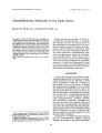
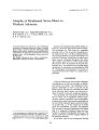
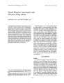
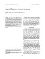
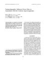
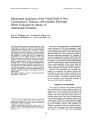
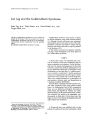
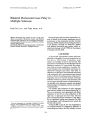
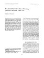
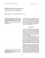
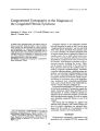
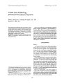
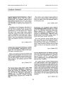
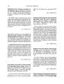
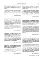
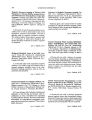
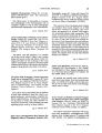
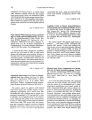
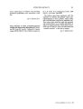
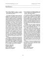
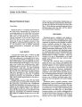
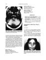
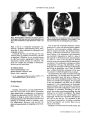
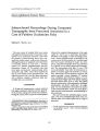
Show Journal of Clinical Neuro- ophthalmology 10( 2): 135- 139, 1990. © 1990 Raven Press, Ltd., New York Computerized Tomography in the Diagnosis of the Congenital Fibrosis Syndrome Saunders L. Hupp, M. D., J. Powell Williams, M. D., and John E. Curran, M. D. A patient with congenital ptosis and inferior rectus fibrosis is described. Marked atrophy of the left inferior rectus muscle was demonstrated by orbital computerized tomography ( 0'). Computerized tomographic findings in this patient and a series of 20 patients with no ocular disease and normal motility indicate that CT is a valuable tool for the evaluation of extraocular muscle size when qualitative comparison is made between corresponding muscles in the same patient. Key Words: Computerized tomography- Congenital fibrosis syndrome- Ptosis. From the Department of Ophthalmology ( S. L. H:) an? the Department of Radiology a. p. W. and J. E. C.), Umverslty of South Alabama College of Medicine, Mobile, Alabama, U. S. A. Address correspondence and reprint requests to Dr. S. L. Hupp, Department of Ophthalmology, University o~ South Alabama College of Medicine, P. O. Box 8448, Mobile, AL 36689, U. s. A. 135 Congenital fibrosis of the extraocular muscles has been described as early as 1840, but the term " congenital fibrosis syndrome" was not used until 1950, when Brown described three sporadic cases ( 1). In this condition, the normal extraocular and! or levator muscle tissue is replaced by fibrosis to a variable extent. One or both inferior rectus muscles are the most frequently affected ( 1-- 6). Generalized fibrosis syndrome is the most severe form of the disorder, with bilateral involvement of all extraocular muscles, including the levator. Often, not all muscles are involved to the same extent. Many alternate names for the condition exist, including congenital ophthalmoplegia, hereditary congenital ophthalmoplegia, ophthalmoplegia imperfecta, and abiotropic ophthalmoplegia externa ( 6). Variants of generalized fibrosis syndrome with incomplete muscular involvement include congenital fibrosis of the inferior rectus with blepharoptosis, strabismus fixus, congenital enophthalmos with ocular muscle fibrosis and blepharoptosis, and the vertical retraction syndrome ( 6). Computerized tomography ( CT) has been a useful tool in the evaluation of extraocular muscle disorders. Numerous reports have described the CT characteristics of various conditions causing enlargement of the extraocular muscles ( 7,8) but few have described muscle atrophy ( 9), and no reports exist describing congenital fibrosis or any of its variants. The clinical and CT findings of a patient with bilateral ptosis, diplopia, and a family history positive for similar complaints are reported here, along with the results of CT examination of the extraocular muscles in 20 normal patients. METHODS Computerized tomography examinations were performed with a Toshiba TCT- 20A scanner with 136 S. L. HUPP ET AL. 2- mm slices obtained in both the axial and positioned coronal planes. Extraocular muscle thickness was evaluated at midorbit in both the axial and coronal views. CASE REPORT A 49- year- old woman reported a 2- month history of binocular vertical diplopia unassociated with trauma or pain. She had been otherwise feeling well with no other weakness or neurologic symptoms. Ocular history is notable for right eye amblyopia and bilateral congenital ptosis more severe 00. She has had two prior ptosis procedures of the right upper eyelid, one as a child and the other as a young adult. Family history is important for congenital ptosis in her father, sister, paternal uncle, and nephew. Amblyopia is also present in her father and nephew. Examination revealed vision correctable to 20/ 40 with central suppression 00 and 20/ 20 with full field as. Color vision was normal au, and no stereopsis was present. One millimeter of enophthalmos was measured as. The upper eyelids were noted to be ptotic with fissures of 8 mm 00 and 9 mm as ( Fig. 1). Levator function was reduced au. The ductions 00 were full, but there was limitation of elevation and depression as with retraction of the globe on extremes of vertical gaze ( Fig. 2). Forced duction testing was abnormal as. The pupil and cranial neurologic examination results were normal. The intraocular examination of both eyes was unremarkable. Computerized tomography scanning of the orbits was performed with 2- mm sections in both the axial and coronal planes. In both projections, marked loss of muscle volume was noted in the left inferior rectus muscle ( 0.5 mm diameter) compared to the right inferior rectus muscle ( 3.0 mm diameter) ( Table 1) ( Fig. 3). Although some asymmetry was present in the horizontal rectus muscles ( Table 1), the size variability was felt to be within normal limits ( Fig. 4). The superior rectusllevator muscle complexes were small ( 3.0- mm diameter) but symmetrical ( Table 1). DISCUSSION Laughlin described certain characteristics of congenital fibrosis, including fibrosis of the extraocular muscles; fibrosis of Tenon's capsule; adhesions between muscles, Tenon's capsule, and globe; inelasticity and fragility of the conjunctiva; absence of elevation or depression of the eyes; little or no horizontal movement; eyes fixed at 20- 30° below FIG. 1. Full frontal view of the patient demonstrating bilateral ptosis. the horizontal; blepharoptosis; chin elevation; and presence since birth ( 2). Poor visual acuity and hyperopic astigmatism may be found in over half the affected individuals ( 10). Additional features described by Letson include autosomal dominant inheritance, exotropia or esotropia, and amblyopia ( 11). Congenital fibrosis of the inferior rectus muscles with blepharoptosis may be unilateral or bilaterally asymmetric. It occurs in either sporadic or autosomal dominant forms with equal sex incidence and often involves the levator as well as the inferior rectus muscles. Strabismus fixus results from involvement of the inferior recti. Extreme tightness of these muscles prevents passive or active vertical movement, and the eyes are often fixed in down gaze, with the patient assuming a chin- up posture ( 3,6). Association with other congenital ocular abnormalities including coloboma, mixed hyperopic astigmatism, amblyopia, and strabismus has been reported in patients with the autosomal dominant form. Nonocular abnormalities such as ventricular septal defect, talipes equinovarus, and facial palsy have been seen in the sporadic cases ( 4,5). TOMOGRAPHY IN CONGENITAL FIBROSIS SYNDROME 137 Histological changes seen in affected musculature indicate that the neural elements are normal. The pathological alterations suggest myopathy rather than neuropathy. Both light and electron microscopy show replacement of skeletal muscle by fibrous tissue and collagen. When ptosis is present, findings include reduction or lack of striations of the levator muscle together with absence of the Z bands and vacuolation of muscle cells, while the smooth muscle cells of Muller appear intact ( 3). Sometimes areas of normal muscle tissue are found enveloped within a thickened and fibrotic capsule although no inflammatory changes are seen ( 5). Anomalous scleral insertion of the involved muscles may be present ( 5). This has been suggested to occur from a defect of maturation at or before the 7th week of gestation. Extraocular muscle anatomy displayed with high- resolution axial and coronal CT scanning of the orbit has been well described ( 12,13). With appropriate techniques, all six of the extraocular muscles can be visualized. The superior rectus and the levator palpebrae muscles are shown on axial sections parallel to or with a positive angulation to the orbitomeatal baseline, an imaginary line connecting the external auditory canal and the ipsilat- FIG. 2. Primary position ( A), upgaze ( B), and downgaze ( C) demonstrating marked limitation of elevation and depression as. TABLE 1. Extraocular ocular muscle diameters ( mm), midorbit LMRB RMR LSR RSR L1R RIR LLR RLR Patient Ob 2.5 3.0 3.0 3.0 0.5 3.0 0.5 1.5 1 2.0 2.0 5.0 5.0 4.0 4.0 1.5 1.5 2 3.0 3.0 8.0 8.0 6.0 6.0 2.0 2.0 3 3.0 3.0 8.0 8.0 8.0 8.0 2.0 2.0 4 3.0 3.0 9.0 9.0 7.0 7.0 1.5 1.5 5 3.0 3.0 7.0 7.0 5.0 5.0 3.0 3.0 6 4.0 4.0 6.0 6.0 8.0 8.0 3.0 3.0 7 4.0 4.0 8.0 8.0 8.0 8.0 4.0 4.0 8 4.0 4.0 9.0 9.0 8.0 8.0 2.0 2.0 9 2.0 2.0 6.0 6.0 5.0 5.0 1.5 1.5 10 4.0 4.0 8.0 8.0 6.0 6.0 2.0 2.0 11 4.0 4.0 10.0 10.0 8.0 8.0 2.0 2.0 12 3.0 3.0 6.0 6.0 6.0 6.0 1.5 1.5 13 3.0 3.0 9.0 9.0 6.0 6.0 3.0 3.0 14 4.0 4.0 7.0 7.0 4.0 4.0 1.5 1.5 15 5.0 5.0 7.0 7.0 5.0 5.0 2.0 2.0 16 3.0 4.0 8.0 8.0 11.0 11.0 4.0 4.0 17 4.0 5.0 4.0 4.0 10.0 10.0 3.0 3.0 18 4.0 3.0 10.0 10.0 10.0 10.0 3.0 3.0 19 4.0 5.0 9.0 7.5 10.0 11.0 6.0 7.0 20 5.0 5.0 8.0 8.0 9.0 8.0 5.0 5.0 Average 3.6 7.6 7.2 2.7 B LMR, left medial rectus muscle; lSR, left superior rectus; L1R, left inferior rectus; RMR, right medial rectus; RSR, right superior rectus; RIR, right inferior rectus; llR, left lateral rectus; and RLR, right lateral rectus. b Patient with congenital fibrosis. J Clin Nroro- ophthalmoI. Vol. 10. No. 2. 1990 138 S. L. HUPP ET AL. a FIG. 3. Coronal ( A) and axial ( B) computerized tomography section of the orbit demonstrating marked asymmetry of the inferior recti. The left inferior rectus muscle is atrophic ( solid arrows) compared to the right ( open arrows). eral lateral canthus. The inferior rectus muscle is well delineated by fat and runs almost parallel to an axial plane 20° below the orbitomeatal baseline. Oblique computer reformations are required to show the oblique muscles in their long axis. In general, muscle size is best evaluated if the plane of section or reformation is perpendicular to its course at the point where the belly of the muscle is thickest ( 8). The CT findings in a number of extraocular muscle disorders have been described, including thyroid ophthalmology, infectious or inflammatory myositis, metastatic or intrinsic tumor, vascular abnormalities, and traumatic hematoma ( 7,8). Recently, the CT appearance of extraocular muscle atrophy has been described in two cases, one unilateral atrophy in a cavernous sinus lesion, the other a bilateral case of probable Kearns- Sayre syndrome. The atrophic muscles were clearly identifiable on both axial and coronal images ( 9). FIG. 4. Axial computerized tomography section through the midorbit demonstrating normal lateral anr1 rT'wrl,, 1 _ cot, ,,;!:; I< H31Iv The assessment of extraocular muscle size has been studied largely with reference to the enlarged musculature seen in Graves' ophthalmopathy. While CT and orbital echography have been employed to obtain measurements of enlarged muscles in this context, no reliable information as to the lower level of normal muscle size is available ( 14- 18). Coronal CT sections cut only one muscle, the medial rectus, perpendicularly ( 13). A true cross- sectional image of the other muscles may only be obtained from computer reformation images at 90° to the plane of the individual muscle ( 12,13). Orbital echography has been demonstrated to be a sensitive tool for the measurement of extraocular muscle size with accuracy approaching within 0.5 mm of muscle diameter ( 18). Echography has an advantage compared to CT in that it is a more accurate and quicker measure of extraocular muscle size and does not expose the patient to radiation. Unfortunately, this technique is limited in availability due to the need for sensitive calibrated instrumentation and experienced technicians ( 18). The CT imaging of extraocular muscles in 20 patients aged 12- 91 years with no eye disease and normal motility was performed at our institution. Muscles were studied in coronal and axial planes with 2- mm slices. The muscles showed a wide variation in cross- sectional diameter between patients: the medial rectus was 2.0- 5.0 mm; lateral rectus, 1.5- 7.0 mm; inferior rectus, 4.0- 11.0 mm; and superior rectus, 4.0- 10.0 mm ( Table 1). Importantly, there was no significant variability between corresponding muscles from one eye to the other in the same patient. The superior rectus ( average diameter, 7.6 mm) is usually the widest muscle, with the inferior rectus ( average diameter, 7.2 mm) second in size. The lateral rectus ( average diame- J Lilli, , r ul" " fliililu. IIlIIi/, ", 11 TOMOGRAPHY IN CONGENITAL FIBROSIS SYNDROME 139 ter, 2.7 mm) is most often the smallest of the four muscles measured. There was no correlation between muscle size and age. SUMMARY Computerized tomography imaging is an important tool to assess both congenital and acquired extraocular muscle disease, either enlargement or atrophy. Determination of abnormal muscle size is best made by qualitative comparison with similar structures in the same patient. In this case, the demonstration of extraocular muscle atrophy was particularly helpful in confirming the diagnosis when combined with the clinical findings and family history. REFERENCES 1. Brown HW. Congenital structural muscle anomalies. In: Allen TH, ( ed.) Strabismus ophthalmic symposium. St. Louis: CV Mosby, 1950: 229. 2. Laughlin RC. Congenital fibrosis of the extraocular muscles. Am J OphthalmoI1956; 41: 432. 3. Harley RD, Rodrigues MM, Crawford JS. Congenital fibrosis of the extraocular muscles. Trans Am Ophthalmol Soc 1978; 76: 197- 226. 4. Harley RD, Rodrigues MM, Crawford JS. Congenital fibrosis of the extraocular muscles. J Pediatr Ophthalmol Strabismus 1978; 15: 34fr- 58. 5. Apt L, Axelrod RN. Generalized fibrosis of the extraocular muscles. Am JOphthalmol 1978; 85: 822- 9. 6. Hiatt RL, Halle AA. General fibrosis syndrome. Ann Ophthaimol 1983; 15: 1103- 9. 7. Haik BG, Saint- Louis LA, Smith ME. Computed tomography of extraocular muscle abnormalities. Int Ophthalmol Clin 1986; 26: 123- 50. 8. Miller MT, Mafee MF. Computed tomography scanning in the evaluation of ocular motility disorders. Radiol Clin North Am 1987; 25: 733- 52. 9. Hansman ML, Peyster RG, Heiman- Patterson T, Greenfield VS. CT demonstration of extraocular muscle atrophy. J Comput Assist Tomogr 1988; 12: 49- 5. 10. Crawford JS. Congenital fibrosis syndrome. Can JOphthalmol 1970; 5: 331-- 6. 11. Letson RD. Surgical management of the ocular congenital fibrosis syndrome. Am Orthop J1980; 30: 97- 101. 12. Unsold R. Computed tomographic anatomy of the orbit. Int Ophthalmol Clin 1982; 22: 45- 80. 13. Unsold R, Newton TH, DeGroot J. CT evaluation of extraocular muscles: anatomic- CT correlations. Graefes Arch Clin Exp OphthalmoI1980; 214: 155- 80. 14. Trokel SL, Hilal SK. Submillimeter resolution CT scanning of orbital diseases. Ophthalmology 1980; 87: 412- 7. 15. Feldon SE, Weiner JM. Clinical significance of extraocular muscle volumes in Graves' ophthalmopathy. Arch Ophthalmol 1982; 100: 1266- 9. 16. Holt JE, O'Conner PS, Douglas JP, Byrne B. Extraocular muscle size comparison using standardized A- scan echography and computerized tomography scan measurements. Ophthalmology 1985; 92: 1351- 5. 17. UhIenbrook D. Computed tomography in Graves' ophthalmopathy-- evaluation regarding the muscle size and density units. Neurosurg Rev 1988; 11: 45- 51. 18. Byrne SF, Glaser JS. Orbital tissue differentiation with standardized echography. Ophthalmology 1983; 90: 1071- 90. J Clin Neuro- ophthalmol, Vol. 10, No. 2, 1990 |