A collection of videos relating to the diagnosis and treatment of eye movement disorders. This collection includes many demonstrations of examination techniques.
Dan Gold, D.O., Associate Professor of Neurology, Ophthalmology, Neurosurgery, Otolaryngology - Head & Neck Surgery, Emergency Medicine, and Medicine, The Johns Hopkins School of Medicine.
A collection of videos relating to the diagnosis and treatment of eye movement disorders.
NOVEL: https://novel.utah.edu/
TO
Filters: Collection: "ehsl_novel_gold"
| Title | Description | Type | ||
|---|---|---|---|---|
| 126 |
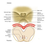 |
Figure 51: Lateral Medullary Lesion Causing Saccadic Dysmetria (Supplement) | Image | |
| 127 |
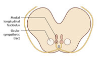 |
Figure 51: Lateral Medullary Lesion Causing Saccadic Dysmetria (Supplement) | Image | |
| 128 |
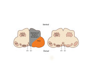 |
Figure 53: Vascular Distribution and Anatomy Relevant to the Lateral Medullary (Wallenberg) Syndrome | This axial section of the medulla highlights those structures that, when damaged, are responsible for the vestibular and ocular motor features of the Wallenberg syndrome. The nucleus prepositus hypoglossi (NPH) and medial vestibular nucleus (MVN) complex is important for horizontal gaze-holding (neu... | Image |
| 129 |
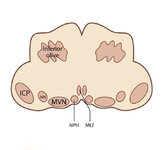 |
Figure 53: Vascular Distribution and Anatomy Relevant to the Lateral Medullary (Wallenberg) Syndrome (Supplement) | Image | |
| 130 |
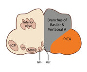 |
Figure 53: Vascular Distribution and Anatomy Relevant to the Lateral Medullary (Wallenberg) Syndrome (Supplement) | Image | |
| 131 |
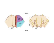 |
Figure 61: Vascular Distribution and Anatomy (Including 6th, 7th, 8th Nerves, MLF) of the Pons | In this axial section of the pons, the proximity of the 7th (VII) and 8th (VIII) fascicles can be appreciated, and a lateral inferior pontine syndrome (anterior inferior cerebellar artery, AICA territory), which could involve both of these fascicles, could cause acute prolonged vertigo accompanied b... | Image |
| 132 |
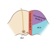 |
Figure 61: Vascular Distribution and Anatomy (Including 6th, 7th, 8th Nerves, MLF) of the Pons (Supplement) | Image | |
| 133 |
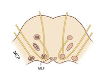 |
Figure 61: Vascular Distribution and Anatomy (Including 6th, 7th, 8th Nerves, MLF) of the Pons (Supplement) | Image | |
| 134 |
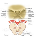 |
Figure 64: The Course of the 3rd (III) Nerve | The 3rd nucleus lies at the ventral border of the periaqueductal gray matter, at the level of the superior colliculus. In between the two nuclei is the midline central caudal nucleus (CCN), which innervates bilateral levator palpebrae muscles (explaining how a unilateral nuclear 3rd can cause bilate... | Image |
| 135 |
 |
Figure 64: The Course of the 3rd (III) Nerve (Supplement) | Image | |
| 136 |
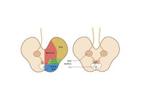 |
Figure 65: Vascular Distribution and Anatomy (Including 3rd Nerve) of the Rostral Midbrain | In this axial section of the midbrain at the level of the superior colliculus, the paired 3rd nuclei are located ventral to the periaqueductal grey, and the midline central caudal nucleus (CCN) is located in between. The fascicles that exit the IIIrd nuclei carry the fibers destined to innervate the... | Image |
| 137 |
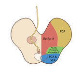 |
Figure 65: Vascular Distribution and Anatomy (Including 3rd Nerve) of the Rostral Midbrain (Supplement) | Image | |
| 138 |
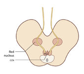 |
Figure 65: Vascular Distribution and Anatomy (Including 3rd Nerve) of the Rostral Midbrain (Supplement) | Image | |
| 139 |
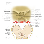 |
Figure 68: The Course of the 4th (IV) Nerve | The 4th nucleus lies at the ventral border of the periaqueductal gray matter, at the level of the inferior colliculus. The fascicles exit the nucleus dorsally and decussate at the anterior medullary velum (anterior floor of the fourth ventricle), and then exit the brainstem dorsally. The peripheral ... | Image |
| 140 |
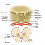 |
Figure 68: The Course of the 4th (IV) Nerve (Supplement) | Image | |
| 141 |
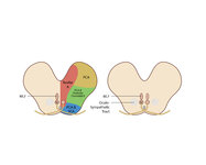 |
Figure 69: Vascular Distribution and Anatomy (Including 4th Nerve) of the Caudal Midbrain | In this axial section of the midbrain at the level of the inferior colliculus, the 4th nuclei are located ventral to the periaqueductal grey, dorsal to the medial longitudinal fasciculus (MLF) and medial to the oculosympathetic tract. Fascicles exit the nucleus dorsally and decussate at the anterior... | Image |
| 142 |
 |
Figure 69: Vascular Distribution and Anatomy (Including 4th Nerve) of the Caudal Midbrain (Supplement) | Image | |
| 143 |
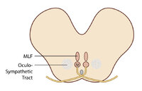 |
Figure 69: Vascular Distribution and Anatomy (Including 4th Nerve) of the Caudal Midbrain (Supplement) | Image | |
| 144 |
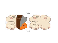 |
Figure 80: Vascular Distribution and Anatomy Relevant to the Medial Medullary Syndrome | This axial section of the medulla highlights those structures that, when damaged, are often responsible for spontaneous upbeat nystagmus (UBN). The nucleus of Roller and nucleus intercalatus normally have an inhibitory influence over the cerebellar flocculus, and when there is a lesion of Roller/int... | Image |
| 145 |
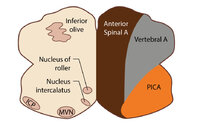 |
Figure 80: Vascular Distribution and Anatomy Relevant to the Medial Medullary Syndrome (Supplement) | Image | |
| 146 |
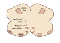 |
Figure 80: Vascular Distribution and Anatomy Relevant to the Medial Medullary Syndrome (Supplement) | Image | |
| 147 |
 |
Five Common Ocular Motor Signs in Cerebellar Disorders - Saccadic Hypermetria, Saccadic Pursuit & VOR Suppression, Gaze-evoked & Rebound Nystagmus | (1) Saccadic hypermetria - an overshoot of the visual target (2) Saccadic smooth pursuit - due to impaired pursuit and low gain, saccades are needed to keep up with the visual target. This gives it a ‘choppy' appearance. (3) Saccadic vestibulo-ocular reflex (VOR) suppression - another... | Image/MovingImage |
| 148 |
 |
Fixation and Gaze Holding | Fixation and gaze-holding: assess for nystagmus or saccadic intrusions by observing the eyes in primary position. Then instruct the patient to look in each position of gaze, and to hold that position to assess for gaze-evoked nystagmus. In doing so, motility can also be evaluated with both eyes view... | Image/MovingImage |
| 149 |
 |
The Gans Maneuver for Right Posterior Canal Benign Paroxysmal Positional Vertigo | This maneuver is recommended for individuals with cervical restrictions or precautions, as the maneuver avoids cervical hyperextension and may reduce cervical pain associated with repositioning maneuvers. The Epley maneuver has higher subjective and objective success rates compared to the Gans maneu... | Text |
| 150 |
 |
The Gans Maneuver for Right Posterior Canal Benign Paroxysmal Positional Vertigo (Video) | This maneuver is recommended for individuals with cervical restrictions or precautions, as the maneuver avoids cervical hyperextension and may reduce cervical pain associated with repositioning maneuvers. The Epley maneuver has higher subjective and objective success rates compared to the Gans maneu... | Image/MovingImage |