Collection of materials relating to neuro-ophthalmology as part of the Neuro-Ophthalmology Virtual Education Library.
NOVEL: https://novel.utah.edu/
TO
- NOVEL966
Filters: Collection: ehsl_novel_novel
| Title | Creator | Description | Subject | ||
|---|---|---|---|---|---|
| 301 |
 |
Introduction to Neuroimaging in Neuro-Ophthalmology | Devin D. Mackay, MD | Introduction to the subject of neuroimaging in the field of neuro-ophthalmology. | Imaging |
| 302 |
 |
The Ocular Examination of the Comatose Patient | John Pula, MD | Description of conducting an ocular examination of a comatose patient. | Examination; Coma |
| 303 |
 |
Lumbar Puncture | Jamison Hofer, DO; Sachin Kedar, MD | Explanation of lumbar puncture procedure. | Lumbar Puncture |
| 304 |
 |
The Mental Status Examination | James R. Bateman, MD, MPH; Victoria S. Pelak, MD | Introduction to the mental status examination. See accompanying videos: Executive function: https://collections.lib.utah.edu/ark:/87278/s6bw1rp1, Limb-Kinetic apraxia: https://collections.lib.utah.edu/ark:/87278/s63c084b, Ideomotor apraxia: https://collections.lib.utah.edu/ark:/87278/s67410xj | Mental Status |
| 305 |
 |
The Clinical Examination of Higher Order Visual Function: Syndrome-Based Approach | Victoria S. Pelak, MD; James R. Bateman, MD, MPH; Brianne Bettcher, PhD | Explanation of higher order visual function examination. See accompanying video, Double simultaneous visual field stimulation: https://collections.lib.utah.edu/ark:/87278/s6gn2h9d | Visual Function |
| 306 |
 |
Ocular Fundus Photography | Devin Mackay, MD | Overview of the various ocular fundus cameras and how they are used, including: Digital mydriatic tabletop fundus camera, Digital nonmydriatic table top fundus camera, Digital handheld nonmydriatic fundus camera and the Smartphone handheld fundus camera. | Digital Fundus Photography; Imaging |
| 307 |
 |
Neuro-Ophthalmic Manifestations of Sellar and Parasellar Masses | Rudrani Banik, MD | Neuroanatomy of the Chiasm. | Parasellar Masses |
| 308 |
 |
Introduction to the Evaluation of Visual Function | Sean Gratton, MD | An introduction to evaluating a patient's visual function. | Visual Function |
| 309 |
 |
Distance Visual Acuity Testing | Sean Gratton, MD | Demonstration of measuring distance visual accuity. | Visual Acuity Testing |
| 310 |
 |
Red Color Desaturation | Sean Gratton, MD | Exploring red color desaturation. | Red Color Desaturation |
| 311 |
 |
Refraction | Sean Gratton, MD | An introduction to refraction. | Refraction |
| 312 |
 |
Ectropion and Entropion | Julie Falardeau, MD; Eric A. Steele, MD | This is a brief PowerPoint presentation describing 2 common disorders of eyelid position: ectropion and entropion. We provide the classification of these 2 disorders as well as clinical photographs | Ectropion; Entropion; Eyelid |
| 313 |
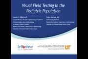 |
Visual Field Testing in the Pediatric Population | Lauren C. Ditta, MD | Visual field testing can be performed in the pediatric population with ease, however the proper technique for testing must be utilized and is tailored to each child's unique clinical situation and age. Confrontation visual field testing is a quick and easy; test that can yield a rough assessment of ... | Visual Field Testing; Pediatric; Confrontation Visual Field; Automated; Perimetry |
| 314 |
 |
Lagophthalmos | Julie Falardeau, MD; John D. Ng, MD | This is a brief PowerPoint presentation for the NANOS Examination Curriculum describing how to evaluate lagophthalmos, discussing the main causes of lagophthalmos and demonstrating few clinical examples. | Lagophthalmos; Eyelid |
| 315 |
 |
Normal Light Reflex and Relative Afferent Pupillary Defect (RAPD) | Marshall Huang, 4th Year Medical Student | A Relative Afferent Pupillary Defect is an examination finding in patients who have an asymmetric pupillary reaction to light when it is shined back and forth between the two eyes. It is most commonly a sign of asymmetric optic nerve disease or damage but can also present in widespread asymmetric r... | Light Reflex; RAPD |
| 316 |
 |
Tonometry | Ore-ofe Adesina, MD | Presentation covering the measurement of intraocular pressure, Tonometry. Covers indentation, applantation and electronic tonometry. | Tonometry; Intraocular Pressure |
| 317 |
 |
Basic Eyelid Measurements | John D. Ng, MD, MS, FACS | Demonstration of basic eyelid measurements. | Eyelid |
| 318 |
 |
Ice Test in Myasthenic Ptosis | Julie Falardeau, MD | Demonstration of ice test. | Ice Test; Myasthenic Ptosis |
| 319 |
 |
Examination of Lymph Nodes | John D. Ng, MD, MS, FACS | Demonstration of lymph node examination. | Exam; Lymph Nodes |
| 320 |
 |
Diffusion Tensor Imaging (DTI) | Devin D. Mackay, MD | Explanation of using diffusion tensor imaging (DTI) in examinations. | Diffusion Tensor Imaging (DTI) |
| 321 |
 |
NExT Introduction | Sachin Kedar, MD, Editor-in-Chief | Transcript of video introduction to the NExT curriculum collection. | NANOS Examination Techniques |
| 322 |
 |
Magnetic Resonance Imaging (MRI) | Devin D. Mackay, MD | Explanation of using magnetic resonance imaging (MRI) in examinations. | Magnetic Resonance Imaging (MRI) |
| 323 |
 |
OJS Reviewer Tutorial | A. Roylance | Tutorial for testing new OJS system for reviews of NOVEL submissions. | OJS Review System |
| 324 |
 |
OJS Author Tutorial | A. Roylance | Tutorial for testing new OJS system for reviews of NOVEL submissions. | OJS Review System |
| 325 |
 |
Video Introduction to the NExT Curriculum Collection | Sachin Kedar, MD, Editor-in-Chief | Introduction to the NANOS NExT ebook. Explains the purpose of the collection. Covers the sections of the outline, and describes the learning levels and objectives. | NANOS Examination Techniques |