Collection of materials relating to neuro-ophthalmology as part of the Neuro-Ophthalmology Virtual Education Library.
NOVEL: https://novel.utah.edu/
TO
- NOVEL981
Filters: Collection: ehsl_novel_novel
| Title | Creator | Description | Subject | ||
|---|---|---|---|---|---|
| 1 |
 |
Uvea | Altuğ Ay; Florian Henri Guillot; Amanda Dean | Uvea topic questions for the NANOS IC | Choroid; Ciliary Dody; Uvea |
| 2 |
 |
Epilepsy Review | Shreya Katwala; Sean Gratton | This submission is a narrated powerpoint review of Epilepsy. It reviews the definition and diagnostic criteria of epilepsy. Common etiologies and types of epilepsy are reviewed. Basic epilepsy managment is also reviewed. | Epilepsy; Focal Epilepsy; Generalized Epilepsy; Provoked Seizures; Seizures |
| 3 |
 |
Wernicke-Korsakoff Syndrome: Vitamin B1 Deficiency | Trinity Dobbs; Sean Gratton | This is a narrated powerpoint presentation about the neurological and neuro-ophthalmic presentations of thiamine deficiency. The clinical manifestations are reviewed. Basic biochemistry is briefly discussed. Management and diagnosis are explored. | B1; Korsakoff; Nystagmus; Thiamine; Wernicke |
| 4 |
 |
Screening Battery for Higher Order Visual Dysfunction Due to Neurodegenerative Disease | Victoria Pelak | Awareness that neurodegenerative disease (NDD) can cause visual brain dysfunction is critical for early detection and proper diagnosis. However, awareness must be combined with knowledge about visual brain pathways and clinical assessment tools that can be used to identify cerebral visual dysfunctio... | Alzheimer's Disease; Dementia with Lewy Bodies; Eye Clinic Rapid Screening Battery For Visual Cortical Dysfunction; Posterior Cortical Atrophy; Visual Cortical Dysfunction |
| 5 |
 |
Traumatic Horner Syndrome | John Robert Mark; Yin Allison Liu | This presentation is a discussion about the presentation of traumatic Horner syndrome (AKA "Oculosympathoparesis") and the emergent next steps that should be taken. The discussion highlights the varying presentations and gives an example of a patient who suffered an injury to the neck resulting in a... | Horner Syndrome; Horners; ICA Dissection; Internal Carotid Artery Dissection; Miosis, Oculosympathetic Paresis; Oculosympathoparesis; Preganglionic; Ptosis; Pupillary Pathology; Second-order Injury; Traumatic Horner Syndrome |
| 6 |
 |
Oculopharyngeal Muscular Dystrophy (OPMD) | Eduardo Gonzalez; Sean Gratton | This is a brief review of Oculopharyngeal Muscular Dystrophy. It includes a review of the background, pathophysiology, epidemiology, diagnosis, and management of the disorder. The key clinical features including ptosis and dysphagia are reviewed in detail. | Chronic Progressive External Ophthalmoplegia; Dysphagia; Muscular Dystrophy; Myasthenia Gravis; Oculopharyngeal Muscular Dystrophy; Ptosis |
| 7 |
 |
A Suspected Case of Wildervanck Syndrome | Hari R. Anandarajah; Tejaswini K. Deshmukh; Ryan D. Walsh | A 5-year-old male with congenital right-sided hearing loss presented to the neuro-ophthalmology clinic for strabismus evaluation. He had longstanding bilateral abduction deficits with associated esotropia, for which he underwent bilateral medial rectus recessions at age three. He had persistent limi... | Cervico-oculo-acoustic Syndrome; Congenital Deafness; Duane Syndrome; Klippel-Feil Cervical Anomaly; Wildervanck Syndrome |
| 8 |
 |
Chronic Migraine | Olivia Liu; Sean Gratton | This narrated powerpoint is a review of Chronic Migraine. This includes a review of the pathophysiology of migraine and what contributes to the chronicity of migraine. Treatments of chronic migraine are also reviewed. | Central Sensitization; Chronic Migraine; Migraine |
| 9 |
 |
Aqueous and Vitreous Humor | Ryung San Lee; Amanda Dean Henderson | Multiple choice test set administered to test comprehension of "Aqueous and vitreous humor" sub-section of NANOS Illustrated Curriculum for Neuro-Ophthalmology. The topics in this submission cover questions related to anatomical and physiologic concepts in aqueous and vitreous humor. More specifical... | Anatomy; Aqueous Humor; Vitreous Humor |
| 10 |
 |
Myelin Oligodendrocyte Glycoprotein Antibody Associated Disease (MOGAD) Revisited | Zahir Sheikh; Michael Gilhooley | The purpose of this presentation is to review the varied clinical manifestations of MOGAD for the neurology and neuro-ophthalmology providers. | Demyelinating; MOG; MOGAD; Myelin |
| 11 |
 |
Ocular Surface, Cornea, Lens | Ryung San Lee; Amanda Dean Henderson | Multiple choice questions set from the NANOS Illustrated Curriculum for Neuro-Ophthalmology. This question set is meant to accompany the video lecture on the anatomy of the ocular surface, cornea, and lens. The ocular surface is a complex topic and pertinent to the comprehensive ophthalmic care prov... | Cornea; Ocular Surface; Uvea |
| 12 |
 |
Trigeminal autonomic cephalalgias (TACs): Paroxysmal hemicrania (PH) and hemicrania continua (HC) | Jihad Yaqoob Ali Al Kharbooshi; Tommy Lik Hang Chan; J. Alexander Fraser | In this narrated slideshow, we describe the pathophysiology, epidemiology, diagnostic criteria, subtypes, workup, and management of hemicrania continua (HC) and paroxysmal hemicrania (PH). | Hemicrania Continua; Paroxysmal Hemicrania; TAC, Trigeminal Autonomic Cephalalgia |
| 13 |
 |
Other Trigeminal Autonomic Cephalalgias (TACs) | Jihad Yaqoob Ali Al Kharbooshi; Tommy Lik Hang Chan; J. Alexander Fraser | In this narrated slideshow, we review the pathophysiology, epidemiology, differential diagnosis, diagnostic criteria, subtypes, and management of the other trigeminal autonomic cephalalgias (cluster headache and the SUNHA syndromes). It is a companion to our earlier slideshow, "Trigeminal autonomic ... | Indomethacin; SUNA; SUNCT; SUNHA; TAC; Trigeminal Autonomic Cephalalgia |
| 14 |
 |
Genetic Causes of Isolated Optic Neuropathy | Kathleen Ho; Devin Mackay | Overview of isolated genetic optic neuropathies and their neuro-ophthalmic manifestations with an illustrative case example and discussion of clinical presentation, relevant biochemistry, and testing. | Genetics; Hereditary; MFN2; NDUFV1; OPA1; OPA3; Optic Atrophy; Optic Neuropathy; POLG; SPG7; WFS1 |
| 15 |
 |
Retinal Vasculitis: Classification, Diagnosis, and Treatment | Jonah Edward Yousif; Alejandra M. Maiz; Thérèse Maria Sassalos; Sangeeta Khanna | This manuscript discusses retinal vasculitis and its classification, diagnosis, and treatment. We first discuss the definition and symptoms of retinal vasculitis followed by its classification as a small-vessel vasculitis compared to large and medium vessel vasculitides. Following this, we describe ... | Fluorescein Angiography; Retina; Retinal Vasculitis; Uveitis; Vasculitis |
| 16 |
 |
Macular Edema | Stephanie Chau Nguyen; James Brian Davis; Amanda Dean Henderson | Macular edema can result from pathologic extravasation of fluid through blood vessels in the macular region. This can be a feature of heritable diseases like dominantly inherited cystoid macular edema or acquired causes such as diabetic retinopathy, hypertensive retinopathy, retinal vein occlusions,... | Anti-VEGF; Diabetes; DME; Macular Edema; OCT |
| 17 |
 |
Generative Adversarial Networks in Ophthalmology | Babajide Olubusayo Owosela; Anjolaoluwa Popoola; Sachin Kedar | In this video we will describe Generative Adversarial Networks (GAN) models and its applications and limitations in Ophthalmology. GAN, a technique within machine learning, enables computers to utilize real data to generate valuable synthetic data, artificially produced information that mimics the s... | Artificial Intelligence; Generative Adversarial Networks; Machine Learning; Synthetic Data |
| 18 |
 |
NANOS Illustrated Curriculum Section B Introduction | Jason Peragallo, MD | Introduction video to the resources available in Section B of the NANOS Illustrated Curriculum. | Afferent visual pathway; Efferent visual pathway, Pupillary visual pathway; Examination |
| 19 |
 |
Paraneoplastic Anti-Hu and Anti-CV2 Positive Ataxic (Cerebellar and Sensory Ganglionopathy) Encephalitis | Anuj Rastogi; Rahul Sharma; Gustavo Saposnik; Alexandra Muccilli | We describe a case of an elderly man with Non-small Cell Lung Cancer, presenting with paraneoplastic Anti-Hu and Anti-CV2 positive encephalitis. He had both cerebellar ataxia and sensory ataxia, likely from the paraneoplastic induced cerebellar degeneration and sensory ganglionopathy, respectively. ... | Anti-CV2; Anti-Hu; Ataxia; Cerebellar Ataxia; Encephalitis; Nystagmus; Sensory Ganglionopathy |
| 20 |
 |
Age Related Macular Degeneration | Riya H. Patel; James Brian Davis; Amanda Dean Henderson | Age-related macular degeneration (AMD) is a degenerative disease of the retina that causes central vision loss, and it is the leading cause of blindness in the developed world. Age is a strong nonmodifiable risk factor for AMD. Patients may have genetic susceptibility to AMD from mutations in genes ... | AMD; AREDS2; Exudative; Geographic Atrophy; Macular Degeneration; Nonexudative |
| 21 |
 |
Lancaster Red-Green Test | Angela Cao; Anne Abel | The Lancaster Red Green Test is used to assess ocular misalignment. This brief video explains how to administer and interpret the test. | Diplopia; Lancaster Red Green Test; Strabismus |
| 22 |
 |
NANOS Illustrated Curriculum Section H Introduction | Sean Gratton, MD; Zoë R. Williams, MD | Introduction video to the resources available in Section H of the NANOS Illustrated Curriculum. | History, Neuro-ophthalmology; Principles of Practice, Neuro-Ophthalmology; Systems of Healthcare; Research, Neuro-ophthalmology |
| 23 |
 |
NANOS Illustrated Curriculum Section D Introduction | Meagan D. Seay, DO; Victoria S. Pelak, MD | Introduction video to the resources available in Section D of the NANOS Illustrated Curriculum. | Lesions, Afferent Visual Pathway; Disorders, Afferent Visual Pathway; Pupillary Anatomy; Disorders, Pupillary Function; Lesions, Efferent Visual Pathway; Ocular Motility; Abnormal Eye Movements; Eyelids; Abnormal Facial Movements |
| 24 |
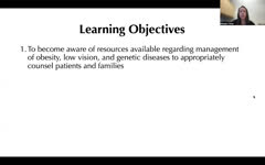 |
NANOS Illustrated Curriculum Section G Introduction | Meagan D. Seay, DO | Introduction video to the resources available in Section G of the NANOS Illustrated Curriculum. | Obesity, Management; Low Vision, Management; Genetic Diseases, Management; Patient Counseling |
| 25 |
 |
Facioscapulohumeral Muscular Dystrophy (FSHD) | Rohith Erukulla; Brooke Johnson | This is an introduction to Facioscapulohumeral Muscular Dystrophy (FSHD) and its causes, presentation, diagnosis, treatment, and ongoing related research. | Dystrophy; Facioscapulohumeral Muscular Dystrophy; FSHD; Myopathy |