AAO-NANOS Neuro-Ophthalmology Clinical Collection: Derived from the AAO-NANOS Clinical Neuro-Ophthalmology collection produced on CD. The images are of selected cases from the NANOS teaching slide exchange, and the CD was produced under the direction of Larry Frohman, MD and Andrew Lee, MD.
The American Academy of Ophthalmology (AAO); The North American Neuro-Ophthalmology Association (NANOS).
NOVEL: https://novel.utah.edu/
TO
Filters: Collection: ehsl_novel_aao_nanos
| Title | Description | Subject | ||
|---|---|---|---|---|
| 326 |
 |
Ocular Manifestations of Congenital/Inherited Diseases | This patient is a 40-year-old man with a history of abetalipoproteinemia (Bassen-Kornweig syndrome), diagnosed at age 9. Neurologic complications have included ataxia, retinal degeneration, peripheral neuropathy, progressive leg weakness, dysarthria, and intermittent bladder incontinence. On his neu... | Bassen-Kornzweig Syndrome |
| 327 |
 |
Ocular Manifestations of Congenital/Inherited Diseases | This patient is a 40-year-old man with a history of abetalipoproteinemia (Bassen-Kornweig syndrome), diagnosed at age 9. Neurologic complications have included ataxia, retinal degeneration, peripheral neuropathy, progressive leg weakness, dysarthria, and intermittent bladder incontinence. On his neu... | Bassen-Kornzweig Syndrome |
| 328 |
 |
Ocular Manifestations of Congenital/Inherited Diseases | This patient is a 40-year-old man with a history of abetalipoproteinemia (Bassen-Kornweig syndrome), diagnosed at age 9. Neurologic complications have included ataxia, retinal degeneration, peripheral neuropathy, progressive leg weakness, dysarthria, and intermittent bladder incontinence. On his neu... | Bassen-Kornzweig Syndrome |
| 329 |
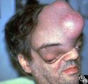 |
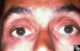
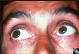
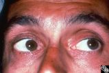
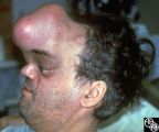
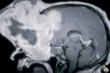
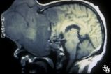
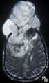
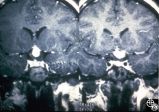
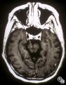
Neuro-Ophthalmic Manifestations of Brain Tumors | The patient is a 45-year-old recluse found to harbor this frontal lobe mass. Remarkably, this patient had only mild bilateral optic neuropathies with visual acuities in the 20/25 range. This right disc was mildly swollen and the left mildly pale. He could not fit into the fundus camera for disc phot... | Meningioma |
| 330 |
 |
Neuro-Ophthalmic Manifestations of Brain Tumors | The patient is a 45-year-old recluse found to harbor this frontal lobe mass. Remarkably, this patient had only mild bilateral optic neuropathies with visual acuities in the 20/25 range. This right disc was mildly swollen and the left mildly pale. He could not fit into the fundus camera for disc phot... | Meningioma |
| 331 |
 |
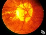
Ocular Manifestations of Congenital/Inherited Diseases | This 21-year-old woman had a 2-year history of blurred vision. A computerized visual field demonstrated a temporal defect OS. MRI confirmed a chiasmal mass lesion. The pathology was consistent with hemangioblastoma. Further workup revealed retinal angiomas and multiple other hemangioblastomas of the... | von Hippel-Lindau Disease |
| 332 |
 |
Neuro-Ophthalmic Manifestations of Brain Tumors | The patient is a 45-year-old recluse found to harbor this frontal lobe mass. Remarkably, this patient had only mild bilateral optic neuropathies with visual acuities in the 20/25 range. This right disc was mildly swollen and the left mildly pale. He could not fit into the fundus camera for disc phot... | Meningioma |
| 333 |
 |
Neuro-Ophthalmic Manifestations of Brain Tumors | The patient is a 45-year-old recluse found to harbor this frontal lobe mass. Remarkably, this patient had only mild bilateral optic neuropathies with visual acuities in the 20/25 range. This right disc was mildly swollen and the left mildly pale. He could not fit into the fundus camera for disc phot... | Meningioma |
| 334 |
 |
Neuro-Ophthalmic Manifestations of Brain Tumors | The patient is a 45-year-old recluse found to harbor this frontal lobe mass. Remarkably, this patient had only mild bilateral optic neuropathies with visual acuities in the 20/25 range. This right disc was mildly swollen and the left mildly pale. He could not fit into the fundus camera for disc phot... | Meningioma |
| 335 |
 |
Neuro-Ophthalmic Manifestations of Brain Tumors | The patient is a 45-year-old recluse found to harbor this frontal lobe mass. Remarkably, this patient had only mild bilateral optic neuropathies with visual acuities in the 20/25 range. This right disc was mildly swollen and the left mildly pale. He could not fit into the fundus camera for disc phot... | Meningioma |
| 336 |
 |
Ocular Manifestations of Congenital/Inherited Diseases | This 21-year-old woman had a 2-year history of blurred vision. A computerized visual field demonstrated a temporal defect OS. MRI confirmed a chiasmal mass lesion. The pathology was consistent with hemangioblastoma. Further workup revealed retinal angiomas and multiple other hemangioblastomas of the... | von Hippel-Lindau Disease |
| 337 |
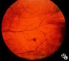 |
Ocular Manifestations of Congenital/Inherited Diseases | This 21-year-old woman had a 2-year history of blurred vision. A computerized visual field demonstrated a temporal defect OS. MRI confirmed a chiasmal mass lesion. The pathology was consistent with hemangioblastoma. Further workup revealed retinal angiomas and multiple other hemangioblastomas of the... | von Hippel-Lindau Disease |
| 338 |
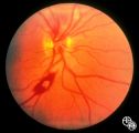 |
Systemic Disorders With Optic Nerve and Retinal Findings | This fondus image shows a white-centered hemorrhage in a leukemia patient with orbital aspergillosis. | Leukemia; Orbital Aspergilosis; Retinal Hemorrhage |
| 339 |
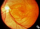 |
Isolated Congenital Optic Disc Anomalies | An optic pit is a small defect in the optic disc that may be asymptomatic in isolation. Patients may develop an associated serous detachment of the macula. The condition is usually unilateral but may be bilateral. A fluorescein angiogram may demonstrate the serous detachment, and laser photocoagulat... | Optic Pit With Serous Macular Detachment |
| 340 |
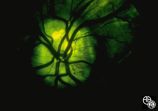 |
Optic Disc Drusen With Autofluorescence | This photograph of optic disc drusen demonstrates autoflourescence with flourescein barrier filters in place. Imaging: flourescein barrier filters. | Optic Disc Drusen; Autofluorescence |
| 341 |
 |
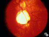
Isolated Congenital Optic Disc Anomalies | Benign tumors of blood vessels (hemangiomas) may occur on the optic nerve and may mimic optic disc edema. Disease/Diagnosis: Optic Nerve Hemangioma. | Optic Nerve Hemangioma |
| 342 |
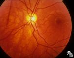 |
Isolated Congenital Optic Disc Anomalies | Patients with hypoplasia of the optic nerve may have normal or subnormal visual acuity or visual field. The condition may be unilateral or bilateral. Optic nerve hypoplasia is usually idiopathic, but maternal diabetes, or maternal use of anti-epileptic drugs or alcohol are predisposing factors. Opti... | Optic Nerve Hypoplasia; Septo-Optic Dysplasia |
| 343 |
 |
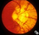
Isolated Congenital Optic Disc Anomalies | Optociliary shunt vessels are venous collaterals that form in response to chronic venous obstruction, shunting the venous blood from the retinal circulation into the choroidal circulation. Although they may be congenital, they may occur in patients with chronic disc edema, following central retinal ... | Optociliary Shunt Vessels |
| 344 |
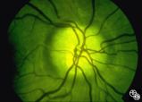 |
Isolated Congenital Optic Disc Anomalies | This optic disc displays multiple drusen. Note the pseudopapilledema here. One can differentiate this from true papilledema in that there is no obscuration of the vessel by the peripapillary nerve fiber layer as they cross the disc margin. This photograph was taken with barrier filters in place, but... | Optic Disc Drusen; Optic Nerve Drusen; Pseudopapilledema |
| 345 |
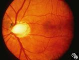 |
Isolated Congenital Optic Disc Anomalies | An optic pit is a small defect in the optic disc that may be asymptomatic in isolation. The pit can be small or large, and central or peripheral. Disease/Diagnosis: Optic Pit. | Optic Pit; Macular Detachment |
| 346 |
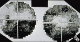 |
Optic Disc Drusen Visual Fields | This is the visual field of patient with optic nerve drusen. Whereas they typically do not cause central field loss, optic disc drusen may cause nerve fiber bundle layer defects and, thus, peripheral field defects, including altitudinal defects (seen inferiorly in the left eye) or arcuate defects (s... | Optic Disc Drusen; Optic Nerve Drusen; Visual Fields |
| 347 |
 |
Retinal Coloboma Underneath a Relatively Normal Optic Nerve | Optic nerve colobomas appear as enlarged, white optic discs that are deeply excavated, often with some sapring of the superior rim. They result from an abnormal fusion of the proximal embryonic fissure. Optic nerve colobomas occur unilaterally or bilaterally with a similar frequency and can result i... | Optic Nerve Coloboma |
| 348 |
 |
Isolated Congenital Optic Disc Anomalies | Patients with midline closure defects may exhibit abnormalities in the optic nerve, choroid, retinal pigment epithelium or retina. Anterior closure defects may result in colobomas of the structures of the anterior segment, such as the iris. Disease/Diagnosis: Coloboma. | Optic Nerve Coloboma |
| 349 |
 |
Peripapillary Staphyloma | Patients with ectasia of the outer layers of the eye may exhibit a posterior protrusion that appears on funduscopy as an area of deep excavation of the retina (posterior staphyloma). When it occurs around the optic disc, as in this case, it is termed a peripapillary staphyloma. This may occur in ass... | Staphyloma |
| 350 |
 |
Motility Disturbances | The patient is a 53-year-old man with diplopia from right oculomotor nerve palsy and left hemiparesis (Weber's syndrome), with associated left lung hilar mass. The spinal tap showed pleocytosis consistent with carcinomatous meningitis. This image demonstrates oculomotor nerve metastatic carcinomatos... | Oculomotor Palsy; Weber; Fascicular Oculomotor (Third) Nerve Palsy |