AAO-NANOS Neuro-Ophthalmology Clinical Collection: Derived from the AAO-NANOS Clinical Neuro-Ophthalmology collection produced on CD. The images are of selected cases from the NANOS teaching slide exchange, and the CD was produced under the direction of Larry Frohman, MD and Andrew Lee, MD.
The American Academy of Ophthalmology (AAO); The North American Neuro-Ophthalmology Association (NANOS).
NOVEL: https://novel.utah.edu/
TO
Filters: Collection: ehsl_novel_aao_nanos
| Title | Creator | Description | ||
|---|---|---|---|---|
| 1 |
 |
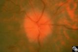
Isolated Congenital Optic Disc Anomalies | Anthony C. Arnold, MD | This patient has known pseudoxanthoma elasticum (an uncommon elastic tissue disorder characterized by plaque-like skin folds [plucked chicken skin] and degeneration of collagen fibers involving multiple systems, including the GI tract and heart), angioid streaks, and optic disc drusen. |
| 2 |
 |
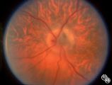
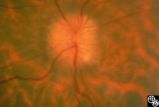
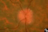
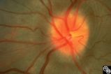
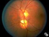
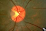
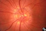
Isolated Optic Neuritis/Neuropathy | Anthony C. Arnold, MD | This 48-year-old man presented with a 1-month history of headache. Both discs had the appearance seen in this image, with prominent peripapillary nerve fiber layer myelination; the disc itself is hyperemic, with dilated, telangiectatic surface vasculature, suggesting true disc edema as well. |
| 3 |
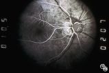 |
Optic Disc Drusen, Fluorescein Angiogram | Anthony C. Arnold, MD | Images 92_64 and 92_67 demonstrate the characteristics of optic disc drusen on flourescein angiography. This image shows the early arteriovenous phase, with irregular dye uptake and focal hypoflourescence superotemporally. Pair with 92_63 and 92_67 |
| 4 |
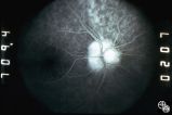 |
Optic Nerve Drusen, Late Fluorescein Angiogram | Anthony C. Arnold, MD | Images 92_64 and 92_67 demonstrate the characteristics of optic disc drusen on flourescein angiography. This image displays the nodular staining of the drusen without leakage. Pair with 92_63 and 92_64. |
| 5 |
 |
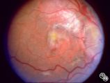
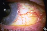
Neuro-Ophthalmic Vascular Disease | Anthony C. Arnold, MD | This 76-year-old woman has a 7-month history of redness and pressure sensation in both eyes that is worse in the morning. She has noted intermittent horizontal diplopia during this time. Angiography demonstrated a right dural cavernous sinus fistula, which was successfully occluded with direct injec... |
| 6 |
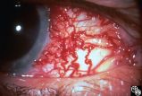 |
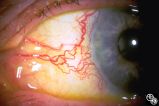
Neuro-Ophthalmic Vascular Disease | Anthony C. Arnold, MD | This 76-year-old woman has a 7-month history of redness and pressure sensation in both eyes that is worse in the morning. She has noted intermittent horizontal diplopia during this time. Angiography demonstrated a right dural cavernous sinus fistula, which was successfully occluded with direct injec... |
| 7 |
 |
Neuro-Ophthalmic Vascular Disease | Anthony C. Arnold, MD | This 76-year-old woman has a 7-month history of redness and pressure sensation in both eyes that is worse in the morning. She has noted intermittent horizontal diplopia during this time. Angiography demonstrated a right dural cavernous sinus fistula, which was successfully occluded with direct injec... |
| 8 |
 |
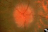
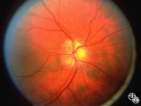
Congenitally Tilted Optic Disc | Anthony C. Arnold, MD | Colobomas or defects of the optic nerve may exhibit spontaneous pulsations. Disease/Diagnosis: Coloboma. |
| 9 |
 |
Isolated Optic Neuritis/Neuropathy | Anthony C. Arnold, MD | This 42-year-old male with pseudotumor cerebri and chronic papilledema demonstrated refractile bodies, which can be seen with chronic optic disc edema. This image exhibits decreased disc edema and resolution of the refractile bodies OD after therapy. Pair with 96_01, 96_02, 96_03, 96_05, and 96_06. |
| 10 |
 |
Isolated Optic Neuritis/Neuropathy | Anthony C. Arnold, MD | This 42-year-old male with pseudotumor cerebri and chronic papilledema demonstrated refractile bodies, which can been seen with chronic optic disc edema. This image demonstrates later recurrence of the refractile bodies with worsening papilledema OD. Pair with 96_01, 96_02, 96_03, 96_04, and 96_06. |
| 11 |
 |
Isolated Optic Neuritis/Neuropathy | Anthony C. Arnold, MD | This 42-year-old male with pseudotumor cerebri and chronic papilledema demonstrated refractile bodies, which can be seen with chronic optic disc edema. This image demonstrates later recurrence of the refractile bodies with worsening papilledema OD. Pair with 96_01, 96_02, 96_03, 96_04, and 96_05. |
| 12 |
 |
Isolated Optic Neuritis/Neuropathy | Anthony C. Arnold, MD | This 42-year-old male with pseudotumor cerebri and chronic papilledema demonstrated refractile bodies, which can be seen with chronic optic disc edema. This image shows the chronic papilledema at presentation, with associated refractile hyaline bodies at the disc periphery in both eyes. Pair with 96... |
| 13 |
 |
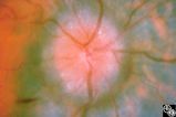
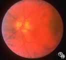
Isolated Optic Neuritis/Neuropathy | Anthony C. Arnold, MD | This image demonstrates Paton's lines in a 34-year-old patient with pseudotumor cerebri and chronic papilledema. |
| 14 |
 |
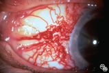
Neuro-Ophthalmic Vascular Disease | Anthony C. Arnold, MD | This 76-year-old woman has a 7-month history of redness and pressure sensation in both eyes that is worse in the morning. She has noted intermittent horizontal diplopia during this time. Angiography demonstrated a right dural cavernous sinus fistula, which was successfully occluded with direct injec... |
| 15 |
 |
Neuro-Ophthalmic Vascular Disease | Anthony C. Arnold, MD | This 76-year-old woman has a 7-month history of redness and pressure sensation in both eyes that is worse in the morning. She has noted intermittent horizontal diplopia during this time. Angiography demonstrated a right dural cavernous sinus fistula, which was successfully occluded with direct injec... |
| 16 |
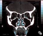 |
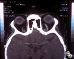
Neuro-Ophthalmic Vascular Disease | Anthony C. Arnold, MD | This coronal CT scan shows the enlarged superior ophthalmic vein in the left orbit. This 76-year-old woman has a 7-month history of redness and pressure sensation in both eyes that is worse in the morning. She has noted intermittent horizontal diplopia during this time. Angiography demonstrated a ri... |
| 17 |
 |
Isolated Optic Neuritis/Neuropathy | Anthony C. Arnold, MD | This 42-year-old male with pseudotumor cerebri and chronic papilledema demonstrated refractile bodies, which can been seen with chronic optic disc edema. This image exhibits decreased disc edema and resolution of the refractile bodies OD after therapy. Pair with 96_01, 96_02, 96_04, 96_05, and 96_06... |
| 18 |
 |
Bridge Coloboma | Anthony C. Arnold, MD | The patient is a 35-year-old asymptomatic woman with a bridge coloboma, also known as double optic disc. Disease/Diagnosis: Coloboma. |
| 19 |
 |
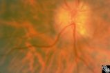
Isolated Congenital Optic Disc Anomalies | Anthony C. Arnold, MD | This image shows drusen that are especially prominent superotemporally. Pair with 92_64 and 92_67. |
| 20 |
 |
Isolated Optic Neuritis/Neuropathy | Anthony C. Arnold, MD | This 42-year-old male with pseudotumor cerebri and chronic papilledema demonstrated refractile bodies, which can be seen with chronic optic disc edema. This image shows the chronic papilledema at presentation, with associated refractile hyaline bodies at the disc periphery in both eyes. Pair with 96... |
| 21 |
 |
Isolated Congenital Optic Disc Anomalies | Anthony C. Arnold, MD | A 10-year-old girl had central visual loss due to this optic pit. Disease/Diagnosis: Optic Pit. |
| 22 |
 |
Isolated Congenital Optic Disc Anomalies | Anthony C. Arnold, MD | This is a photograph of peripheral drusen. The paired image 92_69 demonstrates the typical autofluorescence. |
| 23 |
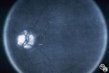 |
Optic Disc Drusen Autofluorescence | Anthony C. Arnold, MD | This case of optic disc drusen demonstrates the typical autofluorescence. Pair with 92_68. Imaging: Autoflourescence? |
| 24 |
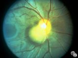 |
Ocular Manifestations of Congenital/Inherited Diseases | Anthony C. Arnold, MD | Note the retinal/optic nerve head astrocytoma in a 12-year-old boy with tuberous sclerosis. Disease/Diagnosis: Tuberous Sclerosis. |
| 25 |
 |
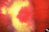
Optic Neuropathies | Daniel M. Jacobson MD | This 28-year-old otherwise-healthy woman was referred to for treatment of what was thought to be optic neuritis OD. Three weeks earlier she had noted a dark inferior scotoma OD that progressed to involve fixation over the next 10-12 days. She experienced photopsias OD at the onset. She had no viral ... |