| OCR Text |
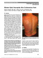
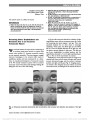
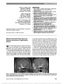
Show Homonymous Ganglion Cell Layer Thinning After Isolated Occipital Lesion: Macular OCT Demonstrates Transsynaptic Retrograde Retinal Degeneration Paolo G. Meier, MD, Philippe Maeder, MD, Randy H. Kardon, MD, PhD, François-Xavier Borruat, MD Abstract: A 48-year-old man was examined 24 months after medial and surgical treatment of an isolated well-circumscribed right occipital lobe abscess. An asymp-tomatic residual left homonymous inferior scotoma was present. Fundus examination revealed temporal pallor of both optic discs, and optical coherence tomography (OCT) revealed mild temporal loss of retinal nerve fiber layer in both eyes. No relative afferent pupillary defect was present. Assessment of the retinal ganglion cell layer demonstrated homonymous thinning in a pattern corresponding to the homonymous visual field loss. There were no abnormalities of the lateral geniculate nuclei or optic tracts on review of the initial brain computed tomography and follow-up mag-netic resonance imaging. We believe our patient showed evidence of transsynaptic retrograde degeneration after an isolated right occipital lobe lesion, and the homonymous neuronal loss was detected on OCT by assessing the retinal ganglion cell layer. Journal of Neuro-Ophthalmology 2015;35:112-116 doi: 10.1097/WNO.0000000000000182 © 2015 by North American Neuro-Ophthalmology Society Transsynaptic retrograde degeneration (TRD) of the visual pathways, that is loss of undamaged retinal ganglion cells occurring upstream from a retrogeniculate lesion, has been convincingly reported in experimental con-ditions (1-3). Some patients with occipital lesions sparing the lateral geniculate nucleus and optic tract can develop TRD with bow tie optic atrophy in the eye contralateral to the lesion and temporal pallor in the fellow eye (4). How-ever, optic nerve changes can be subtle and sometimes over-looked on ophthalmoscopy. Histological evidence of TRD in humans was provided by Beatty et al (5) who reported such a case 40 years after occipital lobectomy. The devel-opment of optical coherence tomography (OCT) allows measurement of the retinal nerve fiber layer (RNFL), and this technology has provided evidence of TRD after isolated occipital lesions (6-8). Additional evidence of TRD with occipital lesions has been demonstrated with magnetic res-onance imaging (MRI), revealing atrophy of the ipsilateral optic tract (9,10). The development of segmentation software of macular OCT provides thickness maps of the retinal ganglion cell layer-inner plexiform layer complex (RGCL-IPL). This approach is advantageous for discerning patterns of homon-ymous loss corresponding to TRD from postgeniculate lesions. We report homonymous thinning of RGC-IPL in the macula of a patient with an isolated occipital lobe abscess producing a corresponding homonymous visual field scotoma within the central 15°. CASE REPORT A 46-year-old man in good health reported a 10-day history of headaches and scintillating scotomata. The patient was afebrile with normal vital signs, and general physical examination was normal. Visual acuity was 20/20 bilaterally but visual field examination revealed a dense left homon-ymous hemianopia (Fig. 1). There was no relative afferent pupillary defect (RAPD). Ophthalmoscopy was normal. Brain computed tomography disclosed a well-demarcated lesion within the right occipital lobe with perifocal edema thought to represent a neoplasm (Fig. 2). Neuro-Ophthalmology Unit, Hôpital Ophtalmique Jules-Gonin (PGM, F-XB), University of Lausanne, Lausanne, Switzerland; Department of Radiology (PGM), CHUV, University of Lausanne, Lausanne, Switzerland; and Department of Veterans Affairs Medical Center (RHK), University of Iowa Hospitals and Clinics, Iowa City, Iowa. The authors report no conflicts of interest. Address correspondence to François-Xavier Borruat, MD, Neuro- Ophthalmology Unit, Hôpital Ophtalmique Jules-Gonin, Avenue de France 15, Lausanne 1004, Switzerland; E-mail: francois.borruat@fa2.ch 112 Meier et al: J Neuro-Ophthalmol 2015; 35: 112-116 Original Contribution Copyright © North American Neuro-Ophthalmology Society. Unauthorized reproduction of this article is prohibited. The patient was treated with intravenous methylpred-nisolone (125 mg 3 times a day) for 48 hours followed by craniotomy. At surgery, a cortical abscess was drained, and bacteriologic analysis revealed Actinomyces Meyeri and Ag-gregatibacter aphrophilus. Systemic intravenous antibiotic therapy (ceftriaxone 2 g twice a day and metronidazole 0.5 g 3 times a day) was given for 6 weeks. The patient was found to have a patent foramen ovale that was surgically closed. Six weeks after surgery, ophthalmic examination revealed partial resolution of the left homonymous hemi-anopia, absence of a RAPD, and no evidence of optic atro-phy. Twelve months after initial presentation, MRI showed right occipital cortical atrophy with mild ipsilateral white matter gliosis (Fig. 3). Review of all MRI studies revealed no evidence of damage to the lateral geniculate nuclei or optic tracts. Two years later, the patient was referred for neuro-ophthalmic evaluation. Visual acuity was 20/15 in each eye, pupils reacted normally, and color visual testing and slit-lamp examination were normal. There was temporal pallor of both optic discs (Fig. 4). Automated visual field testing showed a congruous left inferior homonymous congruous scotoma (Fig. 5). FIG. 1. Automated static perimetry (Octopus, program G1, Haag Streit, Schlieren, Switzerland) reveals a left homonymous hemianopia. FIG. 2. Axial computed tomography shows a right occipital lobe lesion with surrounding edema. FIG. 3. T2 axial magnetic resonance imaging 12 months after draining of brain abcess shows cortical atrophy of the right occipital lobe with moderate gliosis. Meier et al: J Neuro-Ophthalmol 2015; 35: 112-116 113 Original Contribution Copyright © North American Neuro-Ophthalmology Society. Unauthorized reproduction of this article is prohibited. Using spectral-domain OCT (Cirrus 4000 HD-OCT; Carl Zeiss Meditec, Dublin, CA), average RNFL thickness was normal in both eyes (right eye: 95 mm; left eye: 98 mm) with slight thinning of temporal quadrant in each eye. Mac-ular OCT showed no significant thinning of the total retinal thickness. Analysis of the RGC-IPL complex with the Gan-glion Cell Analysis module (Zeiss Cirrus software, version 6.5.0; Zeiss, Dublin, CA) disclosed a moderate homony-mous thinning in both eyes on the total thickness RGC-IPL map and significant to less than the 1% level on the prob-ability map (Fig. 6). The pattern of the RGCL thinning was homonymous, affecting the temporal retina in the right eye and the nasal retina in the left eye. The RGC-IPL thinning respected the vertical meridian. Although the homonymous visual field defect extended to approximately 16°, ganglion cell thinning appeared to extend to approximately 6°. FIG. 4. Two years after initial presentation, color (top) and red-free (bottom) fundus photographs shows temporal pallor of both optic discs. FIG. 5. Two years after initial presentation, automated static perimetry (Octopus, program G1, Haag Streit) discloses a residual left inferior homonymous scotoma. 114 Meier et al: J Neuro-Ophthalmol 2015; 35: 112-116 Original Contribution Copyright © North American Neuro-Ophthalmology Society. Unauthorized reproduction of this article is prohibited. FIG. 6. Optical coherence tomography performed 2 years after initial presentation (Cirrus 4000 HD-OCT; Carl Zeiss Meditec). A. Fundus diagram with the thickness map of RGCL-IPL for right eye (left) and left eye (right). Homonymous thinning of RGCL-IPL complex (i.e., loss of RGCL-IPL in the temporal retina of the right eye and in the nasal retina of the left eye indicated by arrows) is present in a pattern corresponding to the residual homonymous left visual field defect. B. Deviation maps of the RGCL-IPL loss demonstrates the homonymous defect of RGCL-IPL. Note that the extent of the RGCL-IPL thinning on the deviation maps (approximately 6°) does not extend as far peripherally as the visual field defect (16°). Sector maps illustrated the degree of homonymous RGCL-IPL thinning in each eye. C. Macular profiles with horizontal OCT B-scans through the fovea appear normal in both eyes. D. RNFL analysis shows loss of nerve fibers in the temporal quadrant of both optic discs more marked in the left disc. RGCL-IPL, retinal ganglion cell layer-inner plexiform layer complex; OCT, optical coherence tomog-raphy; RNFL, retinal nerve fiber layer. Meier et al: J Neuro-Ophthalmol 2015; 35: 112-116 115 Original Contribution Copyright © North American Neuro-Ophthalmology Society. Unauthorized reproduction of this article is prohibited. DISCUSSION The combination of bilateral optic atrophy and homony-mous visual field loss usually implies the presence of a lesion of the pregeniculate visual pathways. Typically, this results in temporal atrophy of the optic disc ipsilateral to the lesion and a bow tie atrophy of the contralateral eye. This pattern arises from loss of noncrossing fibers in the ipsilateral eye and the loss of crossing fibers in the contralateral eye (4). The presence of an RAPD in the contralateral eye indicates a lesion of the optic tract, whereas the absence of an RAPD signifies a lesion beyond the point where the pupillomotor fibers leave the optic tract. OCT can demonstrate thinning of the RNFL in optic tract lesions, but the pattern of RNFL loss does not readily demonstrate a homonymous nature of the lesion. A bow tie pattern of RNFL loss also is not usually apparent. The availability of software to provide segmentation of the retinal layers in the macula allows the analysis of the RGC-IPL complex. Homonymous thinning of the RGC-IPL in a patient with an optic tract lesion due to neuromyelitis optica has been published (11). Our patient had a right occipital lobe abscess. Both before and after neurosurgical drainage, neuroimaging studies did not detect any abnormality of the optic tracts or lateral geniculate nuclei. The homonymous thinning of the RGC-IPL on OCT exhibited by our patient most likely represents TRD. Interestingly, the peripheral extent of the homonymous ganglion cell-inner plexiform layer thinning (6°) on the probability plot was not as extensive as the visual field defect (16°). Possibly, the central macular fibers near fixation are more likely to show TRD. More than 50 years ago, Van Buren (1) created a focal surgically-induced lesion in 1 occipital lobe of the adult macaque monkey. Forty-eight months later, he demonstrated homonymous thinning of the retina supportive of the concept of TRD. Similar experiments in other nonhuman primates confirmed this observation (2,3). However, documentation of optic atrophy in humans after retrogeniculate lesion is rare, and the concept of TRD in human adults has been challenged (12). However, recent studies with OCT have demonstrated the possibility of TRD in adult humans with acquired retro-geniculate disorders. Jindahra et al (6) demonstrated thinning of RNFL in both eyes in a series of 26 patients with both congenital and acquired homonymous hemianopia. Patients exhibited a significant loss of RNFL mean thickness as com-pared to controls, and those with congenital lesions exhibited greater RNFL loss than individuals with acquired lesions. In a later study (7), the same investigators studied 38 patients with purely acquired retrogeniculate lesions and reported a decelerating rate of RNFL loss of 9.08 mm per log-years. Yet, no thinning of RNFL was detected when small visual field defects were present. Park et al (8) reported a constant pattern of RNFL loss in patients with cerebral infarction with the optic nerve ipsilateral to the lesion losing uncrossed (tem-poral) fibers and the fellow eye losing crossed (nasal) fibers. In a cohort of patients with autosomal-demonstrated optic atrophy, Rönnbäck et al (13) showed that measure-ment of RGCL-IPL complex was more sensitive than RNFL thickness in detecting structural loss. The OCT re-sults in our patient support a similar conclusion in patients with TRD due to a retrogeniculate lesion. REFERENCES 1. Buren JM. Trans-synaptic retrograde degeneration in the visual system of primates. J Neurol Neurosurg Psychiatry. 1963;26:402-409. 2. Cowey A, Alexander I, Stoerig P. Transneuronal retrograde degeneration of retinal ganglion cells and optic tract in hemianopic monkeys and humans. Brain. 2011;134: 2149-2157. 3. Hendrickson A, Warner CE, Possin D, Huang J, Kwan WC, Bourne JA. Retrograde transneuronal degeneration in the retina and lateral geniculate nucleus of the V1-lesioned marmoset monkey. Brain Struct Funct. [published ahead of print October 13, 2013] doi: 10.1007/s00429-013-0659-7. 4. Hoyt WF, Kommerell G. Fundus oculi in homonymous hemianopia [in German]. Klin Monbl Augenheilkd. 1973;162:456-464. 5. Beatty RM, Sadun AA, Smith LEH, Vonsattel JP, Richardson EP. Direct demonstration of transsynaptic degeneration in the human visual system: a comparison of retrograde and anterograde changes. J Neurol Neurosurg Psychiatry. 1982;45:143-146. 6. Jindahra P, Petrie A, Plant GT. Retrograde trans-synaptic retinal ganglion cell loss identified by optical coherence tomography. Brain. 2009;132:628-634. 7. Jindahra P, Petrie A, Plant GT. The time course of retrograde trans-synaptic degeneration following occipital lobe damage in humans. Brain. 2012:135;534-541. 8. Park HYL, Park YG, Cho AH, Park CK. Transneuronal retrograde degeneration of the retinal ganglion cells in patients with cerebral infarction. Ophthalmology. 2013;120:1292-1299. 9. Bridge H, Jindhara P, Barbur J, Plant GT. Imaging reveals optic tract degeneration in hemianopia. Invest Ophthalmol Vis Sci. 2011;52:382-388. 10. Millington RS, Yasuda CL, Jindhara P, Jenkinson M, Barbur JL, Kennard C, Cendes F, Plant GT, Bridge H. Quantifying the pattern of optic tract degeneration in human hemianopia. J Neurol Neurosurg Psychiatry. 2014;85:379-386. 11. Romero RS, Gutierrez I, Wang E, Reder AT, Bhatti MT, Bernard JT, Javed A. Homonymous hemimacular thinning: a unique presentation of optic tract injury in neuromyelitis optica. J Neuroophthalmol. 2012;32:150-153. 12. Miller NR, Newman SA. Transsynaptic degeneration. Arch Ophthalmol. 1981;99:1654. 13. Rönnbäck C, Milea D, Larsen M. Imaging of the macula indicates early completion of structural deficit in autosomal-dominant optic atrophy. Ophthalmology. 2013;120: 2672-2677. 116 Meier et al: J Neuro-Ophthalmol 2015; 35: 112-116 Original Contribution Copyright © North American Neuro-Ophthalmology Society. Unauthorized reproduction of this article is prohibited. |