| OCR Text |
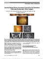
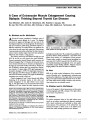
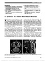
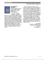
Show Drug-Related Mitochondrial Optic Neuropathies Michelle Y. Wang, MD, Alfredo A. Sadun, MD, PhD Background: There is a group of optic neuropathies of either genetic or acquired origin characterized by similar clinical manifestations with preferential involvement of the papillomacular bundle (PMB). PMB fibers are most suscep-tible to injury as they are small, unmyelinated, and have high-energy demands. These optic neuropathies share a presumed common pathophysiology of mitochondrial dysfunction. Evidence Acquisition: A variety of medications cause optic neuropathy by interfering with mitochondrial function. The evidence linking these therapeutic agents as a cause of mitochondrial optic neuropathy (MON) is well established in some and less certain in others. The differential diagnosis includes other optic nerve disorders producing bilateral, symmetric visual loss, including certain nutritional deficien-cies, toxins, and genetic diseases. Results: Ethambutol, chloramphenicol, linezolid, erythromy-cin, streptomycin, and antiretroviral drugs can cause drug-related MON. In many cases, drug toxicity is dose and duration dependent, and discontinuation of the drug in a timely manner can lead to significant visual recovery. Conclusions: Mitochondrial optic neuropathies are increas-ingly recognized as a spectrum of conditions that reach a similar end point by compromising a common pathway of mitochondrial dysfunction. Clinicians should be aware of drugs that can cause a MON. Prompt recognition of this association is critical in preventing irreversible, profound visual loss. Journal of Neuro-Ophthalmology 2013;33:172-178 doi: 10.1097/WNO.0b013e3182901969 © 2013 by North American Neuro-Ophthalmology Society MITOCHONDRIAL OPTIC NEUROPATHY Mitochondrial optic neuropathy (MON) is increasingly recognized as a major spectrum of optic neuropathies resulting from different genetic and acquired etiologies (1). Clinical Features The clinical presentation of MON is characterized by slowly progressive bilateral loss of central vision, dyschromatopsia, central or cecocentral scotomas, and loss of high spatial frequency contrast sensitivity (2) (Table 1). Patients often describe the visual loss as a central haze or dark cloud. Pain is not a feature of MON. Ophthalmoscopic features during the acute/subacute stage often reveal a hyperemic optic disc and peripapillary retinal nerve fiber layer (PRNFL) swelling (3). With time, temporal pallor of the optic disc develops. There is no relative afferent pupillary defect due in part to symmetric optic nerve involvement. Clinical findings such as poor visual acuity, dyschroma-topsia, and central visual field loss can be explained by selective damage to the papillomacular bundle (PMB). The fibers of the PMB are most susceptible due to their long unmyelinated segment in the retina and their small caliber. Preferential involvement of the PMB is a feature common to a wide range of acquired and genetic mitochondrial optic neuropathies (4,5). Figure 1 summarizes the spectrum of MON. Leber hered-itary optic neuropathy (LHON) and autosomal dominant optic atrophy (ADOA) are 2 well-documented examples due to mitochondrial or somatic DNA mutations. Other examples include nutritional deficiencies, such as lack of folic acid and vitamin B12, and combinations of nutritional defi-ciency and toxicity causing tobacco-alcohol and Cuban epi-demic optic neuropathies. In all instances, MON begins with dysfunction of mitochondrial oxidative phosphorylation (6) and results in impaired function of the PMB. Common Pathway Mitochondria provide the majority of cellular energy by oxidative phosphorylation. During the process, electrons are transferred down a chain of complexes. When electrons do not complete the process, reactive oxygen species are generated as a byproduct. The combination of energy depletion and oxidative stress results in the opening of the mitochondrial permeability transition pore, allowing for leakage of cytochrome c, a key activator for apoptosis (Fig. 2). Department of Ophthalmology, USC School of Medicine, Los Angeles, California. The authors report no conflicts of interest. Address correspondence to Alfredo A. Sadun, MD, PhD, Department of Ophthalmology, USC School of Medicine, 1450 San Pablo St., Los Angeles, CA 90033; E-mail: asadun@usc.edu 172 Wang and Sadun: J Neuro-Ophthalmol 2013; 33: 172-178 State-of-the-Art Review Section Editors: Grant T. Liu, MD Randy H. Kardon, MD, PhD Copyright © North American Neuro-Ophthalmology Society. Unauthorized reproduction of this article is prohibited. The axons of retinal ganglion cells (RGCs) converge toward the optic nerve head and pass through the lamina cribrosa. Myelination of axons begins only posterior to the lamina cribrosa. The fibers of the PMB are anatomically distinct in 2 regards. First, like all axons emanating from RGCs, they run an unmyelinated course through the retinal nerve fiber layer (RNFL). Second, they are narrow in caliber. Membrane potential must be recharged at each node of Ranvier and along any other unmyelinated segment. A number of factors probably lead to selective vulnerability of the PMB axons. These include high energy needs coupled with low energy production, a high surface area to volume ratio, and absence of saltatory conduction due their unmyelinated structure (7). As a part of a compensatory mechanism, mitochondria accumulate within RGC axons. These axonal changes can be detected by optical coherence tomography (OCT) (8,9). Appearance of optic disc pallor usually occurs in later stages, indicating irreversible axonal loss. With cessation of trigger-ing factors, improvement in visual fields and corresponding reduction in the nerve fiber layer thickness can be observed in some acquired cases with resolution of axonal engorgement. Workup and Ancillary Testing The best screening tests for MON are those that measure the functions subserved by the PMB, including visual acuity, color vision, contrast sensitivity at high spatial frequencies, central visual field testing and, possibly, pattern visual-evoked potentials. The extent of visual loss varies widely; however, visual acuity of hand motions, light perception, or no light perception is very rare as non- PMB retinal nerve fibers usually are spared. Color vision loss sometimes may be more profound than visual acuity loss. Contrast sensitivity may be effective in detecting subclinical toxic optic neuropathy (10). Central or cecocen-tral scotomas are the hallmark visual field defects. The red Amsler grid is a particularly useful screening test. OCT is a useful ancillary test in the subacute and chronic stages by measuring the RNFL thickness and confirming thinning involving the PMB beginning in the inferotemporal sector (11-14). In the later stages, OCT demonstrates thinning of RNFL thickness in all quadrants (9). DRUG-INDUCED MITOCHONDRIAL OPTIC NEUROPATHY A number of drugs injure the optic nerve by interfering with mitochondrial oxidative phosphorylation, thereby produc-ing a classic clinical picture of MON. Drugs proven to cause MON by blocking oxidative phosphorylation include ethambutol, chloramphenicol, line-zolid, erythromycin, streptomycin, and antiretroviral drugs (15-17). A number of other drugs that can cause optic neu-ropathy but less strongly associated with mitochondrial dys-function are amiodarone, infliximab, clioquinol, dapsone, quinine, pheniprazine, suramin, and isoniazid (16,18). Ethambutol Worldwide, there are approximately 9.2 million new cases of tuberculosis (TB) each year, and about 55% of these patients take ethambutol to prevent or delay the emergence of drug resistance (19). As a consequence, ethambutol is the FIG. 1. Mitochondrial dysfunction plays a central role in the pathogenesis of many optic nerve diseases, either inherited or acquired. Instead of classifying them as separate enti-ties, mitochondrial optic neuropathies have now been rec-ognized as a spectrum of conditions leading to optic atrophy by a similar pathway. CEON, Cuban epidemic of mitochon-drial optic neuropathy; TAA, tobacco-alcohol amblyopia. TABLE 1. Clinical features of mitochondrial optic neuropathy Fairly symmetric visual impairment Loss of high spatial frequency contrast sensitivity Early and profound loss of color vision Central or centrocecal visual field defects Temporal pallor of the optic discs (delayed) Wang and Sadun: J Neuro-Ophthalmol 2013; 33: 172-178 173 State-of-the-Art Review Copyright © North American Neuro-Ophthalmology Society. Unauthorized reproduction of this article is prohibited. most common cause of toxic optic neuropathy accounting for 100,000 new cases each year (20). Ethambutol is a metal chelator, destroying bacteria by inhibiting arabinosyltransferase, an important enzyme in mycobacterial cell wall synthesis. Due to the similarity between mammalian mitochondrial DNA (mtDNA) and bacterial ribosomes, ethambutol also disrupts oxidative phosphorylation and mitochondrial function by interfering with iron-containing complex I and copper-containing complex IV (17). Copper is a required cofactor for cytochrome c oxidase, an essential com-ponent in the electron transport chain. Ethambutol may reduce the level of copper, thereby interfering with oxidative phos-phorylation. Replacing copper leads to improved RGC surviv-ability in in vivo models of ethambutol optic neuropathy (21). It is interesting that copper deficiency due to malabsorption from bariatric surgery also has been associated with visual loss from optic neuropathy (22,23). Other studies suggest that zinc also might play a role in ethambutol toxicity (21,24), and individuals with reduced serum zinc level may be more suscep-tible to ethambutol ocular toxicity (25,26). A study using cell cultures demonstrated that the chelating effect of ethambutol may inhibit lysosomal activation, resulting in accumulation of zinc in lysosomes with increased lysosomal membrane perme-ability and cell death (27). Ethambutol ocular toxicity has been well documented in the literature shortly after its introduction in 1960s (28-44). The frequency of vision impairment has been reported in 50% of patients at a dose of 60-100 mg/kg/d, 5%-6% at 25 mg/kg/d, and 1% at #15 mg/kg/d (45). Visual loss is typically insidious and symmetrical, occurring typically 2-8 FIG. 2. Accumulation of reactive oxygen species (ROS) leads to a decrease in the electrical potential across the mito-chondrial membrane, which allows for an opening of the mitochondrial permeability transition pore (MPTP), allowing for leakage of cytochrome c (Cyt c) into the cytosol. Cyt c then binds to apoptosis activating factor-1 (APAF-1), which activates procaspase-9, triggering the caspase cascade and apoptosis. FIG. 3. Ethambutol optic neuropathy. A. A 65-year-old woman developed vision of 20/400, right eye, and 20/800, left eye, after beginning ethambutol at a dose of 26.5 mg/kg/d for Mycobacterium avium. Automated visual fields show bilateral cecocentral scotomas with temporal field depression. B. Three months after cessation of ethambutol, visual acuity was 20/60, right eye, and 20/80, left eye, with improvement in visual fields. One year later, vision was 20/30 bilaterally. 174 Wang and Sadun: J Neuro-Ophthalmol 2013; 33: 172-178 State-of-the-Art Review Copyright © North American Neuro-Ophthalmology Society. Unauthorized reproduction of this article is prohibited. months after the initiation of therapy. Central field loss usually is detected (38,39) (Fig. 3) but other patterns such as bitem-poral defects have been described (46-48), and neuroi-maging may be required because these findings suggest chiasmal involvement. Age, hypertension, and renal dis-ease have been reported as risk factors (49). The standard treatment regimen for newly diagnosed cases of TB con-sists of an initial phase lasting 2 months, followed by a continuation phase of 4-6 months. The initial phase usually includes isoniazid, rifampicin, pyrazinamide, and ethambutol. Ethambutol toxicity is duration dependent and dose dependent. The recommended single daily dose for the initial phase in adults is 15-20 mg/kg body weight for 2 months or 20-35 mg/kg body weight 3 times a week (50). The dose is as efficacious when given 3 times weekly as when given daily with the potential advantage of better compliance, reduced cost, and less ocular toxicity (51). Typically, toxic levels of ethambutol occur when dosage is not adjusted according to the patients' weight or renal function. However, vision loss has been reported in 1% of patients taking even the recommended dose (52,53). The Centers for Disease Control has a dosing table for adults based on estimated body weight (54). The World Health Organization has recommended a daily dose of 20 mg/kg (range, 15-25 mg/kg) for children of all ages with drug-susceptible TB (55). A higher range of daily dose (20-30 mg/kg) should be considered only for drug-resistant TB (56). Optic neuropathy is rare with treatment of less than 2 months and often reversible with early withdrawal. How-ever, irreversible damage may occur especially if treatment exceeds 6 months (57,58). After cessation of ethambutol, visual impairment often worsens over a period of months, followed by stabilization and gradual improvement over the next 6 months. Although vision may improve after cessation of the drug, it is not unusual to have permanent visual deficits (59,60). As ethambutol is renally excreted, patients with impaired renal function are at greater risk for toxicity (61). Most cases of vision loss in patients on recommended doses occur in those with poor renal function (58). It is important to individualize the treatment regimen and monitor patients closely for early signs of optic neuropathy. Patients should be educated to withdraw the drug at the onset of any visual symptoms. There is no consensus on the standard of treatment for ethambutol ocular toxicity or specific screening and monitoring recommendations for asymptomatic patients. A baseline ophthalmologic examina-tion, including visual acuity, color vision, and visual fields, should be performed before initiation of ethambutol and repeated at the onset of visual symptoms. During the treatment phase, asymptomatic patients taking the recom-mended doses may be monitored every 1 to 3 months (62). Monthly monitoring may be necessary for patients with increased risks for toxicity, such as diabetes, chronic renal failure, renal TB, alcoholism, old or young age, or coexisting ocular deficits (63). Contrast sensitivity and multifocal ERG also may be helpful tests to detect subclinical changes (64). Pattern visual-evoked responses may demonstrate an increased mean latency of the P100 wave (65), and OCT assists in monitoring RNFL thickness (66). Chloramphenicol In the 1970s, chloramphenicol was frequently used as chronic treatment for children with cystic fibrosis (67). Chloramphenicol inhibits bacterial protein synthesis by binding to the 50S ribosomal subunit, thereby inhibiting mitochondrial protein synthesis as well (68). The inci-dence and severity of optic neuropathy is dose dependent. Prompt cessation of the drug and treatment with vitamin B complex usually leads to substantial recovery of visual function. Transmission electron microscopy of bone mar-row cells of patients taking chloramphenicol has shown swollen mitochondria with disrupted cristae and an abnormally high level of intramitochondrial iron depos-its, confirming the toxic effect of the drug (69). The clinical findings of chloramphenicol optic neuropathy are characterized by hyperemic optic discs with blurred margins, swelling of the PMB, and central scotomas (70). Linezolid Linezolid was first introduced in 2000 to treat methicillin-resistant Staphylococcus aureus and vancomycin-resistant Enterococcus. Linezolid inhibits protein synthesis by binding to 23S ribosomal RNA (rRNA) of the bacterial 50S ribosomal subunit and inhibiting formation of the 70S initiation com-plex. As mitochondrial ribosomes are similar to those of bac-teria, protein synthesis in mitochondria also is disrupted. Linezolid is generally well tolerated when used up to 28 days. Both optic and peripheral neuropathies have been reported in patients taking linezolid for longer periods (71). Linezolid reaches inhibitory concentrations for most gram-positive pathogens within 4 hours after a single oral dose of 600 mg (72). Toxicity has been associated with off label extended therapy of 5-50 months (73-75). Full visual recovery has been reported in some cases after discontinuing the drug (76); however, the peripheral neuropathy is often irreversible. The initial optic disc edema and PRNFL resolves after cessa-tion of the drug (76). In a rat model, linezolid has been shown to induce a dose-dependent and time-dependent decrease in the activity of mitochondrial complex I and complex IV (77). Other Antibiotics Erythromycin binds to the 23S rRNA of the 50S ribosomal subunit, impairing protein synthesis. Erythromycin-induced mitochondrial dysfunction has also been noted to be dose dependent (78). Similarly, streptomycin, an amino-glycoside, better known for toxicities involving the eighth cranial nerve and peripheral nerves, also may cause optic neuropathy (79). Wang and Sadun: J Neuro-Ophthalmol 2013; 33: 172-178 175 State-of-the-Art Review Copyright © North American Neuro-Ophthalmology Society. Unauthorized reproduction of this article is prohibited. Genetic Mitochondrial Dysfunction Predisposes Patients to Greater Toxicity Pre-existing dysfunction in mitochondrial metabolism from genetic causes such as LHON and ADOA likely makes patients more vulnerable to drug-induced MON. The nucleoside analog azidothymidine (AZT), also known as zidovudine, is an important component of highly active anti-retroviral therapy (HAART), in the treatment of HIV. It belongs to a class of drugs known as nucleoside reverse tran-scriptase inhibitors that function by interfering with viral DNA replication. This class of drugs is used not only by retroviral reverse transcriptase but also by the mtDNA poly-merase gamma (80). Therefore, all nucleoside analogue reverse transcriptase inhibitors may induce mitochondrial tox-icity by inhibiting mitochondrial polymerase gamma and mtDNA replication (81,82). Case reports of profound visual loss and color deficien-cies in LHON patients harboring either the 11778 or 14484 mutations after initiation of HAART have been reported, suggesting that antiretroviral therapy may be associated with increased risk in genetically predisposed patients (83,84). Ethambutol and erythromycin have also been suggested to trigger LHON (85-87). Similarly, eth-ambutol has been linked to visual loss in a patient with dominant optic atrophy (DOA) with an OPA1 mutation (88). If possible, these drugs should be avoided in patients harboring genetic mitochondrial defects. DIFFERENTIAL DIAGNOSIS OF DRUG-INDUCED OPTIC NEUROPATHY A careful history usually provides key information allowing identification of MON. A complete list of medications may identify the causal agent. Serum vitamin levels of B1, B2, B12, and folic acid as well as red blood cell pyruvate may be helpful in detecting nutritional causes. A 24-hour urine collection for heavy metal screening may help identify particular toxins. A positive family history of optic nerve disease, presence of telangiectatic vessels around the optic disc, and the sequential involvement of both eyes are very suggestive of LHON. This diagnosis may be confirmed by genetic testing. An autosomal dominant family history and slowly progressive optic neurop-athy starting in late childhood favors DOA. Optic neuritis may occasionally occur bilaterally and produce central or cecocentral scotomas. However, a history of multiple sclerosis, presence of white matter defects on magnetic resonance imaging, and recovery of visual acuity over several weeks support the diagnosis of optic neuritis. Similarly, pituitary tumor or other chiasmal syndromes produce bitemporal visual field loss, which, on occasion, may simulate a cecocentral defect. In such cases, neuro-imaging is warranted. Systemic associations such as combinations of paresthe-sias, ataxia, or hearing loss suggest multifactorial, mixed nutritional optic neuropathies. These cases do not usually result from toxic exposure or single vitamin deficiency (89). THE 5 POSTULATES FOR TOXIC OPTIC NEUROPATHY It is not unusual to find published case reports claiming to describe a new toxic optic neuropathy. Such a collection of anecdotal cases or case series may suggest an association. But establishing an agent to be causal requires a higher standard. We propose the following postulates for establishing toxic optic neuropathy: 1. There should be a strong scientific rationale to explain why there is an optic neuropathy. Why would RGCs or their axons be vulnerable? 2. There should be something resembling a clinical dose-response curve. Higher doses should make the optic neuropathy worse and more likely. 3. Longer duration of exposure is a risk factor. Longer periods of exposure or a higher total dosage should increase the risk. 4. At least some recovery after discontinuation. Stopping the toxin should help, at least a little. 5. Asymmetry should be the exception and explicable. Toxins will not preferentially choose one optic nerve over the other. Generally, the more postulates satisfied, the greater the likelihood of causation. PREVENTIVE MEASURES AND PUBLIC HEALTH CONCERNS With MON, there is often a window of reversibility because mitochondrial dysfunction may lead to functional impairment without immediate axonal loss. Axons may undergo compensatory stages of mitochondrial congrega-tion, slowed axonal transport, and axonal swelling before apoptosis. However, if the injury is long standing and the axons have already suffered irreversible damage as reflected by severe optic atrophy, there will be little or no recovery. Therefore, prompt recognition of drug-induced toxicity and cessation of the medication is critical in preventing irreversible vision loss. REFERENCES 1. Sadun AA. Mitochondrial optic neuropathies. J Neurol Neurosurg Psychiatry. 2002;72:423-425. 2. Sadun AA, Win PH, Ross-Cisneros FN, Walker SO, Carelli V. Leber's hereditary optic neuropathy differentially affects smaller axons in the optic nerve. Trans Am Ophthalmol Soc. 2000;98:223-235. 3. Carelli V, Ross-Cisneros F, Sadun AA. Mitochondrial dysfunction as a cause of optic neuropathies. Prog Retinal Eye Res. 2004;23:53-89. 4. Sadun AA. Acquired mitochondrial impairment as a cause of optic nerve disease. Tr Am Soc. 1998;96:881-923. 176 Wang and Sadun: J Neuro-Ophthalmol 2013; 33: 172-178 State-of-the-Art Review Copyright © North American Neuro-Ophthalmology Society. Unauthorized reproduction of this article is prohibited. 5. Rizzo JF III. Adenosine triphosphate deficiency: a genre of optic neuropathy. Neurology. 1995;45:11-16. 6. Carelli V, Ross-Cisneros FN, Sadun AA. Optic nerve degeneration and mitochondrial dysfunction: genetic and acquired optic neuropathies. Neurochem Int. 2002;40: 573-584. 7. Pan BX, Ross-Cisneros FN, Carelli V, Rue KS, Salomao SR, Moraes-Filho MN, Moraes MN, Berezovsky A, Belfort R Jr, Sadun AA. Mathematically modeling the involvement of axons in Leber's hereditary optic neuropathy. Invest Ophthalmol Vis Sci. 2012;53:7608-617. 8. Zoumalan CI, Agarwal M, Sadun AA. OCT can measure axonal loss in patients with ethambutol-induced optic neuropathy. Graefes Arch Clin Exp Ophthalmol. 2005;243:410-416. 9. Barboni P, Savini G, Valentino ML, Montagna P, Cortelli P, De Negri AM, Sadun F, Bianchi S, Longanesi L, Zanini M, de Vivo A, Carelli V. Retinal nerve fiber layer evaluation by optical coherence tomography in Leber's hereditary optic neuropathy. Ophthalmology. 2005;112:120-126. 10. Salmon JF, Carmichael TR, Welsh NH. Use of contrast sensitivity measurement in the detection of subclinical ethambutol toxic optic neuropathy. Br J Ophthalmol. 1987;71:192-196. 11. Barboni P, Carbonelli M, Savini G, Foscarini B, Parisi V, Valentino ML, Carta A, De Negri A, Sadun F, Zeviani M, Sadun AA, Schimpf S, Wissinger B, Carelli V. OPA1 mutations associated with dominant optic atrophy influence optic nerve head size. Ophthalmology. 2010;117:1547-1553. 12. Ito Y, Nakamura M, Yamakoshi T, Lin J, Yatsuya H, Terasaki H. Reduction of inner retinal thickness in patients with autosomal dominant optic atrophy associated with OPA1 mutations. Invest Ophthalmol Vis Sci. 2007;48:4079-4086. 13. Kim TW, Hwang JM. Stratus OCT in dominant optic atrophy: features differentiating it from glaucoma. J Glaucoma. 2007;16:655-658. 14. Milea D, Sander B, Wegener M, Jensen H, Kjer B, Jørgensen TM, Lund-Andersen H, Larsen M. Axonal loss occurs early in dominant optic atrophy. Acta Ophthalmol. 2010;88:342-346. 15. McMartin KE, Ambre JJ, Tephly TR. Methanol poisoning in human subjects. Role for formic acid accumulation in metabolic acidosis. Am Med. 1980;8:414-418. 16. Sobel RS, Yanuzzi LA. Optic nerve toxicity. A classification. In: Singerman I, Jampol LM, eds. Retinal and Choroidal Manifestations of Disease. Baltimore, MD: Williams & Wilkins, 1991:226-250. 17. Kozak SF, Inderlied CB, Hsu HY, Heller KB, Sadun AA. The role of copper on ethambutol's antimicrobial action and implications for ethambutol-induced optic neuropathy. Diagn Microbiol Infect Dis. 1998;30:83-87. 18. Chan JW, Castellanos A. Infliximab and anterior optic neuropathy: case report and review of the literature. Graefes Arch Clin Exp Ophthalmol. 2010;248:283-287. 19. World Health Organization. Global Tuberculosis Control: Surveillance, Planning, Financing: WHO Report 2008. WHO/ HTM/TB/2008.393. Geneva, Switzerland: World Health Organization, 2008. 20. Sadun AA, Wang MY. Ethambutol optic neuropathy: how we can prevent 100,000 new cases of blindness each year. J Neuroophthalmol. 2008;28:265-268. 21. Yoon YH, Jung KH, Sadun AA, Shin HC, Koh JY. Ethambutol-induced vacuolar changes and neuronal loss in rat retinal cell culture: mediation by endogenous zinc. Toxicol Appl Pharmacol. 2000;162:107-114. 22. Becker DA, Balcer LJ, Galetta SL. The neurological complications of nutritional deficiency following bariatric surgery. J Obes. 2012 2012:608534. 23. Pineles SL, Wilson CA, Balcer LJ, Slater R, Galetta SL. Combined optic neuropathy and myelopathy secondary to copper deficiency. Surv Ophthalmol. 2010;55:386-392. 24. Leopold IH. Zinc deficiency and visual impairment. Am J Ophthalmol. 1978;85:871-875. 25. Delacoux E, Moreau Y, Godefroy A, Evstigneef T. Prevention of ocular toxicity of ethambutol: study of zincaemia and chromatic analysis. J Fr Ophtalmol. 1978;1:191-196. 26. De Palma P, Franco F, Bragliani G, Michetti L, Marescotti A, Pirazzoli G, Fiorenza M. The incidence of optic neuropathy in 84 patients treated with ethambutol. Metab Pediatr Syst Ophthalmol. 1989;2:80-82. 27. Chung H, Yoon YH, Hwang JJ, Cho KS, Koh JY, Kim JG. Ethambutol-induced toxicity is mediated by zinc and lysosomal membrane permeabilization in cultured retinal cells. Toxicol Appl Pharmacol. 2009;235:163-170. 28. Carr RE, Henkind P. Ocular manifestations of ethambutol, toxic amblyopia after administration of an experimental antituberculous drug. Arch Ophthalmol. 1962;67:566-571. 29. Citron K. Ethambutol. A review with special reference to ocular toxicity. Tubercle. 1969;50(suppl):32-36. 30. Leibold JE. Drugs having a toxic effect on the optic nerve. Int Ophthalmol Clin. 1971;11:137-157. 31. Bronte Stewart J, Pettigrew AR, Foulds WS. Toxic optic neuropathy and its experimental production. Trans Ophthalmol Soc UK. 1976;96:355-358. 32. Yoshikawa T, Nagami P. Adverse drug interactions in TB therapy: risks and recommendations. Geriatrics. 1982;37:61-68. 33. Fledelius HC, Petrera JE, Skjødt K, Trojaborg W. Ocular ethambutol toxicity. Acta Ophthalmol. 1987;65:251-255. 34. Kahana LM. Toxic ocular effects of ethambutol. Can Med Assoc J. 1987;137:212-216. 35. Smith JL. Should ethambutol be barred? J Clin Neuroophthalmol. 1987;7:84-86. 36. Kahana LM. Ethambutol and the eye. Lancet. 1988;2:627. 37. Rahman M, Nizam R. Ethambutol toxic neuroretinopathy in Bangladesh. Pakistan J Ophthalmol. 1989;5:37-40. 38. Kumar A, Sandramouli S, Verma L, Tewari HK, Khosla PK. Ocular ethambutol toxicity: is it reversible? J Clin Neuroophthalmol. 1993;13:15-17. 39. Choi SY, Hwang JM. Optic neuropathy associated with ethambutol in Koreans. Korean J Ophthalmol. 1997;11: 106-110. 40. Murray FJ. U.S. Public Health Service experience with ethambutol. International Congress of Chemotherapy, Vienna. 1967;6:33-382. 41. Alvarez KL, Krop LC. Ethambutol-induced ocular toxicity revisited. Ann Pharmacother. 1993;27:102-103. 42. Thomas RJ. Neurotoxicity of antibacterial therapy. South Med J. 1994;87:869-874. 43. Chuenkongkaew W, Samsen P, Thanasombatsakul N. Ethambutol and optic neuropathy. J Med Assoc Thai. 2003;86:622-625. 44. Chan RY, Kwok AK. Ocular toxicity of ethambutol. Hong Kong Med J. 2006;12:56-60. 45. Santaella RM, Fraunfelder FW. Ocular adverse effects associated with systemic medications: recognition and management. Drugs. 2007;67:75-93. 46. Asayama T. Two cases of bitemporal hemianopsia due to ethambutol. Jpn J Clin Ophthalmol. 1969;23:1209-1212. 47. Kho RC, Al-Obailan M, Arnold AC. Bitemporal visual field defects in ethambutol-induced optic neuropathy. J Neuroophthalmol. 2011;31:121-126. 48. Lim SA. Ethambutol-associated optic neuropathy. Ann Acad Med Singapore. 2006;35:274-278. 49. Chen HY, Lai SW, Muo CH, Chen PC, Wang IJ. Ethambutol-induced optic neuropathy: a nationwide population-based study from Taiwan. Br J Ophthalmol. 2012;96:1368-1371. 50. World Health Organization. Treatment of Tuberculosis: Guidelines for National Programmes, 3rd edition. WHO/CDS/ TB/2003.313. Geneva, Switzerland: World Health Organization, 2003. 51. Griffith DE, Brown-Elliott BA, Shepherd S, McLarty J, Griffith L, Wallace RJ Jr. Ethambutol ocular toxicity in treatment regimens for Mycobacterium avium complex lung disease. Am J Respir Crit Care Med. 2005;172:250-253. 52. Barron GJ, Tepper L, Iovine G. Ocular toxicity from ethambutol. Am J Ophthalmol. 1974;77:256-260. Wang and Sadun: J Neuro-Ophthalmol 2013; 33: 172-178 177 State-of-the-Art Review Copyright © North American Neuro-Ophthalmology Society. Unauthorized reproduction of this article is prohibited. 53. Citron KM, Thomas GO. Ocular toxicity from ethambutol. Thorax. 1986;41:737-739. 54. Blumberg HM, Burman WJ, Chaisson RE, Daley CL, Etkind SC, Friedman LN, Fujiwara P, Grzemska M, Hopewell PC, Iseman MD, Jasmer RM, Koppaka V, Menzies RI, O'Brien RJ, Reves RR, Reichman LB, Simone PM, Starke JR, Vernon AA; American Thoracic Society, Centers for Disease Control and Prevention and the Infectious Diseases Society. American Thoracic Society/Centers for Disease Control and Prevention/ Infectious Diseases Society of America: treatment of tuberculosis. Am J Respir Crit Care Med. 2003;167:603-662. 55. World Health Organization. Ethambutol Efficacy and Toxicity. Literature Review and Recommendation for Daily and Intermittent Dosage in Children. Geneva, Switzerland: WHO, 2006. 56. Donald PR, Maher D, Maritz JS, Qazi S. Ethambutol dosage for the treatment of children: literature review and recommendations. Int J Tuberc Lung Dis. 2006;10:1318-1330. 57. Kim U, Hwang JM. Early stage ethambutol optic neuropathy: retinal nerve fiber layer and optical coherence tomography. Eur J Ophthalmol. 2009;19:466-469. 58. Talbert Estlin KA, Sadun AA. Risk factors for ethambutol optic toxicity. Int Ophthalmol. 2010;30:63-72. 59. Tsai RK, Lee YH. Reversibility of ethambutol optic neuropathy. J Ocul Pharmacol Ther. 1997;13:473-477. 60. Woung LC, Jou JR, Liaw SL. Visual function in recovered ethambutol optic neuropathy. J Ocul Pharmaco Ther. 1995;11:411-419. 61. De Vita EG, Miao M, Sadun AA. Optic neuropathy in ethambutol treated renal tuberculosis. J Clin Neuroophthalmol. 1987;7:77-83. 62. Cornblath WT, Clavert PC. Best catch from NANOSNET. II. Monitoring for ethambutol optic nerve toxicity. J Neuroophthal. 2002;22:124-125. 63. Fraunfelder FW, Sadun AA, Wood T. Update on ethambutol optic neuropathy. Expert Opin Drug Saf. 2006;5:615-618. 64. Kandel H, Adhikari P, Shrestha GS, Ruokonen EL, Shah DN. Visual function in patients on ethambutol therapy for tuberculosis. J Ocul Pharmacol Ther. 2012;28:174-178. 65. Menon V, Jain D, Saxena R, Sood R. Prospective evaluation of visual function for early detection of ethambutol toxicity. Br J Ophthalmol. 2009;93:1251-1254. 66. Chai SJ, Foroozan R. Decreased retinal nerve fibre layer thickness detected by optical coherence tomography in patients with ethambutol-induced optic neuropathy. Br J Ophthalmol. 2007;91:895-897. 67. Harley RD, Huang NN, Macri CH, Green WR. Optic neuritis and optic atrophy following chloramphenicol in cystic fibrosis patients. Trans Am Acad Ophthalmol Otolaryngol. 1970;74:1011-1031. 68. Gilman AG, Goodman LS, Rail TW, Murad F, eds. Goodman and Gilman's the Pharmacological Basis of Therapeutics, 7th edition. New York, NY: Macmillan, 1985:1179-1183. 69. Smith U, Smith DS, Yunis AA. Chloramphenicol-related changes in mitochondrial ultrastructure. J Cell Sci. 1970;7:501-521. 70. Godel V, Nemet P, Lazar M. Chloramphenicol optic neuropathy. Arch Ophthalmol. 1980;98:1417-1421. 71. Birmingham MC, Rayner CR, Meagher AK, Flavin SM, Batts DH, Schentag JJ. Linezolid for the treatment of multidrug-resistant, gram-positive infections: experience from a compassionate-use program. Clin Infect Dis. 2003;36:159-168. 72. Fiscella RG, Lai WW, Buerk B, Khan M, Rodvold KA, Pulido JS, Labib S, Shapiro MJ, Blair NP. Aqueous and vitreous penetration of linezolid (Zyvox) after oral administration. Ophthalmology. 2004;111:1191-1195. 73. Rucker JC, Hamilton SR, Bardenstein D, Isada CM, Lee MS. Linezolid-associated toxic optic neuropathy. Neurology. 2006;66:595-598. 74. McKinley SH, Foroozan R. Optic neuropathy associated with linezolid treatment. J Neuroophthalmol. 2005;25:18-21. 75. Saijo T, Hayashi K, Yamada H, Wakakura M. Linezolid-induced optic neuropathy. Am J Ophthalmol. 2005;139:1114-1116. 76. Javaheri M, Khurana RN, O'hearn TM, Lai MM, Sadun AA. Linezolid-induced optic neuropathy: a mitochondrial disorder? Br J Ophthalmol. 2007;91:111-115. 77. De Vriese AS, Coster RV, Smet J, Seneca S, Lovering A, Van Haute LL, Vanopdenbosch LJ, Martin JJ, Groote CC, Vandecasteele S, Boelaert JR. Linezolid-induced inhibition of mitochondrial protein synthesis. Clin Infect Dis. 2006;42:1111-1117. 78. Yu-Wai-Man P, Griffiths PG, Chinnery PF. Mitochondrial optic neuropathies-disease mechanisms and therapeutic strategies. Prog Retin Eye Res. 2011;30:81-114. 79. Walker GF. Blindness during streptomycin and chloramphenicol therapy. Br J Ophthalmol. 1961;45:555-559. 80. Lewis W, Dalakas MC. Mitochondrial toxicity of antiviral drugs. Nat Med. 1995;1:417-422. 81. Samuels DC. Mitochondrial AZT metabolism. IUBMB Life. 2006;58:403-408. 82. Scruggs ER, Dirks Naylor AJ. Mechanisms of zidovudine-induced mitochondrial toxicity and myopathy. Pharmacology. 2008;82:83-88. 83. Shaikh S, Ta C, Basham AA, Mansour S. Leber hereditary optic neuropathy associated with antiretroviral therapy for human immunodeficiency virus infection. Am J Ophthalmol. 2001;131:143-145. 84. Mackey DA, Fingert JH, Luzhansky JZ, McCluskey PJ, Howell N, Hall AJ, Pierce AB, Hoy JF. Leber's hereditary optic neuropathy triggered by antiretroviral therapy for human immunodeficiency virus. Eye (Lond). 2003;17:312-317. 85. Ikeda A, Ikeda T, Ikeda N, Kawakami Y, Mimura O. Leber's hereditary optic neuropathy precipitated by ethambutol. Jpn J Ophthalmol. 2006;50:280-283. 86. Seo JH, Hwang JM, Park SS. Antituberculosis medication as a possible epigenetic factor of Leber's hereditary optic neuropathy. Clin Experiment Ophthalmol. 2010;38:363-366. 87. Luca CC, Lam BL, Moraes CT. Erythromycin as a potential precipitating agent in the onset of Leber's hereditary optic neuropathy. Mitochondrion. 2004;4:31-26. 88. Guillet V, Chevrollier A, Cassereau J, Letournel F, Gueguen N, Richard L, Desquiret V, Verny C, Procaccio V, Amati-Bonneau P, Reynier P, Bonneau D. Ethambutol-induced optic neuropathy linked to OPA1 mutation and mitochondrial toxicity. Mitochondrion. 2010;10:115-124. 89. Miller NR. Toxic and deficiency optic neuropathies. In: Miller NR, ed. Walsh and Hoyt's Clinical Neuro-ophthalmology, Vol. 1, 6th edition. Baltimore, MD: Williams & Wilkins, 2005:448-463. 178 Wang and Sadun: J Neuro-Ophthalmol 2013; 33: 172-178 State-of-the-Art Review Copyright © North American Neuro-Ophthalmology Society. Unauthorized reproduction of this article is prohibited. |