| OCR Text |
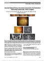
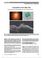
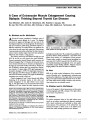
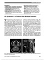
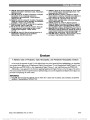
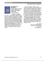
Show Diagnosis of Traumatic Optic Neuropathy: Application of Diffusion Tensor Magnetic Resonance Imaging Uttam K. Bodanapally, MBBS, Shanmuganathan Kathirkamanathan, MD, Elena Geraymovych, MD, Stuart E. Mirvis, MD, Andrew Y. Choi, MD, Alan B. McMillan, PhD, Jiachen Zhuo, PhD, Robert K. Shin, MD Background: Using diffusion tensor imaging, we evaluated the directional diffusivities of the optic nerve in patients with traumatic optic neuropathy (TON). Methods: Our study consisted of 12 patients with unilat-eral TON, 6 patients with severe traumatic brain injury (comparison group A), and 6 patients with normal conven-tional brain magnetic resonance imaging (comparison group B). The contralateral optic nerve in patients with TON also was evaluated (comparison group C). Two trauma radiologists, blinded to the clinical diagnosis, indepen-dently obtained the directional diffusivities. The intra-orbital optic nerve was divided into anterior and posterior segments to evaluate intersegmental differences in direc-tional diffusivities. Results: The mean axial diffusivity (AD) in both optic nerve segments and the mean diffusivity (ADC) in the posterior segment on the affected side were significantly lower and differentiated subjects with TON from those in comparison groups A and B. Area under the receiver operating charac-teristic curve was 0.762, 0.746, and 0.737 for posterior AD, anterior AD, and posterior ADC, respectively. The mean AD, mean diffusivity, and radial diffusivity were lower in the affected nerves in comparison to the contralateral nerve (comparison group C), but the values did not reach statistical significance. Conclusion: Decreased AD and mean diffusivity in the posterior segment of the optic nerve may serve as a bio-marker of axonal damage in patients with TON and merits further investigation as a predictor of initial visual acuity and potential visual recovery. Journal of Neuro-Ophthalmology 2013;33:128-133 doi: 10.1097/WNO.0b013e318283c3ed © 2013 by North American Neuro-Ophthalmology Society Traumatic optic neuropathy (TON) is a devastating acute injury of the optic nerve that causes disruption of visual function and leads to lifelong disability. TON is reported to have a high prevalence in young men in their early 30s (1). It is thought that the most common sites of nerve injury are at the foramina of the optic canal, the canalicular segment of the optic nerve, and under the falci-form dural fold (2). The concept of primary and secondary injury in TON has been proposed by Walsh (3). It is presumed that the primary injury occurs because of irreversible contusion necrosis from shearing of retinal ganglion cell axons at the time of impact. Secondary injury occurs because of destruc-tive postinjury metabolic and biochemical changes, resulting in optic nerve edema within the confines of the inflexible optic canal and exacerbates the ischemia and apoptosis (4). The diagnosis of TON primarily is based upon clinical findings. Conventional magnetic resonance imaging (MRI) and multidetector computed tomography often have normal optic nerve imaging findings in patients with TON. In their current form, these neuroimaging techniques are unable to consistently demonstrate optic nerve injury. This limitation led us to evaluate functional imaging techniques such as diffusion tensor imaging (DTI) in patients with TON. The human optic nerve is a white matter tract emanating from ganglion cells located in the retina. Diffusion tensor imaging offers a potential means to evaluate white matter injury. The cylindrical anatomy and symmetry of white matter tracts that run in the optic nerve makes it possible to Departments of Radiology (UKB, SK, SEM, AYC, ABM, JZ) and Ophthalmology and Visual Sciences (EG, RKS), University of Mary-land Medical Center, Baltimore, Maryland; and Department of Radiology (AYC), Air Force School of Aerospace Medicine, Wright- Patterson AFB, Ohio. Disclosures: The authors report no conflicts of interest. Supplemental digital content is available for this article. Direct URL citations appear in the printed text and are provided in the full text and PDF versions of this article on the journal's Web site (www. jneuro-ophthalmology.com). Address correspondence to Uttam K. Bodanapally, MBBS, Department of Radiology, University of Maryland Medical Center, 22S Greene Street, Baltimore, MD 21201; E-mail: ubodanapally@ umm.edu 128 Bodanapally et al: J Neuro-Ophthalmol 2013; 33: 128-133 Original Contribution Copyright © North American Neuro-Ophthalmology Society. Unauthorized reproduction of this article is prohibited. obtain directional diffusivities. Such measurements are helpful to predict axonal integrity and myelin disruption. Diffusion tensor imaging measurements are based upon changes in Brownian motion (diffusion) of water in white matter, influenced by barriers presented by axonal membranes and myelin sheaths. This technique measures the preferential directions of water diffusion across multiple spatial directions in the presence of a magnetic gradient. Diffusion of water in tissues is anisotropic (directionally dependent) or isotropic (directionally independent). Fractional anisotropy (FA) meas-ures the fraction of diffusivity that can be ascribed to anisotropic diffusion. A value of 0 is equivalent to diffusion that is same in all directions of 3-dimensional space as occurs in cerebrospinal fluid (CSF), which has no barriers to diffusion. A value of 1 is seen in maximum anisotropy, and diffusion is unidirectional. The architecture of the parallel white matter tracts, and their myelin sheaths facilitate diffusion of water molecules preferentially along the direction of the nerve fibers, hence the FA is closer to a value of 1. When there is damage to axonal membranes, diffusion at the injury site becomes unrestricted and isotropic, resulting in a decrease in FA value. Axial diffusivity (AD) represents water diffusion parallel to the axon fibers. Axonal injury results in decreased preferential diffusion along the fiber tracts, and AD decreases. Diffusion perpendicular to axonal fibers is denoted as radial diffusivity (RD). Myelin damage results in increased water diffusion in a perpendicular direction and increases RD. Mean diffusivity (ADC) is the average of diffusivities in all the directions. This measurement fails to account for the interfering effects of local barriers and calculates the value as if all the diffusion rates are solely because of Brownian motion. Mean diffusivity decreases in acute injury because of cellular swelling in cytotoxic edema. The intracellular compartment, bounded by cell membranes, organelles, and protein-rich environment, results in restriction of water movement. In addition, the water diffusion in extracellular spaces also becomes more restricted because of cell swelling. Both of these effects result in net decrease in mean diffusivity. We hypothesize that the directional diffusivity measure-ments [AD, mean diffusivity (ADC), RD and FA] may detect structural changes within white matter tracts of the optic nerve in patients with TON and provide neuro-imaging support for this clinical diagnosis. METHODS Our study was compliant with the Health Insurance Portability and Accountability Act, and permission was obtained from our institutional review board. The study was conducted at a level 1 trauma center. The inclusion criteria for this retrospective study were 1) presence of decreased visual acuity associated with relative afferent pupillary defect compatible with clinical diagnosis of TON, 2) history of blunt cranial trauma, 3) acquisition of diffusion tensor images as part of the MRI protocol of the brain (#15 days after trauma), and 4) age $18 years and older, regardless of sex. Comparison Groups Comparison group A: 6 age- and sex-matched patients with severe traumatic brain injury (TBI) with postresuscitation Glasgow coma scale (GCS) # 6T (T = tracheal intubation). Comparison group B: 6 age- and sex-matched patients with normal GCS, without relative afferent pupillary defect or abnormalities on conventional MRI sequences, who were scanned for reasons unrelated to head trauma. Comparison group C: contralateral optic nerves in study group patients with unilateral TON. MRI PROTOCOL AND REGION-OF-INTEREST ANALYSIS Magnetic Resonance Imaging All imaging was performed on a 1.5T Avanto scanner (Siemens Medical Solutions, Erlangen, Germany) with parallel imaging capability. For MRI protocol, see Supplemental Digital Con-tent 1, http://links.lww.com/WNO/A77. Diffusion gradients were sensitized in 6 or 12 collinear directions at an effective b value of 1,000 s/mm2. REGION-OF-INTEREST ANALYSIS All diffusion data were processed by using Trackvis software (A. A. Martinos Center for Biomedical Imaging, Depart-ment of Radiology, Massachusetts General Hospital, Bos-ton, MA). Maps of directional diffusivities were calculated using the AFNI tool "3dcalc" (5) automated by a MATLAB script (The Mathworks, Inc, Natick, MA). The region of interest (ROI) was manually placed over the optic nerve on the non-diffusion-weighted (b0) image, which was defined in the axial map and adjusted in the coronal and sagittal images obtained by multiplanar reconstruction using the Trackvis software (Fig. 1). To avoid CSF partial volume artifacts, the ROI mostly included voxels at the center of the optic nerve. The orbital optic nerve was divided into anterior and posterior segments on axial b0 images at approximately 10 mm behind the globe. The division was made to evaluate intersegmental differences in directional diffusivities as the posterior segment is thought to be more prone to injury. Regions of interest were selected indepen-dently by 2 radiologists blinded to the clinical status of the patients to include 10-15 voxels longitudinally in each seg-ment. Data were extracted using the AFNI tool "3dROI-stats" to calculate the mean and standard deviation of directional diffusivities, for each respective ROI automated by a MATLAB script. The average of the directional diffu-sivities obtained from each set of ROIs selected by the 2 radiologists was used for analysis. Bodanapally et al: J Neuro-Ophthalmol 2013; 33: 128-133 129 Original Contribution Copyright © North American Neuro-Ophthalmology Society. Unauthorized reproduction of this article is prohibited. Statistical Analysis Statistical analysis was performed using JMP software (versions 9 and 10; SAS Institute, Cary, NC). Analysis was conducted using Welch t test for unequal variances. For comparisons between each group, Welch t test was used to assess the difference in directional diffusivities for the ante-rior and posterior segments in the 4 comparison groups. Receiver operating characteristic (ROC) curve analysis was used to evaluate the usefulness of directional diffusivities in the diagnosis of TON. A P value of less than 0.05 was accepted as a statistically significant difference. RESULTS Demographics The ophthalmology database from May 2008 to December 2009 at the University of Maryland Medical Center revealed 44 patients with a diagnosis of TON. Twelve of these 44 patients with unilateral TON were evaluated with DTI and formed the basis of this study. Imaging was performed in these patients for evaluation of associated TBI. Demo-graphic, clinical, and imaging characteristics of the study group and comparison groups are given in Tables 1 and 2. Group Differences in Directional Diffusivities Table 3 summarizes the directional diffusivities of the optic nerve in both the anterior and posterior segments using the Welch t test. Each comparison group is correlated separately with the study group. Low Axial Diffusivity as a Predictor of TON The mean AD in both the posterior and the anterior segments of the optic nerve differentiated subjects with TON from those without TON in comparison group A (posterior segment, P = 0.012; anterior segment, P = 0.05) and compar-ison group B (posterior segment, P = 0.036; anterior segment, P = 0.027) (Fig. 2A, B). The mean AD in both the segments on the injury side were lower than in comparison group C, but the values were not statistically significant. Receiver oper-ating characteristic curve analysis for posterior AD determined an area under curve (AUC) of 0.762 and for anterior AD an AUC of 0.746 (see Figures E2 and E3, Supplemental Dig-ital Content 2 and 3, http://links.lww.com/WNO/A69 and http://links.lww.com/WNO/A70). Low Mean Diffusivity in the Posterior Segment as a Predictor of TON The mean of mean diffusivity in the posterior segment of the optic nerve differentiated subjects with TON from those without TON in comparison group A (P = 0.027) and comparison group B (P = 0.038) (Fig. 2C). Area under the ROC curve determined a discrimination ability TABLE 1. Characteristics of 12 patients with unilateral traumatic optic neuropathy Age, mean (range), y 35 (22-69) Sex (M:F) 7:5 Time to imaging, mean (range), d 7 (1-15) Side of TON, right:left 5:7 Diagnosis per brain MRI Normal structural MRI 2 Primary brain contusions 7 DAI 3 Type of injury MVC 9 Assault 2 Fall 1 DAI, diffuse axonal injury; MRI, magnetic resonance imaging; MVC, motor vehicle collision; TON, traumatic optic neuropathy. FIG. 1. A. Axial b0 averaged image showing the optic nerves. B. The region of interest (ROI) is placed at the central section of the optic nerve segments (anterior and posterior) on the b0 averaged magnified axial image using the Trackvis software. 130 Bodanapally et al: J Neuro-Ophthalmol 2013; 33: 128-133 Original Contribution Copyright © North American Neuro-Ophthalmology Society. Unauthorized reproduction of this article is prohibited. of 0.737 between TON and comparison group A and group B (see Figure E4, Supplemental Digital Content 4, http://links.lww.com/WNO/A71). Posterior RD and Anterior Mean Diffusivity The mean RD values in the posterior and anterior segments of the affected nerves were lower than the comparison group A and group B showing a trend toward statistical signifi-cance (0.05 , P , 0.1). The mean anterior RD and FA values in both the segments of the nerve showed no significant differences. There were no statistically significant differences in directional diffusivities between the comparison groups A and B. DISCUSSION The DTI results in our study likely correspond to alteration in optic nerve microstructure and may act as a noninvasive biomarker of axonal damage caused by TON. We found that DTI may help in identifying TON patients with area under ROC curves ranging from 0.737 to 0.762 for the signifi-cantly different directional diffusivities (6). The pattern of damage observed in our sample showed anterior-posterior gradient of decrease in mean diffusivity and AD, that is, greater deterioration of diffusion values in the posterior seg-ment of the optic nerve. This is consistent with greater pro-pensity of damage to the posterior segment of the optic nerve near the optic canal. TABLE 3. Correlations between diffusion parameters in traumatic optic neuropathy patients and comparison groups Groups Compared TON (95% CI), mm2/ms Group A (95% CI), mm2/ms Posterior AD 2.09 (1.74-2.45) 2.58 (2.45-2.71), P = 0.012 Anterior AD 2.29 (1.92-2.66) 2.66 (2.55-2.77), P = 0.05 Posterior ADC 1.41 (1.16-1.66) 1.71 (1.6-1.83), P = 0.027 Anterior ADC 1.61 (1.34-1.89) 1.85 (1.78-1.92), P = 0.09 Posterior RD 1.07 (0.86-1.28) 1.28 (1.15-1.41), P = 0.08 Anterior RD 1.27 (1.03-1.53) 1.44 (1.38-1.51), P = 0.17 Posterior FA 0.44 (0.37-0.5) 0.45 (0.41-0.49), P = 0.73 Anterior FA 0.39 (0.33-0.45) 0.4 (0.38-0.42), P = 0.83 Groups Compared Group B (95% CI), mm2/ms Group C (95% CI), mm2/ms Posterior AD 2.5 (2.31-2.7), P = 0.036 2.25 (2-2.5), P = 0.42 Anterior AD 2.74 (2.57-2.91), P = 0.027 2.42 (2.12-2.72), P = 0.55 Posterior ADC 1.71 (1.55-1.86), P = 0.038 1.54 (1.34-1.74), P = 0.38 Anterior ADC 1.90 (1.76-2.02), P = 0.067 1.7 (1.48-1.91), P = 0.6 Posterior RD 1.31 (1.17-1.45), P = 0.05 1.18 (0.99-1.38), P = 0.39 Anterior RD 1.46 (1.35-1.58), P = 0.15 1.34 (1.14-1.53), P = 0.68 Posterior FA 0.42 (0.4-0.44), P = 0.6 0.43 (3.66-4.49), P = 0.8 Anterior FA 0.41 (0.39-0.43), P = 0.52 0.4 (0.34-0.47), P = 0.78 Statistically significant differences (P , 0.05) are highlighted in bold. AD, axial diffusivity; ADC, near diffusivity; CI, confidence interval; FA, fractional anisotropy; RD, radial diffusivity; TON, traumatic optic neuropathy. TABLE 2. Characteristics of patients in comparison groups A and B Comparison Group A (n = 6) Comparison Group B (n = 6) Age, mean (range), y 38 (20-53) 37 (19-58) Sex (male:female) 4:2 4:2 Reason for performing MRI TBI = 6 Syncope = 2; tumor = 2; right hand tingling = 1; seizure (interictal) = 1 Primary brain contusions associated with DAI 4 0 Isolated DAI 2 0 GCS, mean (range) 5T (3T-7T) 15T Time to MRI, mean (range), d 6 (1-15) 1 (1-2) DAI, diffuse axonal injury; GCS, Glasgow coma scale; MRI, magnetic resonance imaging; TBI, total brain injury. Bodanapally et al: J Neuro-Ophthalmol 2013; 33: 128-133 131 Original Contribution Copyright © North American Neuro-Ophthalmology Society. Unauthorized reproduction of this article is prohibited. Pathological findings in TON include hemorrhage, demyelination, focal necrosis, and axonal damage (7). Closed-space edema, contusion necrosis, and infarction because of vascular thrombosis or spasm have all been impli-cated from autopsy studies (8). The decrease in axial and mean diffusivity found in our patients with TON could be because of axolemma damage, leading to axonal swelling (9). Axonal swelling also reduces space between neighboring axons, resulting in decreased RD (10). We found a similar reduction in posterior segment RD values although not to the level of statistical significance. Cytotoxic edema from either contusion or acute ischemia within the optic nerve from vascular thrombosis or spasm also may explain the decrease in radial and mean diffusivity values (10,11). Ischemia alone fails to explain the changes in direc-tional diffusivities in TON. In the affected optic nerve, AD, which is a surrogate for axonal injury, showed a disproportion-ate decrease in value compared with mean diffusivity and RD values. Hence, a combination of axonal damage because of contusion and nerve ischemia from vascular compression may be a plausible explanation for the diffusion changes in TON. There were no significant differences in FA values between the comparison groups. This is in contrast to the findings of Yang et al (12) who found a significant decrease in FA value in patients with TON. The same study also reported a significant increase in mean diffusivity values, in contrast to our results. Yang et al explained the increase in mean diffusivity from ischemic demyelination or necrosis of nerve fiber bundles within the short period (mean time from injury to examination, 5.2 days), when in fact acute ischemia should decrease the mean diffusivity values (10,11). Our observations showed a significant decrease in AD and posterior mean diffusivity values between injured nerve and optic nerves in comparison groups A and B. The reason for a nonstatistically significant reduction in AD and mean diffusivity between the affected optic nerve and the contralateral unaffected nerve in patients with TON remains unclear. Possible explanations include 1) presence of bilateral asymmetric optic nerve injury (13); 2) secondary damage of the contralateral optic nerve from associated TBI, mediated by various toxins released from dying cells result-ing in oxidative stress, excitotoxic damage, and apoptosis (14); and 3) secondary transsynaptic degeneration of the contralateral optic nerve, caused by injury to the visual cortex or optic radiations from TBI (15). A major limitation of our study was that it was retrospective, without a uniform orbital MRI protocol. Other limitations include the small number of patients, the inherent difficulty in imaging the optic nerve because of its size, mobility, and surrounding air in the paranasal sinuses. Interpretation of diffusion tensor anisotropy and selection of ROI were complicated by a variety of factors including artifacts and partial volume averaging, which may have influenced the measured directional diffusivities (10). There were time varia-tions between the injury and acquisition of MRI as some of the patients were critically ill. Although time variations and differ-ences in age and sex may have influenced the results, time to imaging after trauma, age and sex were matched between the groups. ACKNOWLEDGMENT The authors thank Brigitte Pocta for reviewing the article. REFERENCES 1. Lee V, Ford RL, Xing W, Bunce C, Foot B. Surveillance of traumatic optic neuropathy in the UK. Eye. 2010;24:240-250. FIG. 2. Scatterplots of diffusivity data. A. Posterior axial diffusivity (AD). B. Anterior axial diffusivity. C. Posterior mean dif-fusivity. Diffusivity = mm2/sec; small horizontal lines = average diffusivities and associated 95% confidence intervals. TBI, traumatic brain injury; TON, traumatic optic neuropathy. 132 Bodanapally et al: J Neuro-Ophthalmol 2013; 33: 128-133 Original Contribution Copyright © North American Neuro-Ophthalmology Society. Unauthorized reproduction of this article is prohibited. 2. Steinsapir KD, Goldberg RA. Traumatic optic neuropathy: a critical update. Compr Ophthalmol Update. 2005;6: 11-21. 3. Walsh FB. Pathological-clinical correlations: I. Indirect trauma to the optic nerves and chiasm. II. Certain cerebral involvements associated with defective blood supply. Invest Ophthalmol. 1966;5:433-449. 4. Steinsapir KD, Goldberg RA. Traumatic optic neuropathy. Surv Ophthalmol. 1994;38:487-518. 5. Cox RW. AFNI: software for analysis and visualization of functional magnetic resonance neuroimages. Comput Biomed Res. 1996;29:162-173. 6. Hosmer DW, Lemeshow S. Applied Logistic Regression. Wiley Series in Probability and Statistics, 2nd edition. New York, NY: Wiley, 2000. 7. Nau HE, Gerhard L, Foerster M, Nahser HC, Reinhardt V, Joka T. Optic nerve trauma: clinical, electrophysiological and histological remarks. Acta Neurochir (Wien). 1987;89:16-27. 8. Cockerham KP. Traumatic optic neuropathy. In: Thach AB, ed. Ophthalmic Care of the Combat Casualty. Washington, DC: Office of The Surgeon General, TMM Publications, 2003:395- 403. 9. Mac Donald CL, Dikranian K, Bayly P, Holtzman D, Brody D. Diffusion tensor imaging reliably detects experimental traumatic axonal injury and indicates approximate time of injury. J Neurosci. 2007;27:11869-11876. 10. Alexander AL, Lee JE, Lazar M, Field AS. Diffusion tensor imaging of the brain. Neurotherapeutics. 2007;4:316-329. 11. Sorensen AG, Wu O, Copen WA, Davis TL, Gonzalez RG, Koroshetz WJ, Reese TG, Rosen BR, Wedden VJ, Weisskoff RM. Human acute cerebral ischemia: detection of changes in water diffusion anisotropy by using MR imaging. Radiology. 1999;212:785-792. 12. Yang QT, Fan YP, Zou Y, Kang Z, Hu B, Liu X, Zhang GH, Li Y. Evaluation of traumatic optic neuropathy in patients with optic canal fracture using diffusion tensor magnetic resonance imaging: a preliminary report. ORL J Otorhinolaryngol Relat Spec. 2011;73:301-307. 13. Crompton MR. Visual lesions in closed head injury. Brain. 1970;93:785-792. 14. Levin LA. Axonal loss and neuroprotection in optic neuropathies. Can J Ophthalmol. 2007;42:403-408. 15. Hoyt CS. Brain injury and the eye. Eye. 2007;21: 1285-1289. Bodanapally et al: J Neuro-Ophthalmol 2013; 33: 128-133 133 Original Contribution Copyright © North American Neuro-Ophthalmology Society. Unauthorized reproduction of this article is prohibited. |