| OCR Text |
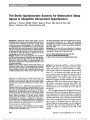
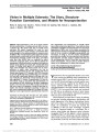
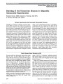
Show Protective Effect of Bax Ablation Against Cell Loss in the Retinal Ganglion Layer Induced by Optic Nerve Crush in Transgenic Mice Nitza Goldenberg-Cohen, MD, Olga Dratviman-Storobinsky, MSc, Shimrit Dadon Bar El, MSc, Yelena Cheporko, MD, Edith Hochhauser, PhD Background: Bax expression is a prerequisite for retinal ganglion cell (RGC) apoptosis. Experimental studies have reported Bax protein upregulation following optic nerve transection. The stimuli that trigger apoptosis share a common executioner proteolysis cascade, including caspase-3 and poly-(adenosine diphosphate ribose) poly-merase cleavage. This study sought to elucidate the role of the mitochondrial apoptotic pathway in RGCs using a Bax transgenic knockout mouse model. Methods: The right optic nerves of 26 C57BL mice, 7 Bax2/2, 7 Bax+/2, and 12 Bax+/+, were subjected to crush injury and analyzed for apoptosis and neuronal cell loss on days 1, 3, and 21. Levels of Bax, Bcl-2, and caspase-3 messenger RNA expression were determined with real-time polymerase chain reaction. Results: Multiple apoptotic cells were detected in the retinas of the Bax +/+ and Bax +/2 mice at days 1 and 3, but not in the Bax2/2 mice. The Bax/Bcl-2 ratio was higher in the Bax+/+ than in the Bax+/2 mice on day 1 (1.33 and 0.83, respectively), with a trend toward an increase on day 3 (1.47 and 1.66, respectively); Bax/Bcl- X showed the same elevation on day 1 in the wild-type mice (1.34) but decreased on day 3 (0.8). Bax gene expression was undetectable in the Bax2/2 mice. Cas-pase- 3 gene expression was higher in the Bax+/+ than in the Bax+/2 mice on day 1 and dropped toward baseline on day 3. The opposite trend was noted in the Bax2/2 mice. Conclusion: The lack of apoptosis combined with the re-duction in proapoptotic genes in the Bax2/2 mice after injury compared to the Bax+/+ and Bax+/2 mice suggests that Bax plays a crucial role in the induction of apoptosis. Suppression of Bax expression may reduce retinal cell loss. Journal of Neuro-Ophthalmology 2011;31:331-338 doi: 10.1097/WNO.0b013e318227e4fb 2011 by North American Neuro-Ophthalmology Society Ischemic injury causes cell death by mechanisms of apo-ptosis and necrosis. The Bax gene has been found to be associated with neuronal cell apoptosis following traumatic brain injury (1) and global brain ischemia (2), whether ge-netically or chemically induced. At 24 hours after trauma, Bax-null mice had fewer TdT-mediated dUTP nick end-labeling (TUNEL)-positive cells than wild-type mice, and at 21 days after injury, there was a higher percentage of surviving neurons in the hippocampal region (1). Accordingly, the introduction of Bax channel inhibitors prevented cytochrome C release from mitochondria, inhibited the decrease in mitochondrial mem-brane potential, and protected cells against apoptosis (2). These investigators did not affect the conformational activation of Bax or its translocation and insertion into the mitochondrial membrane in cells undergoing apoptosis (2). Using a different model of transgenic mice, others found that overexpression of Bax was associated with an increased number of apoptotic nuclei, especially in the cerebral cortex (3). The retrograde degeneration of retinal ganglion cells (RGCs) as a consequence of optic nerve lesions fulfills the criteria of apoptosis (4). Studies in animal models dem-onstrated that transection of the optic nerve is followed by upregulation of Bax protein expression in the RGCs and that the induction of Bax expression is a prerequisite for Pediatric Unit (NG-C), Department of Ophthalmology, Schneider Children's Medical Center of Israel, Petah Tiqwa, Israel; The Krieger Eye Research Laboratory (NG-C, OD-S, SDBE) and Laboratory of Cardiac Research (YC, EH), Felsenstein Medical Research Center, Petah Tiqwa, Israel; and Sackler School of Medicine (NG-C, EH), Tel Aviv University, Tel Aviv; Israel. Supported by the Zanvyl and Isabelle Krieger Fund, Baltimore, MD; Eldor-Metzner Clinician Scientist Award, Chief Scientist, Israel Ministry of Health (grant no. 3-3741 for N.G.-C.). Presented in part at the Rabin Medical Center Research Day, 2008, and at the annual meeting of the Israel Society for Eye and Vision Research, Neve Ilan, Israel, March 2009. N. Goldenberg-Cohen and O. Dratviman-Storobinsky contributed equally to this study. The authors report no conflicts of interest. Address correspondence to Nitza Goldenberg-Cohen, MD, The Krieger Eye Research Laboratory, Felsenstein Medical Research Center, Beilinson Campus, Petah Tiqwa 49100, Israel; E-mail: ncohen1@gmail.com Goldenberg-Cohen et al: J Neuro-Ophthalmol 2011; 31: 331-338 331 Basic Science in Neuro-Ophthalmology Copyright © North American Neuro-Ophthalmology Society. Unauthorized reproduction of this article is prohibited. RGC apoptosis (5). Bax deficiency protected RGCs after axonal injury by optic nerve crush (6). The specific molecular pathways involved in transient is-chemia- induced apoptosis in the retina are still unclear. Early in vitro studies found that various stimuli that trigger the apoptotic process in retinal cells share a common executioner proteolysis cascade that includes caspase-3 and poly-(adenosine diphosphate ribose) polymerase cleavage. Furthermore, re-gardless of the stimulus, Bax expression increased during ap-optosis, whereas Bcl-2 expression decreased. Treatment with caspase inhibitors blocked the apoptotic cell death (7). More recent in vitro and in vivo studies demonstrated that purified RGC damage induced by hypoxia involves a Bax-dependent apoptotic pathway (8). A rat model of central artery occlusion was characterized by apoptotic changes in the inner and outer retinal cell layers (9). Using Western blot analysis, the authors found that mitochondrial translocation of Bax from the cytoplasm started at 3 hours and peaked at 6 hours after damage induction. The translocation was accompanied by cytosolic accumulation of cytochrome C and cleavage of caspase-9. Previous studies evaluated the expression of Bax, Bcl-2, Bcl- X, and caspase-3 genes in the rat retina (9,10). The murine optic nerve crush model simulates the clinical features of traumatic-ischemic damage to the retina. Prompted by our earlier findings that apoptosis is involved in RGC death (11), we sought to elucidate the role of the mitochondrial apoptotic pathway using a Bax transgenic knockout mouse model. A better understanding of ischemia-induced retinal apoptosis may have implications for the development of antiapoptosis agents targeted at the mitochondrial pathway for the treatment of retinal ischemic diseases. METHODS Experimental Animals All protocols were conducted in accordance with the As-sociation for Research in Vision and Ophthalmology Statement for the Use of Animals in Ophthalmic and Vision Research and were approved and monitored by the Animal Care Committee of Rabin Medical Center. The animals were housed under a 14-hour light/10-hour dark cycle with standard chow and water ad libitum. Seven C57BL/6J Bax2/2 (Baxtm1Sjk knockout) congenic mice, 7 C57BL Bax+/2 (heterozygous) mice (inbred), and 12 C57BL/6 Bax+/+ (wild-type) mice (from the colony) aged 10 to 12 weeks were kindly provided by Professor Daniel Offen (Neuroscience Laboratory, Felsenstein Medical Re-search Center, Petah Tiqwa, Israel) (Table 1). Mice geno-type was determined by polymerase chain reaction (PCR) as previously described (12), using the following primers: Bax in, GTT GAC CAG AGT GGC GTA GG; Bax ex, TGA TCA GAA CCA TCA TG; and Neo rev, CCG CTT CCA TTG CTC AGC GG. Induction of Crush Injury All 26 mice used in the study were anesthetized with ketamine (80 mg/kg)/xylazine (4 mg/kg) supplemented with topical anesthesia (0.5% proparacaine hydrochloride). Optic nerve crush was performed as previously described (11). In brief, the right optic nerve was crushed by applying forceps at 2.5 to 3.0 mm posterior to the globe for 7 seconds; this procedure was performed 3 times. The left (untreated) eye served as a control. Subsets of mice in each group were euthanized at days 1, 3, and 21 after injury (Table 1); the last time pointwas selected on the basis of findings that maximal RGC loss occurs 21 days after injury induction (11,13). Paraffin sections of the eyes were analyzed by light microscopy. Histological Examination Seven mice were used for histological analysis on day 21 after injury induction. Following euthanization, both the eyes were enucleated and embedded in paraffin, and 5-mm-thick sagittal sections were stained with hematoxylin and eosin and examined under a light microscope. Total retinal thickness was measured at various fields of each section (3 sections in a slide), in a total of 5 to 8 slides. Meas-urements were performed in triplicate at 180-mm intervals, from the internal to the external limiting membrane. The mean number of cells in the retinal ganglion layer was calculated in a 340 magnification field of 3 consecutive sections per slide. Each section was sampled in at least 8 different areas. Apoptosis Assays Longitudinal cross-sections (5 mm thick) from the paraffin-embedded eyes of 8 mice euthanized on days 1 and 3 were prepared for in situ TUNEL assay (Roche Diagnostics GmbH, Roche Applied Science, Mannheim, Germany); staining was performed with the fluorescein-tagged apo-ptosis detection system. Results were analyzed with a fluo-rescence microscope (Fluoview X; Olympus, Tokyo, Japan) at 580 nm wavelength. The mean number of TUNEL-positive cells per slide was determined in 3 consecutive sections per slide, 1 slide for every 10 sections of 5-mm thickness (total 8 slides per eye), with attention to the in-ternal retinal layers. Findings were compared between the TABLE 1. Number of mice used for the histological and molecular analyses Bax2/2 Bax+/2 Bax+/+ Total Apoptosis assay (days 1 and 3) 2 2 4 8 H&E (day 21) 2 2 3 7 Molecular analysis (days 1 and 3) 3 3 5 11 Total 7 7 12 26 H&E, hematoxylin and eosin staining. 332 Goldenberg-Cohen et al: J Neuro-Ophthalmol 2011; 31: 331-338 Basic Science in Neuro-Ophthalmology Copyright © North American Neuro-Ophthalmology Society. Unauthorized reproduction of this article is prohibited. injured (right) and control (left) eyes of the individual mice and between the injured eyes of the knockout mice and the 2 control groups (Bax+/+ and Bax+/2). Complementary DNA Preparation Retinal tissues were dissected in a total of 11 mice eutha-nized on days 1 and 3 after crush injury and snap-frozen in liquid nitrogen. Total RNA was isolated using TRIzol reagent (Invitrogen, Life Technologies, Carlsbad, CA), according to the manufacturer's protocol, and then reverse transcribed into complementary DNA (cDNA) using ran-dom hexamers (Amersham Biosciences, Buckinghamshire, United Kingdom) and Moloney murine leukemia virus reverse transcriptase (Promega, Madison, WI). Apoptosis-Related Gene Expression Two-stage real-time quantitative polymerase chain reaction (RT-QPCR) was applied to evaluate the expression of Bax, Bcl- 2, Bcl-X, and caspase-3 genes after crush injury. The primers list will be provided upon request. Primer efficiency was cal-culated. We used mouse beta actin (ACTB) to normalize the cDNA input levels because it is known to remain stable under ischemic conditions (14). Analyses were done with the Se-quence Detection System (Prism 7000; Applied Biosystems, Foster City, CA). Reactions were performed in a 20-mL vol-ume containing 4 mL of cDNA, 0.5 mM each of the forward and reverse primers, and buffer included in the Master Mix (SYBR Green I; Applied Biosystems). Cycling conditions consisted of an initial denaturation step of 95 C for 10minutes followed by 50 cycles of 1 minute at 95 C and 1 minute of annealing and extension at 60 C. Triplicate RT-QPCRs were performed for each gene to minimize individual tube vari-ability, and an average was taken at each time point. Threshold cycle efficiency corrections were calculated, and melting curves were obtained using cDNA for each individual gene PCR assay; standard curves were obtained using untreated mouse cDNA for each gene PCR assay. The results were quantified by the comparative threshold cycle (Ct) method (22DDCt method) (15), where DDCt ¼ DCt ðsampleÞ DCt ðreference geneÞ: Statistical Analysis Group differences in histological and molecular findings were analyzed by Student t test. RESULTS Cell Loss in the RGC Layer Following crush injury, the number of cells in the RGC layer (RGCs and amacrine) decreased significantly in the wild-type and heterozygous Bax mice, by 44% and 23%, respectively (Fig. 1, Table 2). The knockout mice had an increased number of cells in the RGC layer even under normal conditions (left eye) (Fig. 1). After crush injury, the number of cells decreased by 16%, but it was still above the mean of the wild-type mice. Retinal Thickness Retinal thickness in the wild-type mice remained constant in the left control eye (mean 6 SD, 219 6 36 mm; range, 206- 225 mm; 17% thinning) at 21 days after injury but decreased significantly in the treated eye (mean 6 SD, 182 6 30 mm; range, 175-200 mm; P, 0.05). There was only a mild change in retinal thickness after injury in the Bax-knockout mice (mean 6 SD, 329 6 30 mm vs 338 6 31 mm of the left control eye; range, 3%), and the final value was significantly higher for the treated wild-type mice (P, 0.05). The retinas of the heterozygous mice were thicker than those of the wild-type mice, measuring 333634 mm in the left eye (range, 284-369 mm) and 299 6 31 mm in the right eye (range, 247-384 mm; 11% postinjury thinning). Cell Apoptosis Maximal apoptosis of the cells in the retinal ganglion layer was detected on days 1 or 3 after injury in the heterozygous mice (12% and 20%, respectively) and the wild-type mice (15% and 35%, respectively). In the transgenic knockout mice, no apoptotic cells were detected at these time points (Fig. 2, Table 2). Apoptotic Gene Expression Bax expression in the wild-type mice was elevated on day 1 and returned to baseline on day 3; in the heterozygous mice, it remained low on days 1 and 3; in the knockout mice, it was undetectable (Table 3). Levels of Bcl-2 and Bcl-X ex-pression in the wild-type mice were elevated on day 1 and then returned to baseline (Bcl-X) or below (Bcl-2). In the heterozygous mice, Bcl-2 levels were below baseline, like the Bax levels. The Bax/Bcl-2 ratio was higher in the wild-type group than that in the heterozygous group on day 1, with a trend for an increase in both the groups on day 3. The Bax/Bcl-X ratio increased on day 1, but decreased to 0.8 on day 3. Caspase-3 expression was higher on day 1 in the wild-type group than in the heterozygous group and decreased toward baseline on day 3. The knockout mice showed an opposite trend, with a decrease in caspase-3 to below baseline on day 1 and its return toward baseline thereafter. DISCUSSION Optic nerve crush damage leads to retinal cell death. Ap-optosis is known to play a key role in cell death after retinal ischemia. Apoptosis has been described in various models of ischemic ocular injuries, including crush (10,11), glaucoma (6), retinal ischemia (9,16), ischemic optic neuropathy Goldenberg-Cohen et al: J Neuro-Ophthalmol 2011; 31: 331-338 333 Basic Science in Neuro-Ophthalmology Copyright © North American Neuro-Ophthalmology Society. Unauthorized reproduction of this article is prohibited. (13,17), and photoreceptor light damage (18). Studies in transgenic animals suggest that the mitochondrion-medi ated apoptosis pathway is involved in ischemia-related cell death. This pathway is triggered by activation of proa-poptotic members of the Bcl-2 family of genes (2,10). Accordingly, Bcl-2, Bax, and Bcl-X, as well as activated caspases, may be necessary players in the control of pro-grammed cell death in the RGC layer (19). Studies have reported an upregulation of Bax in RGCs after injury (5); increased survival of injured RGCs when Bax or caspase-3 was inhibited (19); and prevention of RGC death by ab-lation of the Bax protein (19). In line with these findings, the present study showed that survival of retinal cells of the inner retina, especially in the RGC layer, is increased in Bax-knockout mice subjected to crush injury compared to wild-type and heterozygous Bax mice and that Bax ablation protects the RGCs from apoptosis. It has been proposed that the ratio of Bcl-2 to Bax or other members of the Bcl-2 family may govern the sensi-tivity of cells to apoptotic stimuli. Therefore, we in-vestigated the relationship of the Bax (apoptosis promotor)/ Bcl-2 (apoptosis inhibitor) ratio and the Bax/Bcl-X (apo-ptosis inhibitor) ratio and compared the findings with TUNEL staining for apoptosis. On day 1, both the Bax/Bcl- 2 and Bax/Bcl-X ratios were upregulated. On day 3, the Bax/Bcl-2 ratio remained high, but the Bax/Bcl-X returned to baseline, in agreement with our histological finding of cell preservation. This suggests that the level of Bcl-X may better reflect the apoptosis state of the RGC layer than the level of Bcl-2 (10). It is of note that we did not find sig-nificant changes in these ratios. In a previous ischemia study (20), the Bax/Bcl-2 and Bax/Bcl-X ratios were not modified early after injury, but both were downregulated in the recovery phase, 24 hours later. FIG. 1. RGC loss 21 days after crush injury in Bax2/2 and Bax+/+ mice (340). A. Left (control) eye of Bax+/+ mouse without injury. Note the normal retinal thickness and continuity of the RGC monolayer. B. Bax+/+, 21 days after crush injury demonstrates RGC loss and thinning of the retina (arrows). C. Left (control) eye of Bax+/2 mouse without injury reveals increased retinal thickness and continuity of the RGC monolayer. D. Bax+/2, 21 days after crush injury reveals RGC loss and thinning of the retina (arrows). E. Left (control) eye of Bax2/2 mouse with increased retinal thickness and multiple nuclei in the RGC layer (compared to A). F. Bax2/2 mouse, 21 days after crush injury. There is a minor loss of RGCs (16%) without retinal thinning. RGC number remains high compared with normal retina. 334 Goldenberg-Cohen et al: J Neuro-Ophthalmol 2011; 31: 331-338 Basic Science in Neuro-Ophthalmology Copyright © North American Neuro-Ophthalmology Society. Unauthorized reproduction of this article is prohibited. Some investigators have suggested that in ganglion cells and other cell types of the retina, Bcl-X serve as the major antiapoptotic gene (10), being at least 16-fold more abundant than Bcl-2. Although Bcl-X may better reflect the antiapoptotic state, our results showed only a mild differ-ence in the level of expression between Bcl-2 and Bcl-X at 3 days after optic nerve crush injury. In our study, RT-QPCR analysis performed 1 day after induction of ischemia yielded increased levels of Bax, Bcl-2, and Bcl-X in the wild-type mice, which decreased by day 3 (to a lesser extent for Bcl-X). Similarly, in a previous report on ischemia-induced apoptosis (20), a time-dependent decrease was noted in Bax, Bak, and Bcl-X messenger RNA (mRNA) expression, but there was no alteration in Bcl-2 mRNA. Although we did not measure protein levels, the earlier study (20) reported that the levels of the proapoptotic Bax and Bak proteins remained unchanged, whereas the Bcl-2 and Bcl-X proteins were significantly upregulated. Caspase-3 expression was associated with an increase in cell loss in the wild-type group. At 1 day after injury, caspase-3 levels increased in the wild-type group, and to a lesser extent in the heterozygous group, but there was no change in the knockout group. These results are similar to those reported earlier in ischemic retinas 24 hours after reperfusion (20). It is noteworthy that caspase-3 mRNA expression may not reflect the level of the activated protein. An earlier study examined the retinas of Bax-knockout rd mice for cell death during development, with specific at-tention to photoreceptor degeneration (21). Although Bax deficiency had no effect on photoreceptor and retinal de-generation, the total number of RGCs rose to 226% of normal. Our findings of an increase in retinal thickness and cell number in the retina are in line with this report. However, in our study, the increase in retinal thickness was associated with an increased number of nuclei in all retinal layers, including the outer nuclear layer. The sham-operated Bax-null mice had a significantly greater nuclear volume in the retina than the wild-type mice, similar to the findings reported by Vecino et al (22) in developing retina and the findings of Tehranian et al (1) of increased brain volume in Bax2/2 mice. We noted a mean retinal thickness of 241 mm (range, 216-257 mm) in the Bax2/2 mice compared to a mean retinal thickness of 219 mm in the wild-type mice (range, 206-225 mm). After injury, values in the inner nuclear layer were reduced, but not in the Bax-knockout mice, where there was no evidence of cell loss. Isenmann et al (4) reported rapid elevation of Bax protein expression in rat retinas following crush injury. Levels peaked at 3 days and remained upregulated for at least 1 week thereafter. The same group (5) also reported upregulation of the Bax protein in rat retinas as early as 2 hours following axotomy, again peaking at 3 days after lesion induction. However, these findings were based on immunofluorescent studies, before it was understood that during apoptosis, the Bax protein translocates and concentrates in the mitochondria. TABLE 2. Histological changes in the retina after crush injury Cell Count in the RGC Layer, 21 Days, Mean (6SD) (n = 7) Cell Loss in the RGC Layer (%) Retinal Thickness, Mean (6SD) (n = 7) Apoptotic Cells, % (n/RGC in Field) (n = 8)* Group Crush (RE) Control (LE) Crush (RE) Crush (RE) Control (LE) Day 1 Day 3 Wild-type 14.42* (62.53) 25.77* (62.36) 44.0 (611.32) 252 (624) 297 (644) 43 (10/23) 64 (18/28) Bax+/2 25.96 (60.29) 34.03 (60.96) 23.5 (61.83) 299 (631) 335 (634) 15 (3/20) 20 (4/23) Bax2/2† 32.35* (64.46) 38.45 (62.85) 15.9 (62.50) 329 (630) 338 (631) 0 (0/59) 0 (0/49) Retinal sections analyzed for apoptosis were from the central retina: 50 mm diameter around the optic nerve. *P , 0.05, when comparing cell loss between crush and control of the wild-type and also when comparing crush of Bax2/2 to wild-type mice. †The number of RGCs in the Bax2/2 mice is increased. No apoptosis was detected by TUNEL staining on days 1 and 3. On day 21, there was a 15.9% cell loss in the RGC layer, yet the number of cells remained significantly higher than that in Bax+/+ mice. Only in the Bax+/+ was retinal thinning detected. No change in the retinal thickness was detected in the Bax2/2 mice. LE, left eye; RE, right eye. Goldenberg-Cohen et al: J Neuro-Ophthalmol 2011; 31: 331-338 335 Basic Science in Neuro-Ophthalmology Copyright © North American Neuro-Ophthalmology Society. Unauthorized reproduction of this article is prohibited. Thus, staining may give the appearance of an increase in ex-pression because of the aggregation phenomenon. Since the report of Isenmann et al (4), there has been at least 1 in-dependent study describing an increase in Bax mRNA ex-pression in the axotomy paradigm (23) and 2 studies that failed to detect any significant increase (24,25). In the present study, Bax mRNA levels were increased in the wild-type group as early as 1 day after injury. Semaan et al (24) performed gene dosage experiments in mice, yielding a single wild-type Bax allele. Their findings indicated that genetic background influences the cell death phenotype, including an RGC cell line. This study supports FIG. 2. Apoptosis assay 1 day and 3 days after crush injury in Bax2/2, Bax+/2, and Bax+/+ mice (340). Upper panel: Apoptosis was detected in the RGC layer on day 1 (A-C, TUNEL-positive cells stained in red, arrows) and day 3 (D-F, TUNEL-positive cells stained in red, arrows) in the Bax+/+ (C, F) and Bax+/2 (B, E) mice but not in the Bax2/2 mice (A, D). Lower panel: The graphs summarize apoptosis on day 1 and 3. Wt, wild type (Bax+/+). Control assay (left eye) of Bax2/2 and Bax+/+ mice. 336 Goldenberg-Cohen et al: J Neuro-Ophthalmol 2011; 31: 331-338 Basic Science in Neuro-Ophthalmology Copyright © North American Neuro-Ophthalmology Society. Unauthorized reproduction of this article is prohibited. the assumption that quantitative expression of the Bax gene is important in neuronal susceptibility to damaging stimuli. Further support was provided by the elevated Bax mRNA expression in the susceptible species. In the present study, Bax mRNA expression levels were elevated in the wild-type mice at day 1 after crush injury and not in the Bax+/2 mice. Both the groups, on day 3, showed almost same (baseline) levels of Bax expression. The decreased apoptosis rate in the inner retina of the transgenic Bax-knockout mice corresponds to observations in a glaucoma model (6) and in ischemic brain (2), liver (26), and heart (12) tissues in other transgenic mouse models. The greater tolerance of the RGCs in the Bax-knockout mice after crush injury also agrees with earlier studies of ischemic ocular injury (20). In wild-type mice in these studies, the number of TUNEL-positive cells reached maximum at 24 to 72 hours after injury (8,11,13,16,17). In contrast, following light damage, Bax2/2 mice showed no reduction in the number of TUNEL-positive photoreceptor nuclei at 24 hours or 7 days (18). The higher percentage of surviving cells in the RGC layer in our knockout group at 21 days following crush parallels the results reported in the hippocampal region in a transgenic mouse model of neuronal posttraumatic damage (1). Together, these findings support the notion that deletion of the Bax gene ameliorates RGC death after crush injury. The finding that apoptosis is a significant mechanism of cell death in various eye diseases (ischemia, trauma, or glaucoma, as many other conditions) may have important implications for the development of new treatments that specifically block or interfere with RGC apoptosis. Al-though this means preventing the result of the disease, not managing its cause, in many patients, ganglion cell death has already been stimulated by the time of diagnosis and continues to progress after conventional treatment. REFERENCES 1. Tehranian R, Rose ME, Vagni V, Pickrell AM, Griffith RP, Liu H, Clarle RS, Dixon CE, Kochanete PM, Graham SH. Disruption of bax protein prevents neuronal cell death but produces cognitive impairment in mice following traumatic brain injury. J Neurotrauma. 2008;25:755-767. 2. Hetz C, Vitte PA, Bomvrun A, Rostoytseva TK, Montessuit S, Hiver A, Schwartz MK, Church DJ, Korsmeyer J, Martinon JC, Antonsson B. Bax channel inhibitors prevent mitochondrion-mediated apoptosis and protect neurons in a model of global brain ischemia. J Biol Chem. 2005;280:42960-42970. 3. Teles AV, Ursheno RP, Dorta DJ, Lopes GS, Itsu YT, Smaili SS. Increase in bax expression and apoptosis are associated in Huntington's disease progression. Neurosci Lett. 2008; 438:59-63. 4. Isenmann S, Wahl C, Krajewski S, Reed JC, Bahr M. Up-regulation of Bax protein in degenerating retinal ganglion cells precedes apoptotic cell death after optic nerve lesion in the rat. Eur J Neurosci. 1997;9:1763-1772. 5. Isenmann S, Engel S, Gillardon F, Bahr M. Bax antisense oligonucleotides reduce axotomy-induced retinal ganglion cell death in vivo by reduction of Bax protein expression. Cell Death Differ. 1999;6:673-682. 6. Libby RT, Li Y, Savinora OV, Barter J, Smith RS, Nickells RW, John SW. Susceptibility to neurodegeneration in a glaucoma is modified by Bax gene dosage. PLoS Genet. 2005;1:17-26. TABLE 3. Relative changes in apoptosis-related gene expression after crush injury Group Apoptosis Gene Expression Bax Bcl-2 Bcl-x Bax/Bcl-2 Bax/Bcl-x Caspase-3 Day 1 Day 3 Day 1 Day 3 Day 1 Day 3 Day 1 Day 3 Day 1 Day 3 Day 1 Day 3 Wild-type (n = 5) 2.05 6 0.9 0.97 6 0.1 1.54 6 0.6 0.66 6 0.1 1.52 6 0.9 1.2 6 0.4 1.33 1.47 1.34 0.8 1.94 6 0.1 1.14 6 0.01 Bax+/2 (n = 3) 0.66 0.85 6 0.4 0.79 0.51 6 0.3 - - 0.83 1.66 - - 1.12 1.11 6 0.06 Bax2/2 (n = 3) - - 0.32 0.76 6 0.5 0.24 0.51 60.3 - - - - 0.31 0.94 6 0.3 Goldenberg-Cohen et al: J Neuro-Ophthalmol 2011; 31: 331-338 337 Basic Science in Neuro-Ophthalmology Copyright © North American Neuro-Ophthalmology Society. Unauthorized reproduction of this article is prohibited. 7. Tezel G, Wax MB. Inhibition of caspase activity in retinal cell apoptosis induced by various stimuli in vitro. Invest Ophthalmol Vis Sci. 1999;40:2660-2667. 8. Chen YN, Uamada H, Ma OW,Matsuyama S, AiharaM, ArailM. Hypoxia-induced retinal ganglion cell death and the neuroprotective effects of beta-adrenergic antagonists. Brain Res. 2007;1148:28-37. 9. Zhang Y, Cho CH, Atchaneeyasakul EO, McFarland T, Appuleuttan B, Stout JT. Activation of the mitochondrial apoptotic pathway in a rat model of central retinal artery occlusion. Invest Ophthalmol Vis Sci. 2005;46:2133-2139. 10. Levin LA, Schlamp CL, Spieldoch RL, Geszvain M, Nickells RW. Identification of the bcl-2 family of genes in the rat retina. Invest Ophthalmol Vis Sci. 1997;38:2545-2553. 11. Dratviman-Storobinsky O, Hasanreisoglu M, Offen D, Barhum Y, Weinberger D, Goldenberg-Cohen N. Progressive damage along the optic nerve following induction of crush injury or rodent anterior ischemic optic neuropathy in transgenic mice. Mol Vis. 2008;14:2171-2179. 12. Hochhauser E, Kivity S, Offen D,Maulok N, Ofani H, Barhum Y, Pannet H, Shrey-Vays Y, Shainberg G, Goldshtaub Y, Tobar A, Vidne BA. Bax ablation protects against myocardial ischemia-reperfusion injury in transgenic mice. Am J Physiol Heart Circ Physiol. 2003;284:H2351-H2359. 13. Bernstein SL, Guo Y, Slater BJ, Puche A, Kelman SE. Neuron stress and loss following rodent anterior ischemic optic neuropathy in double-reporter transgenic mice. Invest Ophthalmol Vis Sci. 2007;48:2304-2310. 14. Goldenberg-Cohen N, Dadon S, Avraham BC, Kramer M, HasanreisogluM, Eldar I, Weinberger D, Bahar I.Molecular and histological changes following central retinal artery occlusion in a mouse model. Exp Eye Res. 2008;87:327-333. 15. Giulietti A, Overbergh L, Valckx D, Decallone B, Bouillon R, Mathieu C. An overview of real-time quantitative PCR: applications to quantify cytokine gene expression. Methods. 2001;25:386-401. 16. Kaneda K, Kashii S, Kurosawa T, Kaneko S, Akaike A, Honda Y, Minami M, Satoh M. Apoptotic DNA fragmentation and upregulation of Bax induced by transient ischemia of the rat retina. Brain Res. 1999;815:11-20. 17. Goldenberg-Cohen N, Guo Y, Margolis F, Cohen Y, Miller MR, Bernstein SL. Oligodendrocyte dysfunction after induction of experimental anterior optic nerve ischemia. Invest Ophthalmol Vis Sci. 2005;46:2716-2725. 18. Hahn P, Lindstein T, Lyobarsky A, Ying GS, Pugh EN Jr, Thompson CS, Dinaief JL. Deficiency of Bax and Bak protects photoreceptors from light damage in vivo. Cell Death Differ. 2004;11:1192-1197. 19. Cellerino A, Bahr M, Isenmann S. Apoptosis in the developing visual system. Cell Tissue Res. 2000;301:53-69. 20. Produit-Zengaffinen N, Pournaras CJ, Schorderet DF. Retinal ischemia-induced apoptosis is associated with alteration in Bax and Bcl-x(L) expression rather than modifications in Bak and Bcl-2. Mol Vis. 2009;15:2101-2110. 21. Mosinger Ogilvie J, Deckworth TL, Knudson CM, Korsmeyer SJ. Suppression of developmental retinal cell death but not of photoreceptor degeneration in Bax-deficient mice. Invest Ophthalmol Vis Sci. 1998;39:1713-1720. 22. Vecino E, Hernandez M, Garcia M. Cell death in the developing vertebrate retina. Int J Dev Biol. 2004;48: 965-974. 23. Napankangas U, Lindqvist A, Lindholm D, Hallbook F. Rat retinal ganglion cells upregulate the pro-apoptotic BH3-only protein Bim after optic nerve transection. Brain Res Mol Brain Res. 2003;120:30-37. 24. Semaan SJ, Li Y, Nickells RW. A single nucleotide polymorphism in the Bax gene promoter affects transcription and influences retinal ganglion cell death. ASN Neuro. 2010;2:e00032. 25. McKinnon SJ, Lehman DM, Kerrigan-Baumrind ZA, Merges CA, Pease ME, Kerrigan DF, Ranson NL, Tahzib NG, Reitsamer HA, Levkovitch-Verbin H, Quigley HA, Zack DJ. Caspase activation and amyloid precursor protein cleavage in rat ocular hypertension. Invest Ophthalmol Vis Sci. 2002; 43:1077-1087. 26. Ben-Ari Z, Pappo O, Cheporko Y, Yasovich H, Offen D, Shainberg A, Leshem D, Sulkes J, Vidne BA, Hochhauser E. Bax ablation protects against hepatic ischemia/ reperfusion injury in transgenic mice. Liver Transpl. 2007; 13:1181-1188. 338 Goldenberg-Cohen et al: J Neuro-Ophthalmol 2011; 31: 331-338 Basic Science in Neuro-Ophthalmology Copyright © North American Neuro-Ophthalmology Society. Unauthorized reproduction of this article is prohibited. |