| OCR Text |
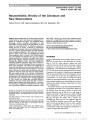
Show Leber Hereditary Optic Neuropathy: Some New Observations Nancy J. Newman, MD Clinical medical science classically progresses from the single case report, to multiple case series, to molecular diagnosis, to an expanded phenotype. Such has been the case with Leber hereditary optic neuropathy (LHON), a disorder first described in the mid-19th century by astute German clinical ophthalmologists as a syndrome of bilateral subacute optic neuropathies in otherwise healthy young men, often with a characteristic funduscopic appearance (1,2). Subsequent large case series in the premolecular era better defined the hereditary nature of the disorder and expanded the clinical phe-notype (3-5). With the discovery of the causal LHON mitochondrial DNA point mutations at the end of the 20th century came further expansion of the demographic and clinical profile of this disease, sometimes with unusual and unexpected observations (6-9). In this issue of the Journal, 2 such interesting observations are reported (10,11). In the Photo Essay by van Westen et al (10), MRI performed several months after visual loss in 2 male members of the same molecularly confirmed 11778 LHON pedigree showed enlargement of the anterior visual pathways without enhancement, and increased T2 signal, not only in the optic nerves and chiasm but also in the optic tracts, extending to the lateral geniculate bodies. Follow-up MRIs months later showed persistent bright T2 signal in normal-sized or atrophied anterior visual pathways. Although most MRIs are reportedly normal in the acute and chronic phases of LHON (6,7), increased T2 signal of the optic nerves has been previously noted on MRIs of LHON patients months to years after visual loss (12,13), less often in the chiasm (14,15), but never before in the optic tracts. Given that the pathology of LHON ultimately involves the entire length of the retinal ganglion cell axon (16), these findings should not be surprising. The bright T2 signal likely reflects axonal degeneration and ultimately gliosis. Why this has not been previously noted may reflect the usual timing of diagnostic imaging early in the acute phase of visual loss before degeneration can occur, combined with low imaging quality and lack of specific views of the optic tracts. Of course, an alternative explanation is that we just haven't looked care-fully- perhaps now we will! In the second article in this issue of the Journal dealing with LHON, Mnatsakanyan et al (11) report brachial plexus peripheral nerve abnormalities in a single patient with LHON visual loss associated with the 3460 mitochondrial DNA mutation, and no antemortem symptoms of peripheral neuropathy. Brachial plexus specimens obtained at necropsy and evaluated by light and electron microscopy showed various stages of extensive axonal degeneration of the large heavily myelinated fibers, clearly abnormal when compared to similar specimens from age-matched controls without known neurologic disease. Histopathology of muscle samples showed variable fiber size consistent with a neurogenic myopathy. Although it is possible that the patient had an unrecognized alternative cause for brachial plexopathy (such as cervical disc disease), good clinical documentation in this patient did not suggest prior neu-rologic disease. Additionally, although only one brachial plexus and no other peripheral nerves were sampled from the patient, the extent of neurodegeneration as demonstrated in both the brachial plexus and the muscles innervated suggested a widespread phenomenon. Morphometric analysis failed to Departments of Ophthalmology, Neurology, and Neurological Surgery, Emory University School of Medicine, Atlanta, Georgia. Supported in part by a departmental grant (Department of Ophthalmology) from the Research to Prevent Blindness, Inc, New York, New York, and by core grant P30-EY06360 (Department of Ophthalmology) from the National Institute of Health, Bethesda, Maryland. Dr. Newman is a recipient of a Research to Prevent Blindness Lew R. Wasserman Merit Award. The author reports no conflicts of interest. Address correspondence to: Nancy J. Newman, MD, Neuro-Ophthalmology Unit, Emory Eye Center, 1365-B Clifton Road, NE, Atlanta, GA 30322; E-mail: ophtnjn@emory.edu Newman: J Neuro-Ophthalmol 2011; 31: 3-5 3 Editorial Copyright © North American Neuro-Ophthalmology Society. Unauthorized reproduction of this article is prohibited. demonstrate a preferential vulnerability of smaller axons, which has been previously suggested to occur in the optic nerves of LHON patients (17). Although never previously demonstrated histopatho-logically, clinical peripheral nerve involvement in LHON has been rarely reported, as have other nonophthalmologic neurologic manifestations, usually designated within the so-called Leber's plus phenotype (18-20). Other syndromic disorders resulting from mitochondrial DNA mutations, such as mitochondrial encephalopathy with lactic acidosis and stroke-like episodes (MELAS), mitochondrial neuro-gastrointestinal encephalopathy (MNGIE), and chronic progressive external ophthalmoplegia (CPEO), have pe-ripheral neuropathy more commonly demonstrated, both clinically and histopathologically. Similarly, other optic neuropathies in which mitochondrial dysfunction has been proposed as a causal mechanism have had peripheral neu-ropathy as a more consistent clinical feature, such as the Cuban epidemic neuropathy in which more patients had clinically manifest peripheral neuropathy than optic neu-ropathy (21). Dominant optic atrophy patients have also recently been shown to manifest other neurologic features, including both clinical and subclinical peripheral neuro-pathy (22). Additionally, various forms of the heredi-tary peripheral neuropathies, including some forms of Charcot-Marie-Tooth disease linked to mitochondrial dysfunction, also include optic neuropathy in their clinical phenotype (23). It should not be so surprising that a mitochondrial DNA mutation that is present in every cell and in every tissue in the body should have more consequences than just optic neuropathy. Indeed, one of the paradoxes in LHON is that the sole tissue to be clinically affected in the vast majority of patients is specifically the optic nerve. As the authors suggest (11), the answer may lie in variable tissue reliance on mitochondrial function and the different regenerative capability of tissues. Studies such as this single case report should help further our understanding of the tissue-specific pathophysiology of LHON. Clinical observation has always been the cornerstone of neuro-ophthalmological practice. It is fitting that in a genetically defined disorder, such as LHON, astute clinical, radiographic, and histopathologic observation of single cases, still plays an important role in the advancement of knowledge of this disease. These new observations should help us better understand the pathophysiology of LHON and other similar disorders, hopefully with the ultimate goal of generating targeted therapeutic options. REFERENCES 1. Von Graefe A. Ein ungewohnlicher fall von hereditare amaurose. Archiv Fuer Ophthalmol. 1858;4:266-268. 2. Leber T. Uber hereditare und congenitalangelegte sehnervenleiden. Albrecht von Graefes Archiv fuer Ophthalmol. 1871;17:249-291. 3. Lundsgaard R. Leber's disease: a genealogic, genetic and clinical study of 101 cases of retrobulbar optic neuritis in 20 Danish families. Acta Ophthalmol. 1944;21(Suppl): 300-306. 4. Van Senus AHC. Leber's disease in the Netherlands. Doc Ophthalmol. 1963;17:1-163. 5. Seedorf T. The inheritance of Leber's disease. A genealogical follow-up study. Acta Ophthalmol. 1985;63: 135-145. 6. Newman NJ, Lott MT, Wallace DC. The clinical characteristics of pedigrees of Leber's hereditary optic neuropathy with the 11778 mutation. Am J Ophthalmol. 1991;111:750-762. 7. Mackey D, Buttery RG. Leber hereditary optic neuropathy in Australia. Aust N Z J Ophthalmol. 1992;20: 177-184. 8. Riordan-Eva P, Sanders MD, Govan GG, Sweeney MG, Da Costa J, Harding AE. The clinical features of Leber's hereditary optic neuropathy defined by the presence of a pathogenic mitochondrial DNA mutation. Brain. 1995; 118:319-337. 9. Hotta Y, Fujiki K, Hayakawa M, Nakajima A, Kanai A, Mashima Y, Hiida Y, Shinoda K, Yamada K, Oguchi Y. Clinical features of Japanese Leber's hereditary optic neuropathy with 11778 mutation of mitochondrial DNA. Jpn J Ophthalmol. 1995;39:96-108. 10. Van Westen D, Hammar B, Bynke G. Magnetic resonance findings in the pregeniculate visual pathways in Leber hereditary optic neuropathy. J Neuroophthalmol. 2011;31: 48-51. 11. Mnatsakanyan L, Ross-Cisneros FN, Carelli V, Wang MY, Sadun AA. Axonal degeneration in peripheral nerves in a case of Leber hereditary optic neuropathy. J Neuroophthalmol. 2011;31:6-11. 12. Mashima Y, Oshitari K, Imamura Y, Momoshima S, Shiga H, Oguchi Y. Orbital high resolution magnetic resonance imaging with fast spin echo in the acute stage of Leber's hereditary optic neuropathy. J Neurol Neurosurg Psychiatry. 1998;64:124-127. 13. Inglese M, Rovaris M, Bianchi S, Mantia L, Mancardi G, Ghezzi A, Montagna P, Salvi F, Filippi M. Magnetic resonance imaging, magnetization transfer imaging, and diffusion weighted imaging correlates of optic nerve, brain, and cervical cord damage in Leber's hereditary optic neuropathy. J Neurol Neurosurg Psychiatry. 2001;70: 444-449. 14. Phillips PH, Vaphiades M, Glasier CM, Gray LG, Lee AG. Chiasmal enlargement and optic nerve enhancement on magnetic resonance imaging in Leber hereditary optic neuropathy. Arch Ophthalmol. 2003;121:577-579. 15. Batioglu F, Atilla H, Eryilmaz T. Chiasmal high signal on magnetic resonance imaging in the atrophic phase of Leber hereditary optic neuropathy. J Neuroophthalmol. 2003;23: 28-30. 16. Adams JH, Blackwood W, Wilson J. Further clinical and pathological observations on Leber's hereditary optic atrophy. Brain. 1966;89:15-26. 17. Sadun AA, Win PH, Ross-Cisneros FN, Walker SO, Carelli V. Leber's hereditary optic neuropathy differentially affects smaller axons in the optic nerve. Trans Am Ophthalmol Soc. 2000;98:223-232. 18. Newman NJ. Leber's hereditary optic neuropathy: new genetic considerations. Arch Neurol. 1993;50: 540-548. 19. Nikoskelainen EK, Marttila RJ, Huoponen K, Juvonen V, Lamminen T, Sonninen P, Savontaus ML. Leber's ‘‘plus'': neurological abnormalities in patients with Leber's hereditary optic neuropathy. J Neurol Neurosurg Psychiatry. 1995;59:160-164. 20. Bouillot S, Martin-Negrier ML, Vital A, Ferrer X, Lagueny A, Vincent D, Coquet M, Orgogozo JM, Bloch B, Vita C. Peripheral neuropathy associated with mitochondrial 4 Newman: J Neuro-Ophthalmol 2011; 31: 3-5 Editorial Copyright © North American Neuro-Ophthalmology Society. Unauthorized reproduction of this article is prohibited. disorders: 8 cases and review of the literature. J Peripher Nerv Syst. 2002;7:213-220. 21. The Cuba Neuropathy Field Investigation Team. Epidemic optic neuropathy in Cuba: clinical characterization and risk factors. N Engl J Med. 1995;333:1176-1182. 22. Yu-Wai-Man P, Griffiths PG, Gorman GS, Lourenco CM, Wright AF, Auer-Grumbach M, Toscano A, Musumeci O, Valentino ML, Caporali L, Lamperti C, Tallaksen CM, Duffey P, Miller J, Whittaker RG, Baker MR, Jackson MJ, Clarke MP, Dhillon B, Czermin B, Stewart JD, Hudson G, Reynier P, Bonneau D, Marques W Jr, Lenaers G, McFarland R, Taylor RW, Turnbull DM, Votruba M, Zeviani M, Carelli V, Bindoff LA, Horvath R, Amati-Bonneau P, Chinnery PF.Multi-system neurological disease is common in patients with OPA1 mutations. Brain. 2010;133:771-786. 23. Zu¨chner S, De Jonghe P, Jordanova A, Claeys KG, Guergueltcheva V, Cherninkova S, Hamilton SR, Van Stavern G, Krajewski KM, Stajich J, Tournev I, Verhoeven K, Langerhorst CT, de Visser M, Baas F, Bird T, Timmerman V, Shy M, Vance JM. Axonal neuropathy with optic atrophy is caused by mutations in mitofusin 2. Ann Neurol. 2006;59: 276-281. Newman: J Neuro-Ophthalmol 2011; 31: 3-5 5 Editorial Copyright © North American Neuro-Ophthalmology Society. Unauthorized reproduction of this article is prohibited. |