| Title |
Traumatic disruption of the optic chiasm. |
| Creator |
Segal, Laura; An, Jella Angela; Gans, Mark |
| Affiliation |
McGill University Health Centre, Montreal, Quebec, Canada |
| Abstract |
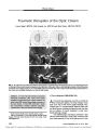
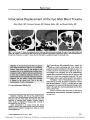
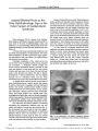
A 27-year-old man developed a persistent bitemporal hemianopia after severe head trauma sustained in a high-speed motor vehicle accident. The initial brain MRI revealed hemorrhagic contusion of the optic chiasm. A brain MRI performed 4 weeks later demonstrated complete chiasmal transection, a phenomenon rarely documented with imaging. |
| Subject |
Accidents, Traffic; Adult; Hemianopsia; Humans; Magnetic Resonance Imaging; Male; Optic Chiasm; Visual Acuity; Visual Fields |
| Format |
application/pdf |
| Publication Type |
Journal Article |
| Collection |
Neuro-Ophthalmology Virtual Education Library: Journal of Neuro-Ophthalmology Archives: https://novel.utah.edu/jno/ |
| Publisher |
Lippincott, Williams & Wilkins |
| Holding Institution |
Spencer S. Eccles Health Sciences Library, University of Utah |
| Rights Management |
© North American Neuro-Ophthalmology Society |
| Setname |
ehsl_novel_jno |
| ID |
226367 |
| Reference URL |
https://collections.lib.utah.edu/ark:/87278/s6vm7jb7/226367 |