| OCR Text |
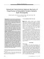
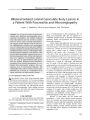
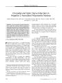
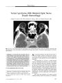
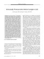
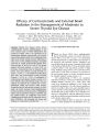
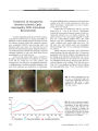
Show ORIGINAL CONTRIBUTION Peripapillary Nerve Fiber Layer Thickness Measured by Optical Coherence Tomography in Patients With No Light Perception From Long- Standing Nonglaucomatous Optic Neuropathies Carmen K. M. Chan, MRCP, MRCOphth and Neil R. Miller, MD Background: The residual peripapillary retinal nerve fiber layer thickness ( PRNFLT) corresponding to no light perception vision from long- standing nonglaucomatous optic neuropathies has not been documented. Such a benchmark would be useful information because PRNFLT is being used as an indicator of the visual recovery potential in patients with optic neuropathies. Methods: By means of optical coherence tomography ( OCT) using a fast RNFL thickness protocol, we determined the PRNFLT in 8 patients with no light perception ( NLP) for at least 1 year from acquired nonglaucomatous optic neuropathies. All patients underwent an assessment of visual acuity, color vision, visual field, pupillary reactions to light stimulation, and ophthalmoscopy. Results: Four of the 8 patients had a normal fellow eye. The average PRNFLT in the 4 normal eyes was 97.90 | Jim ( range 94.82- 100.89 | jtm), whereas the average PRNFLT in 8 of the 9 eyes with NLP was 45.42 | jim ( range 37.65- 51.46 | Jim). Conclusions: Eyes with long- standing NLP vision from nonglaucomatous optic neuropathies retain a residual PRNFLT of about 45 | Jim as measured by OCT. This should be taken into consideration when using PRNFLT to assess visual prognosis in patients with poor vision from various optic neuropathies. (/ Neuro- Ophthalmol 2007; 27: 176- 179) Neuro- Ophthalmology Unit ( NRM), Wilmer Eye Institute, The Johns Hopkins Hospital, Baltimore, Maryland; and the Department of Ophthalmology and Visual Sciences ( CKMC), The Chinese University of Hong Kong and the Hong Kong Eye Hospital, Hong Kong, China. Dr. Chan has received financial support from the Li Po Chun Charitable Trust Fund and the Hong Kong Eye Hospital, Hong Kong. Address correspondence to Neil R. Miller, MD, Maumenee 127, Wilmer Eye Institute, Johns Hopkins Hospital, 600 North Wolfe Street, Baltimore, MD 21287; E- mail: nrmiller@ jhmi. edu Optical coherence tomography ( OCT) is a noninvasive method that has been used to assess macular and optic disc morphology as well as peripapillary retinal nerve fiber layer thickness ( PRNFLT). In particular, some authors have recommended using PRNFLT to assess the potential for visual recovery in patients with optic neuritis ( 1). Theoretically, its use may also be extended to optic neuropathies from other causes, such as trauma and compression. Because OCT demonstrates anatomical structures quite well, many authors have attempted to correlate the thickness of the PRNFL with visual function. The normal average PRNFLT is age dependent ( 2) and is in the range of 98- 104 | jim as measured with the fast RNFL thickness function of the Stratus optical coherence tomograph ( 3,4). In eyes with impaired vision from optic neuropathy, the PRNFLT is reduced. For example, Trip et al ( 5) demonstrated that in 25 eyes with previous optic neuritis, with an average visual acuity of logMar + 0.23 ( Snellen equivalent 20/ 30), the average PRNFLT was 68.7 | jm. Fisher et al ( 6) showed that in patients with multiple sclerosis and optic atrophy, visual function scores were linearly correlated with PRNFLT, in that for every 1 line decrease in low- contrast letter acuity or contrast sensitivity score, the mean PRNFLT decreased by 4 | jim However, because the PRNFL not only contains visual axons but also supports glial cells and blood vessels, even complete optic nerve atrophy would not be expected to result in complete loss of the PRNFL as measured by OCT. Indeed glia constitute at least 18% of the primate RNFL ( 7). OCT may be detecting the glial content together with the sheaths of dead axons thickened by gliosis. Glial proliferation was demonstrated in a histologic study of eyes blind from glaucoma ( 8). In addition, there may be persisting but nonfunctional retinal ganglion cell axons. In a histologic study of enucleated eyes blind from optic neuropathy, 24 ( 42%) of 57 eyes had residual axons numbering at least 5% of normal values ( 9). This issue is extremely important if one is going to use PRNFLT to predict the potential for visual recovery in patients with various optic neuropathies such 176 J Neuro- Ophthalmol, Vol. 27, No. 3, 2007 Nerve Fiber Layer Thickness J Neuro- Ophthalmol, Vol. 27, No. 3, 2007 TABLE 1. Baseline demographics of study patients with long- standing nonglaucomatous primary optic atrophy resulting in no light perception vision in at least Case 1 2 3 4 5 6 7 8 Gender M F F F F F F F * Case 2: Both Age ( years) 21 34 40 51 52 55 72 73 one eye Cause of optic neuropathy Optic nerve glioma, s/ p radiotherapy Traumatic optic neuropathy* Unknown ( patient also has an optic neuropathy of unknown cause in the fellow Spheno- orbital meningioma, s/ p surgery and radiotherapy Optic neuritis and multiple sclerosis Spheno- orbital meningioma, s/ p surgery Optic nerve sheath meningioma, s/ p surgery Suprasellar meningioma, s/ p surgery and eyes affected ( NLP). We were unable to satisfactorily capture an OCT : NLP, no light perception; OCT, optical coherence tomography; OD, right eye; OS, left eye) radiotherapy image eye; from the ] s/ p, status NLP eye OS OS* OD OS OD OS OD OD ight eye because of post. Duration of NLP ( years) 4 12 9 16 1 1 14 14 nystagmus. as those caused by compression or trauma. Although a recent study reported that the average PRNFLT as determined with OCT in 17 eyes blind from glaucoma was 44.93 ± 4.95 | Jim ( 10), it is unclear whether this value is applicable to patients with nonglaucomatous optic neuropathies. In this study, we determined the minimal PRNFLT in eyes with no light perception ( NLP) from long- standing nonglaucomatous optic neuropathies. METHODS This prospective study was conducted at the Neuro-ophthalmology Unit of the Wilmer Eye Institute, The Johns Hopkins Hospital, with institutional review board approval. Patients with NLP from an acquired nonglaucomatous lesion of one or both optic nerves were included in the study Specifically, the duration of documented NLP had to be at least 1 year, and the patient had to have an absent pupillary reaction to direct light stimulation and no intraocular pathologic condition that could explain the visual acuity or affect the PRNFLT. Eight such patients were identified. OCT was performed on both eyes of each patient using a Stratus optical coherence tomograph 3.0 ( Carl Zeiss, Meditec, Dublin, CA) with software version 4.0.4. A fast RNFL thickness ( 3.4) scan acquisition protocol was used and the results were analyzed with the RNFL thickness average analysis ( both eyes) function. In scanning the blind eye, the external fixation target was used to allow the patients TABLE 2 Case 1 2 3 4 5 6 7 8 Average . Average Average NLP PRNFLT in the 8 study : PRNFLT of eye (| xm) 45.74 42.43 49.66 37.65 44.32 51.46 48.62 43.49 45.42 * Not applicable as fellow eye was PRNFLT, peripapillary nerve fiber Fellow eyes ( with NLP) eye normal? Y N N Y N Y Y N abnormal, layer thickness; NLP, no light and fellow eyes Average fellow PRNFLT of eye (| xm) 96.25 Unable to measure 36.02 94.82 50.78 100.89 99.62 56.15 perception. NLP eye PRNFLT (% of normal) 47.5 Not applicable* Not applicable* 39.7 Not applicable* 51.0 48.8 Not applicable* 46.8 177 J Neuro- Ophthalmol, Vol. 27, No. 3, 2007 Chan and Miller Signjl Strength ( MJX 10) 7 FIG. 1. Optical coherence tomography images of the right eye ( normal) of Case 4, demonstrating the ISNT rule ( inferior > superior > nasal > temporal). to fixate with their sighted fellow eye if necessary. A signal by an uneven cornea due to concurrent trigeminal and facial strength of at least 5 was achieved in all patients except Case nerve palsy causing keratopathy, but the data acquisition was 4 ( see below) in whom the signal strength was compromised determined to be adequate for analysis. 100 120 140 160 180 200 220 240 MAS INF TEMP 52 OS Signal Strength ( Max 10) 7 100 120 140 160 180 200 220 240 NAS INF TEMP 0 20 40 60 TEMP SUP Microns 300 200 100 0 0 20 40 60 80 100 120 140 160 180 200 220 240 TEMP SUP NAS INF TEMP 34 N X T 40 Signal Strength ( Max 10) FIG. 2. Optical coherence tomography images of Case 3. Visual acuity in the right eye is no light perception ( NLP) and in the left eye is 20/ 200. Note that the peripapillary nerve fiber layer thickness is greater in the right eye ( NLP eye) than in the left eye ( 20/ 200 eye). For a possible explanation, see text. 178 © 2007 Lippincott Williams & Wilkins Nerve Fiber Layer Thickness J Neuro- Ophthalmol, Vol. 27, No. 3, 2007 The visual function of the fellow eyes was determined by Snellen visual acuity at distance, Jaeger visual acuity at near, color vision using Hand- Rand- Rittler pseudo-isochromatic plates, visual field examination using Humphrey perimetry, and ophthalmoscopy. RESULTS Baseline demographic data are summarized in Table 1 and the PRNFLT results are summarized in Table 2. The average PRNFLT of the 8 eyes with NLP was 45.42 | jim ( range 37.65- 51.46 | Jim). The average PRNFLT of the 4 normal eyes was 97.90 | Jim ( range 94.82- 100.89 | Jim). For Case 3, a woman with bilateral optic neuropathy thought to be inflammatory or ischemic in origin, the PRNFLT was actually thicker in the NLP eye than in the fellow eye ( which had 20/ 200 best- corrected visual acuity and inferior visual field loss). Normal eyes should show a characteristic configuration for disc rim thickness of inferior > superior ^ nasal s temporal, termed the ISNT rule ( 11) ( Fig. 1). In the 4 normal eyes in this study, the average PRNFLTs in the inferior, superior, nasal, and temporal quadrants were 126.0, 114.8, 86.8, and 64.0 | Jim, respectively, obeying the ISNT rule. This pattern was lost in the 8 NLP eyes, which averaged PRNFLTs of 44.8, 52.5, 44.1, and 40.1 | Jim in those four quadrants, respectively. DISCUSSION This small case series demonstrates that the minimal average PRNFLT in eyes with NLP from long- standing, nonglaucomatous optic neuropathies is approximately 45 | Jim, a figure consistent with the average PRNFLT of 44.93 | JLm found in glaucomatous blind eyes by Sihota et al ( 10). Three possible explanations exist as to why the PRNFLT thickness in such cases is 45%- 50% of the average thickness in normal eyes. First, OCT may be detecting the glial content together with the sheaths of dead axons thickened by gliosis. Second, there may be persisting but nonfunctional retinal ganglion cell axons ( 9). It is possible that these residual retinal ganglion cells project to the pretectal nuclei/ superior colliculus/ hypothalamus and do not directly participate in the formation of visual images. Third, the residual thickness may be an artifact generated by the built- in software of the OCT. None of these mechanisms would explain why the PRNFLT in the seeing eye of Case 3 was thinner than that of the fellow eye with NLP ( Fig. 2). Because the underlying cause for her optic neuropathy has not yet been determined it is possible that she had other intraocular pathologic conditions that could have contributed to her loss of vision and reduction of PRNFLT. One possibility is that over the 9 years that the patient's nonseeing eye had been blind gliosis had occurred thus thickening the PRNFL compared with the fellow sighted eye which had not yet developed substantial gliosis. Our findings should be taken into consideration in the interpretation of OCT findings in patients when one is attempting to determine the prognosis for visual recovery in patients with nonglaucomatous optic neuropathies. REFERENCES 1. Costello F, Coupland S, Hodge W, et al. Quantifying axonal loss after optic neuritis with optical coherence tomography. Ann Neurol 2006; 59: 963- 9. 2. Jaffe GJ, Caprioli J. Optical coherence tomography to detect and manage retinal disease and glaucoma. Am J Ophthalmol 2004; 137: 156- 69. 3. Budenz DL, Michael A, Chang RT, et al. Sensitivity and specificity of the StratusOCT for perimetric glaucoma. Ophthalmology 2005; 112: 3- 9. 4. Paunescu LA, Schuman JS, Price LL, et al. Reproducibility of nerve fiber thickness, macular thickness, and optic nerve head measurements using StratusOCT. Invest Ophthalmol Vis Sci 2004; 45: 1716- 24. 5. Trip SA, Schlottmann PG, Jones SJ, et al. Retinal nerve fiber layer axonal loss and visual dysfunction in optic neuritis. Ann Neurol 2005; 58: 383- 91. 6. Fisher JB, Jacobs DA, Markowitz CE, et al. Relation of visual function to retinal nerve fiber layer thickness in multiple sclerosis. Ophthalmology 2006; 113: 324- 32. 7. Ogden TE. Nerve fiber layer of the primate retina: thickness and glial content. Vision Res 1983; 23: 581- 7. 8. Pavlidis M, Stupp T, Naskar R, et al. Retinal ganglion cells resistant to advanced glaucoma: a postmortem study of human retinas with the carbocyanine dye Dil. Invest Ophthalmol Vis Sci 2003 ; 44: 5196- 205. 9. Cursiefen C, Holbach LM, Schlotzer- Schrehardt U, et al. Persisting retinal ganglion cell axons in blind atrophic human eyes. Graefes Arch Clin Exp Ophthalmol 2001; 239: 158- 64. 10. Sihota R, Sony P, Gupta Y et al. Diagnostic capability of optical coherence tomography in evaluating the degree of glaucomatous retinal nerve fiber damage. Invest Ophthalmol Vis Sci 2006; 47: 2006- 10. 11. Harizman N, Oliveira C, Chiang A, et al. The ISNT rule and differentiation of normal from glaucomatous eyes. Arch Ophthalmol 2006; 124: 1579- 83. 179 |