| OCR Text |
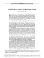
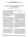
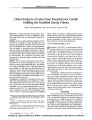
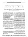
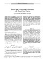
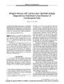
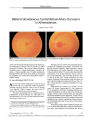
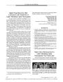
Show ORIGINAL CONTRIBUTION Early Resolution of Third Nerve Palsy Following Endovascular Treatment of a Posterior Communicating Artery Aneurysm Joji Inamasu, MD, Yoshiki Nakamura, MD, Ryoichi Saito, MD, Yoshiaki Kuroshima, MD, Shigeo Ohba, MD, and Kiyoshi Ichikizaki, MD Abstract: A 69- year- old man underwent successful endovascular treatment of a posterior communicating artery aneurysm that had caused a third nerve palsy. Pupil size became normal within 10 days and ptosis and ocular ductions became normal within 3 weeks of the procedure. Based on the reported recovery rates of third nerve palsy after aneurysmal clipping, recovery may occur more rapidly in patients who undergo endovascular treatment. Further data are necessary to substantiate this hypothesis. ( JNeuro- Ophthalmol 2002; 22: 12- 14) solated third nerve palsy is occasionally caused by an intracranial aneurysm ( 1,2), and this sign usually heralds impending rupture of the aneurysm, which in turn causes a catastrophic event, subarachnoid hemorrhage ( SAH). The standard treatment of patients with a cerebral aneurysm presenting with third nerve palsy has been surgery ( 3- 5), but reversal of the neuro- ophthalmologic deficits after surgery takes several months ( 4,5). Endovascular treatment, which has recently been introduced into neurosurgical practice, may shorten the recovery time ( 6). We report a patient with an internal carotid- posterior communicating artery ( IC-PC) aneurysm who presented with acute- onset third nerve palsy and subsequent SAH. The patient was successfully treated with an endovascular procedure, with resolution of the third cranial nerve deficits within 3 weeks after onset. CASE REPORT A 69- year- old man presented with complaints of sudden- onset headache and vomiting. A brain computed The Department of Neurosurgery, National Tokyo Medical Center, Tokyo, Japan Address correspondence to Joji Inamasu, MD, Department of Neurosurgery, National Tokyo Medical Center, Higashigaoka 2- 5- 1, Meguro- ku, Tokyo 152- 8902, Japan tomography scan revealed an SAH in the basal cistern and right sylvian fissure. Cerebral angiography showed a right middle cerebral artery aneurysm and a left IC- PC aneurysm ( Fig. 1). The right middle cerebral artery aneurysm was considered responsible for the SAH, and the patient underwent surgical clipping of the aneurysm. The postoperative course was uneventful until postoperative day 12, when the patient's left pupil was observed to be dilated. Neither ptosis nor ocular movement disturbance was noted at that time. Angiography was repeated and revealed growth of the left IC- PC aneurysm, which had a multilobulated shape ( Fig. 2A). Embolization of the aneurysm was scheduled for the next day, but the aneurysm bled before the embolization procedure, and the patient became comatose. Emergency embolization was performed, and the aneurysm was fully packed with Guglielmi detachable coils of appropriate length and shape ( Fig. IE). One day after embolization, the patient regained consciousness, but ptosis was noted on the left side, and the left pupil remained dilated. Extraocular muscle movements, particularly supraduction and infraduc-tion, were also moderately impaired. Pupils became equal with normal direct light reactions by day 10 after embolization. The ptosis and extraocular movement disturbance had completely resolved by 3 weeks after embolization. Magnetic resonance imaging performed 4 weeks after the embolization showed a thrombosed IC- PC aneurysm in the subarachnoid cistern, visualized as a low- intensity mass on the Tl- weighted images and as a low- to isointensity mass on the T2- weighted images ( Fig. 3). The patient was discharged home with minimal cognitive deficit 2 months after the embolization procedure. DISCUSSION Cerebral aneurysms causing third nerve palsy are for the most part IC- PC aneurysms, and they have been treated surgically, as have aneurysms in other locations ( 2- 5). Reversal of third nerve palsy after surgery, if it is to occur, takes between 1 and 3 months ( 4,5). In as many as two thirds of patients, the oculomotor palsy never resolves 12 J Neuro- Ophthalmol, Vol. 22, No. 1, 2002 Copyright © Lippincott Williams & Wilkins. Unauthorized reproduction of this article is prohibited. ENDO VASCULAR TREA TMENT OF THIRD NER VE PALSY JNeuro- Ophthalmol, Vol. 22, No. 1, 2002 FIG. 1. A: Right carotid angiogram, showing a ruptured right middle cerebral artery aneurysm. B: Left carotid angiogram, showing an unruptured aneurysm at the junction of the left internal carotid and posterior communicating arteries. FIG. 3. T1- weighted ( A) and T2- weighted ( B) magnetic resonance imaging performed 4 weeks after embolization, showing the thrombosed left internal carotid- posterior communicating artery junction aneurysm in the subarachnoid cistern { arrow). completely ( 4). Recent developments in interventional neuroradiology techniques have prompted the widespread use of Guglielmi detachable coils in the treatment of cerebral aneurysms, with the aim of preventing rebleeding from ruptured aneurysms. Whether endovascular treatment is effective in eliminating manifestations due to mass effect has been a matter of controversy, however ( 7), and when and how third nerve palsy caused by a cerebral aneurysm improves after endovascular treatment has rarely been reported. Recently, Birchall etal. ( 6) reported that patients with third nerve palsy caused by IC- PC aneurysms recovered FIG. 2. A: Angiogram following development of third nerve palsy, showing enlargement of the left internal carotid- posterior communicating artery junction aneurysm. B: Angiography following embolization, showing complete obliteration of the aneurysm with Guglielmi detachable coils. within 2 to 3 weeks after endovascular treatment, suggesting earlier resolution of such deficits by endovascular treatment than by surgery. Our case, in which the third nerve palsy was cured completely within 3 weeks after aneurysmal embolization, is consistent with their findings. Unlike clipping, endovascular treatment does not remove the mass effect of an aneurysm immediately. In our case, magnetic resonance imaging performed 1 month after embolization continued to show a thrombosed aneurysmal mass in the subarachnoid space ( Fig. 3). However, the loss or decrease of aneurysmal pulsatility afforded by Guglielmi detachable coil embolization may be more important to early resolution of third nerve palsy caused by cerebral aneurysms than anatomic detachment of the third nerve from an adjacent and adherent cerebral aneurysm by clipping ( 6). Moreover, manipulation of the third nerve during clipping surgery may cause additional structural damage to the third nerve, resulting in a possible delay in postoperative recovery. The pattern of reversal of the third nerve deficits following embolization was unique in our patient, since the mydriasis was the first clinical sign to recover- within 10 days of treatment. Ptosis and the ocular movement disturbance improved more slowly, taking another 2 weeks. This recovery pattern contrasts with that usually seen in surgically treated patients, in whom ptosis is the first clinical sign to recover, with the mydriasis usually persisting for several months ( 3,4). The reason for this finding is unclear, and further accumulation of data will be essential to determine whether other endovascularly treated patients with third nerve palsy have a similar recovery pattern. Nevertheless, this observation leads us to speculate that delayed recovery of pupillary function in surgically treated patients may at 13 Copyright © Lippincott Williams & Wilkins. Unauthorized reproduction of this article is prohibited. JNeuro- Ophthalmol, Vol. 22, No. 1, 2002 Inamasu et al. least in part be attributable to the anatomic distribution of the fibers innervating the pupillary muscles, which are located in the outer layer of the third nerve and are likely to be more vulnerable to additional injuries inflicted during surgical manipulation ( 3,4). REFERENCES 1. Renowden SA, Harris KM, Hourihan MD. Isolated atraumatic third nerve palsy: clinical features and imaging techniques. Br J Radiol 1993; 66: 1111- 7. 2. McFadzean RM, Teasdale EM. Computerized tomography angiography in isolated third nerve palsies. JNeurosurg 1998; 88: 679- 84. 3. Kasner SE, Liu GT, Galetta SL. Neuro- ophfhalmologic aspects of aneurysms. Neuroimaging Clin North Am 1997; 7: 679- 92. 4. Giombini S, Ferraresi S, Pluchino F. Reversal of oculomotor disorders after intracranial aneurysm surgery. Acta Neurochir 1991 ; 112: 19- 24. 5. Kyriakides T, Aziz TZ, Torrens MJ. Postoperative recovery of third nerve palsy due to posterior communicating aneurysms. Br J Neu-rosurg 1989; 3: 109- 11. 6. Birchall D, Khangure MS, McAuliffe W. Resolution of third nerve paresis after endovascular management of aneurysms of the posterior communicating artery. AJNR 1999; 20: 411- 3. 7. Kurokawa R, Saito S, Nakamura Y, et al. Ruptured vertebral artery-posterior inferior cerebellar artery aneurysm associated with facial nerve paresis successfully treated with interlocking detachable coils: case report. Neurol Med Chir ( Tokyo) 1999; 39: 863- 6. 14 © 2002 Lippincott Williams & Wilkins Copyright © Lippincott Williams & Wilkins. Unauthorized reproduction of this article is prohibited. |