| OCR Text |
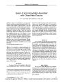
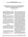
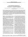
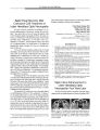
Show ORIGINAL CONTRIBUTION Photophobia as the Presenting Visual Symptom of Chiasmal Compression Aki Kawasaki, MD, and Valerie A. Purvin, MD Five patients with a chief visual complaint of photophobia were subsequently found to have compressive lesions of the optic chiasm. Visual acuity and visual field deficits were often subtle. Magnetic resonance imaging scanning revealed large suprasellar masses, including three pituitary adenomas, a craniopharyngioma, and a clivus chordoma. Photophobia resolved in all patients following treatment of the tumors. A compressive lesion of the optic chiasm should be considered in patients who experience persistent photophobia unexplained by ocular abnormalities. ( JNeuro- Ophthalmol 2002; 22: 3- 8) ost lesions of the anterior visual pathway cause a decrease in vision, often described as blurring, dimming, or darkening. Less commonly, patients experience an excess of vision such as flashes, scintillations, or twinkling. These symptoms are collectively referred to as photopsias and occur, for example, in 30% of patients with idiopathic demyelinating optic neuritis [ 1,2]. Photopsias have also been described, less commonly, in patients with compressive lesions of the optic nerves and chiasm [ 3,4]. Photophobia can also be considered a form of excess vision in which patients experience ordinary light and visual stimuli as excessively bright, causing discomfort and pain. Photophobia is most commonly associated with anterior segment disease of the eye. It frequently accompanies meningeal irritation and migraine. To our knowledge, photophobia has been associated with compressive lesions of the anterior visual pathway in only a few instances [ 4,5]. In this small case series, we describe five patients whose sole or chief visual symptom was photophobia and who Hopital Ophtalmique Jules Gonin ( AK), Lausanne, Switzerland, and Midwest Eye Institute, Neuro- ophthalmology section, and Indiana University Medical Center, Departments of Neuro- ophthalmology, Neurology, and Ophthalmology ( VAP), Indianapolis, Indiana, USA. Address correspondence to Valerie A. Purvin, MD, Midwest Eye Institute, 201 Pennsylvania Parkway, Indianapolis, IN 46280- 1381, USA. Presented at the 27th annual North American Neuro- ophthalmology Society meeting, Rancho Mirage, California, USA, February 2001. Supported by the Midwest Eye Foundation, Indianapolis, Indiana, USA. were subsequently found to have compressive lesions of the optic chiasm. CASE REPORTS Case 1 A 19- year- old man was noted to have excessively large hands and feet. Acromegaly due to a pituitary tumor was diagnosed by radiographic and endocrinologic examination. He was then referred for neuro- ophthalmic consultation. He reported a 1- year history of photophobia and epiphora, often interfering with outdoor sports. He had not previously worn sunglasses on a regular basis but found that it was necessary in the past year, to maintain adequate vision. He had no specific complaints indoors except that bright light exposure often caused tearing and excessive blinking ( Table 1). Best- corrected visual acuity was 20/ 20 in both eyes ( OU) and the patient identified 17 of 17 pseudoisochro-matic plates. Goldmann perimetry revealed minimal superior bitemporal depression of the central He isopter ( Fig. 1). There was very mild temporal pallor of the optic discs. The remainder of the findings of the neuro-ophthalmic examination were normal. Magnetic resonance imaging ( MRI) showed a large pituitary mass extending into the sphenoid sinus and right cavernous sinus and compressing the optic chiasm ( Fig. 2). The patient underwent transsphenoidal subtotal resection of a pituitary adenoma, followed by radiation therapy and maintenance treatment with bromocriptine. Following treatment, he reported resolution of photophobia. Case 2 A 22- year- old woman noted acute onset of photophobia consisting of progressive intolerance of ordinary light over 2 weeks, causing her to wear sunglasses continuously, outdoors and indoors. She also developed intermittent headaches and slight blurriness of vision in her OS. Shortly thereafter, she experienced spontaneous and continuous " twinkling" and " bright spots" in the central vision of both eyes, which impaired her schoolwork. There was a history of amblyopia in her OD since childhood. Medical history J Neuro- Ophthalmol, Vol. 22, No. 1, 2002 3 Copyright © Lippincott Williams & Wilkins. Unauthorized reproduction of this article is prohibited. JNeuro- Ophthalmol, Vol. 22, No. 1, 2002 Kawasaki and Purvin TABLE 1. Case Age,}' Gender Primary symptom Other visual symptoms Nonvisual symptoms Duration of photosensitivity ( until diagnosis) Past ocular history 1 19 male Sensitivity to light T tearing - - iy - 2 22 Female " Can't stand light" Slight blur OS twinkling ( later) Headache 3 w Amblyopia OD 3 43 Male " Glarey," " Too bright" - Headache ( lw prior) 4 mo Congenital nystagmus, 4 41 Male " Glare," " Heat waves" Filmy vision OS ( later) Headache ( later) iy - 5 42 Female T Sensitivity to light - Headache 1 w - Past medical history Visual acuity ( initial) Visual field defects ( initial) Ancillary tests Diagnosis Pre- operative MRI OD: 20/ 20 OS: 20/ 20 Minimal superior bitemporal depression ( lie isopter only) None Pituitary adenoma Sellar/ suprasellar mass with extension into sphenoid and cavernous sinuses, chiasmal compression Migraine OD: 20/ 50 ( baseline) OS: 20/ 20~ 2 Mild central depression OD, small central scotoma OS ERG Pituitary adenoma Sellar/ suprasellar mass, pituitary hemorrhage, chiasmal compression congenital toxoplasmic chorioretinitis T cholesterol, hypertension OD: 20/ 60 ( 20/ 40 baseline) OS: 20/ 400 ( baseline) Peripheral and paracentral loss OD, extensive nasal loss to fixation from chorioretinal scarring RSFA; ultrasound Craniopharyngioma Suprasellar cystic mass, chiasmal compression Hypertension, sleep apnea OD: 20/ 20 OS: 20/ 40" Enlarged physiologic blindspots encroaching on fixation OU RSFA Clivus chordoma Sellar mass with sphenoid and invasion of ethmoid and cavernous sinuses, chiasmal compression Migraine OD: 20/ 20 OS: 20/ 20 Pituitary adenoma Sellar/ suprasellar mass, pituitary hemorrhage, chiasmal compression RSFA- rapid sequence fluorescein angiogram; ERG- electroretinogram; MRI- Magnetic resonance imaging. was unremarkable. She had been taking the same oral contraceptive for 3 years. Best- corrected visual acuity was 20/ 50 OD ( baseline acuity) and 20/ 20- 2 OS. The patient identified all Ishihara color plates with OU, slowly because she found they were " too bright." Brightness contrast was symmetric. Photo-stress time was 10 seconds in OU. Pupils were 4 mm OU in dim light and briskly reactive to light without a relative afferent pupillary defect. External, motility, and biomicro-scopic examination findings were normal. Intraocular pressures were 16 mm OU. Funduscopy revealed mildly crowded discs without edema, hemorrhages, or pallor. The maculas were normal. Goldmann perimetry showed mild enlargement of the blind spot and temporal depression of the central He isopter in the OD and a steeply marginated, small central scotoma in the OS ( Fig. 3). MRI showed a 4 © 2002 Lippincott Williams & Wilkins Copyright © Lippincott Williams & Wilkins. Unauthorized reproduction of this article is prohibited. PHOTOPHOBIA AND CHIASMAL COMPRESSION JNeuro- Ophthalmol, Vol 22, No. 1, 2002 FIG. 1. Case 1. Goldmann perimetry in a 19- year- old man with acromegaly shows a subtle superior bitemporal defect affecting only the central isopter in OU. pituitary mass elevating the intracranial optic nerves and chiasm. There were inhomogeneous signal characteristics within the mass consistent with pituitary apoplexy ( Fig. 4). Results of the endocrinologic evaluation were normal. The patient underwent transsphenoidal resection of a hemorrhagic pituitary adenoma and within 1 month reported significant improvement of her photopsias and photophobia. Her headache and visual blur also resolved. Case 3 A 43- year- old man experienced sudden onset of photophobia- a sense that everything was too bright. This occurred against a background of headache attributed to sinusitis for which he was treated with antibiotics. He continued to have recurrent headaches and persistent visual disturbance for the next 3 months, when he sought neuro-ophthalmic consultation. His prior ocular history was significant for chronically decreased vision more in the OS than the OD due to congenital toxoplasmic chorioretinitis and congenital nystagmus. He was not certain whether his vision had actually worsened recently or whether he just had trouble seeing because of excessive glare and photophobia. Visual acuity was 20/ 60 OD and 20/ 400 OS ( previously documented acuities of 20/ 40 OD and 20/ 400 OS). He failed to identify 2 of 15 Ishihara color plates with the OD and identified only the test color plate with the OS. There was a 2+ left relative afferent pupillary defect. Goldmann perimetry in the OD showed two areas of predominantly peripheral loss ( one temporal and one inferonasal) plus several paracentral scotomas. In the OS there was complete loss of the nasal field not respecting the vertical meridian with extension to involve fixation. The patient exhibited continuous, pendular, horizontal, conjugate, rapid small-amplitude nystagmus with a gaze- evoked component to either side. Ophthalmoscopy showed bilateral disc pallor, extensive peripheral chorioretinal scarring OS, and attenuation of retinal vessels in both eyes. An MRI scan revealed a cystic suprasellar mass with rim enhancement that was elevating the chiasm and spreading the optic tracts ( Fig. 5). The mass was subtotally excised via craniotomy and histologically diagnosed as a craniopharyngioma. The patient's postoperative course was complicated by diabetes insipidus and fever of unknown origin, but his photophobia resolved. Case 4 A 41- year- old man reported a 1- year history of intermittent " heat waves" and " glarey" vision. He had no headaches at that time and did not seek medical advice. Because of increasing photophobia to sunlight and stadium lights, he had begun wearing sunglasses during football practice or any daytime outdoor activity and at nighttime football games 3 months prior to initial examination. Several weeks later, the patient noted blurry vision in his OS and daily headaches, leading to consultation with his ophthalmologist and subsequently to neuro- ophthalmic referral. Initial visual field testing demonstrated enlargement of the physiologic blindspot in OU. Given the patient's report of seeing continuous heat waves, a diagnosis of acute zonal occult outer retinopathy was entertained. On a trial of oral prednisone, he experienced improvement of vision but precipitous decline upon stopping the medication. Repeat perimetry showed a dense temporal defect in OU that did not respect the vertical meridian ( Fig. 6). An MRI scan revealed a large central skull base mass filling the sphenoid sinus and invading the ethmoid sinuses, cavernous sinuses, and suprasellar cistern, and compressing the optic chiasm ( Fig. 7). Subtotal resection via a transsphenoidal approach yielded a histologic diagnosis of clivus chordoma. Postoperatively, his symptoms of seeing heat waves and experiencing glare and photosensitivity improved markedly; visual acuity returned to 20/ 20 OU. Tumor recurrence 8 months later was again accompanied by photophobia. He subsequently underwent two additional surgeries for tumor regrowth, followed by proton beam irradiation. Case 5 A 42- year- old woman noted sudden onset of severe headache and increased sensitivity to light. Although she had a history of migraine headaches in the past, she was certain that these symptoms were unlike her usual migraines, especially the photophobia, which was persistent rather than episodic. Her neuro- ophthalmic examination and Goldmann visual field results were normal. MRI revealed a pituitary mass with bright T2 signal consistent with apoplexy. There was parasellar extension and chiasmal contact without distortion by this tumor. Endocrine evaluation disclosed low Cortisol and thyroid levels. Following replacement therapy, the patient noted improvement of headaches and photophobia. 5 Copyright © Lippincott Williams & Wilkins. Unauthorized reproduction of this article is prohibited. JNeuro- Ophthalmol, Vol 22, No. 1, 2002 Kawasaki and Purvin FIG. 2. Case 1. Sagittal ( A) and coronal ( B) T1 - enhanced MRI scan reveals a large sellar mass with invasion of the sphenoid sinus, suprasellar extension, and mild distortion of the optic chiasm. Histologic diagnosis was pituitary adenoma. DISCUSSION Our five patients with chiasmal compressive lesions ( three pituitary adenomas, one craniopharyngioma, one cli-vus chordoma) all reported an increased sensitivity and intolerance to ordinary light, often described as objects looking " too bright" or " glarey." The duration of the photophobia from onset to diagnosis ranged from 1 week to 1 year ( see Table 1). Two patients experienced photopsias as well- peripheral heatwaves ( Case 4) or central sparkling ( Case 2). The only other visual symptom at onset was mild blurring of vision in one eye of one patient ( Case 2); an additional patient ( Case 4) developed blurred vision in one eye 9 months after onset of photophobia. Four patients had headaches. Examination showed subtle visual dysfunction. FIG. 3. Case 2. Goldmann perimetry in a 22- year- old woman at presentation shows temporal depression and enlargement of the physiologic blindspot in the OD and a steeply marginated relative central scotoma in the OS. There was no loss of visual acuity in 7 of 10 eyes, one line of loss in 1 eye, two lines of loss in 1 eye, and three lines in 1 eye. Visual field testing results were normal in one patient. Two patients had a bitemporal visual field defect; one FIG. 4. Case 2. Sagittal Tl- unenhanced MRI scan reveals a sellar and suprasellar mass causing mild chiasmal compression. The inhomogeneous signal within the mass is consistent with previous pituitary hemorrhage ( apoplexy). Histologic diagnosis was pituitary adenoma. 6 © 2002 Lippincott Williams & Wilkins Copyright © Lippincott Williams & Wilkins. Unauthorized reproduction of this article is prohibited. PHOTOPHOBIA AND CHIASMAL COMPRESSION JNeuro- Ophthalmol, Vol 22, No. 1, 2002 FIG. 5. Case 3. T1- enhanced MRI scan in coronal ( A) and sagittal ( B) planes shows a large cystic sellar mass with rim enhancement and distortion of the optic chiasm. Histologic diagnosis was craniopharyngioma. patient had mild central defects. The fifth patient had extensive preexisting field losses from congenital chorioretinal scarring. The MRI scans always showed large lesions involving the sellar region. Following surgical decompression, photophobia remitted in all five patients. Lesions that compress the optic chiasm typically produce progressive dimming and loss of vision. Especially in the early stages, when visual acuity is normal or nearly normal, visual symptoms are nonspecific, including such descriptions as " foggy," " dim," " hazy," or " fuzzy." Other less common visual symptoms include loss of depth perception, objects appearing and disappearing from view, and intermittent diplopia. FIG. 6. Case 4. Goldmann perimetry in a 41- year- old man with a 1 - year history of glarey vision and heat waves demonstrates marked enlargement of the physiologic blindspot bilaterally. The defects are predominantly temporal but do not respect the vertical meridian. Positive visual phenomena may also be associated with compressive lesions of the optic nerves and chiasm. Safran and colleagues [ 4] prospectively obtained a detailed symptom history from 45 consecutive patients with optic FIG. 7. Case 4. Coronal T1- enhanced MRI scan obtained 6 weeks after the initial evaluation reveals a large central skull base tumor invading the sphenoid and the ethmoid sinuses and elevating the optic chiasm. Histologic diagnosis was clivus chordoma. 7 Copyright © Lippincott Williams & Wilkins. Unauthorized reproduction of this article is prohibited. JNeuro- Ophthalmol, Vol. 22, No. 1, 2002 Kawasaki and Purvin nerve or chiasmal diseases such as optic neuritis, ischemic optic neuropathy, and compressive lesions. They divided symptoms of positive visual phenomena into two groups: photopsias and photophobia. They defined symptoms such as sparks, flashes, or colored lights as " photopsias" and sensations of dazzling or glaring light, a general visual discomfort in bright light, and an awareness of improved visual function in conditions of dim illumination as " photophobia." They found that 30 of 62 visually symptomatic eyes had photopsias or photophobia or both. Of 13 eyes affected by a mass lesion ( pituitary adenoma, meningioma, glioma, aneurysm), 9 eyes had photopsia, photophobia, or both. Only three patients had onset of photopsia or photophobia before onset of visual loss. These three patients were subsequently found to have compressive lesions, two patients with pituitary adenoma compressing the optic chiasm ( similar to three of our five patients) and one patient with a unilateral perioptic meningioma. Walsh and Hoyt [ 5] observed photophobia in one patient with a hypophyseal tumor and in another patient with a recurrent craniopharyngioma. Such small numbers of reported cases suggest that photophobia is a rare symptom of chiasmal compression. Photophobia, an intolerance to ordinary light such that pain may result, is more frequently encountered in diseases of the anterior structures of the eye or meninges. Photoreceptor disease is also associated with photophobia. The mechanism for the light intolerance in cases of corneal disruption, iritis, meningitis, and migraine remains unclear and is presumably related to irritation of the trigeminal afferent pathway. The mechanism producing photophobia is even less clear in our patients. One possibility is release of blood or cystic contents from the suprasellar tumor causing meningeal irritation, thus producing headache and photophobia. This is a plausible explanation for Cases 2 and 5, whose MRI scans were consistent with previous pituitary apoplexy, and Case 3, whose tumor, a craniopharyngioma, contained cystic areas. Such a ' chemical meningitis' would be less likely in the remaimng two patients, especially Case 1 who had no accompanying headache. Case 4 did experience headache, but only after the onset of the photophobia. In these two patients, and perhaps the others as well, photophobia may be related to stretching of pain- sensitive structures at the base of the brain, those associated with meninges or blood vessels, transmitted via trigeminal afferents and processed centrally. Because photosensitivity and photopsias are more commonly associated with ocular disease such as inflammation, corneal disruption, or photoreceptor degeneration, examiners may dismiss the symptoms as psychogenic if the eye examination is normal or mistakenly attribute them to ocular abnormalities. In fact, two of our patients who experienced photopsias as well as photophobia were initially suspected to have an outer retinal inflammatory syndrome, and retinal studies were undertaken before neuroimaging. We advise clinicians to consider compressive lesions of the anterior visual pathway in patients who complain of photophobia, even when visual acuity or results of visual field evaluations are normal. REFERENCES 1. Beck RW, Optic Neuritis Study Group. The Optic Neuritis Treatment Trial. Arch Ophthalmol 1988; 106: 1051- 53. 2. Davis FA, Bergen D, Schauf D, et al. Movement phosphenes in optic neuritis: A new clinical sign. Neurology 1976; 26: 1100^ 4. 3. Weinberger LM, Grant EC. Visual hallucinations and the neuro-optical correlates. Arch Ophthalmol 1940; 23: 166. 4. Safran AB, Kline LB, Glaser JS. Positive visual phenomena in optic nerve and chiasm disease: photopsias and photophobia. In Glaser JS, ed. Neuro- ophthalmology. Symposium of the Bascom Palmer Eye Institute. Vol X. St. Louis: CV Mosby, 1977: 225- 31. 5. Walsh FB, Hoyt WF. Clinical Neuro- ophthalmology. 3rd ed. Baltimore: Williams & Wilkins, 1969: 413- 6. 8 © 2002 Lippincott Williams & Wilkins Copyright © Lippincott Williams & Wilkins. Unauthorized reproduction of this article is prohibited. |