| OCR Text |
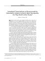
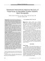
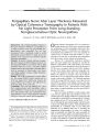
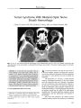
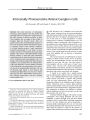
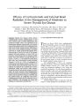
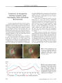
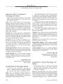
Show ORIGINAL CONTRIBUTION Primary Sinonasal Undifferentiated Carcinoma Presenting With Bilateral Retrobulbar Optic Neuropathy Madhura A. Tamhankar, MD, Nicholas J. Volpe, MD, Laurie A. Loevner, MD, James N. Palmer, MD, and Michael Feldman, MD, PhD Abstract: A 43- year- old man presented with acute bilateral visual loss. Ophthalmologic examination revealed no light perception in the right eye and a visual acuity of 20/ 50 in the left eye with a right afferent pupillary defect. Ophthalmoscopic examination was normal. Brain MRI showed an intracranial but extra- axial mass in the floor of the anterior cranial fossa extending along the olfactory groove and into the sinonasal vault. Endoscopic biopsy showed a high- grade neoplasm consistent with sinonasal undifferentiated carcinoma. This case report highlights an unusual clinical presentation for this rare and aggressive neoplasm. (/ Neuro- Ophthalmol 2007; 27: 189- 192) Sinonasal undifferentiated carcinoma ( SNUC) is a rare aggressive neoplasm of the nasal cavity and paranasal sinuses ( 1). Presenting signs and symptoms typically include nasal obstruction, epistaxis, rhinorrhea, proptosis, and facial pain. Although visual loss from orbital invasion can occur, acute visual loss due to direct compression of intracranial visual pathways without orbital invasion is highly unusual. We report such an occurrence for the first time. CASE REPORT A previously healthy 43- year- old man presented with acute loss of vision in the right eye noted upon awakening. The patient denied any associated symptoms such as pain with eye movement, transient visual obscurations, or diplopia. There was no history of epistaxis, nasal obstruction, facial pain, or rhinorrhea. He did report mild Division of Neuro- ophthalmology ( MAT, NJV), Scheie Eye Institute, and the Departments of Radiology ( LAL), Otorhinolaryngology ( LAL, JNP), and Pathology ( MF), Hospital of the University of Pennsylvania, University of Pennsylvania School of Medicine, Philadelphia, Pennsylvania Address correspondence to Nicholas J. Volpe, MD, Division of Neuro- ophthalmology, Scheie Eye Institute, 51 North 39th Street, Philadelphia, PA 19104- 2689; E- mail: nickvolp@ mail. med. upenn. edu headaches in the frontal region 2 weeks before presentation. Past medical history included a right facial palsy due to Lyme disease 8 months before presentation with complete recovery after a course of intravenous antibiotics. Ophthalmologic examination revealed no light perception in the right eye and a visual acuity of 20/ 50 in the left eye. There was a right afferent pupillary defect. Results of intraocular pressures, ocular motility, and the anterior segment examination were normal in both eyes. Ophthalmoscopic examination revealed normal- appearing optic nerves bilaterally without evidence of swelling or pallor. Visual field testing showed a dense inferior visual field defect in the left eye that also involved the superior and temporal quadrants. Results of the patient's neurologic and general examinations were within normal limits. Brain MRI revealed an intracranial extra- axial mass lesion along the floor of the anterior cranial fossa extending along the olfactory groove and involving the sinonasal vault ( Fig. 1). Dural enhancement was noted along the under-surface of the frontal lobes bilaterally and along the falx cerebri in the anterior interhemispheric fissure. The mass extended posteriorly along the planum sphenoidale into the ruberculum sella and around the right anterior clinoid process. Enhancement was also present around both the prechiasmatic optic nerves. Diagnostic considerations included a dural- based malignancy such as a meningioma, lymphoma, esthesioneuroblastoma, or metastatic tumor. The patient was treated with intravenous dexameth-asone. Over the next 24 hours, vision in the left eye continued to decline to 20/ 200. Accordingly, the patient underwent endoscopic surgical biopsy of the mass in the sinonasal vault. An ill- defined mass was identified along the roof of the right ethmoid. It extended through the cribriform plate and into the adjacent dura and the olfactory bulb. Biopsies were obtained from the roof of the right ethmoid cavity and anterior skull base as well as from the left ethmoid sinus. A frozen section was consistent with carcinoma. A free temporalis fascia graft was placed over the medial ethmoid roof and skull base for reconstruction and prevention of a cerebrospinal fluid leak. The lesion was considered unresectable because of extensive intracranial spread and involvement of prechiasmatic optic nerves. J Neuro- Ophthalmol, Vol. 27, No. 3, 2007 189 J Neuro- Ophthalmol, Vol. 27, No. 3, 2007 Tamhankar et al FIG. 1. Brain MRI performed when patient presented with right eye visual loss. A. Postcontrast T1 coronal image shows a dural- based mass (*) involving the anterior cranial fossa, groove, and anterior falx. There is a right- to- left midline shift of the cerebral hemispheres. B. Postcontrast T1 coronal image shows extension of the mass along the planum sphenoidale ( arrow) with involvement of the right anterior clinoid process and the tuberculum sellae. C. Postcontrast T1 coronal image shows increased signal intensity around the prechiasmatic optic nerves, with the right greater than the left ( arrow). D. T2 coronal image shows abnormally high signal intensity within the frontal lobe white matter, consistent with reactive edema. Pathologic examination ( Fig. 2) showed irregular sheets and nests of small- to intermediate- sized tumor cells, with high nuclear/ cytoplasmic ratios, hyperchromatic nuclei, absence of nucleoli, and extensive mitoses. There was nuclear molding and a focal crush artifact. Squamous or glandular differentiation, fibrillary stroma, ganglion cells, calcifications, or rosettes were not identified. The initial morphologic impression suggested a small cell neuroendocrine carcinoma. Immunostains showed that the tumor cells were positive for low molecular weight cytokeratin CAM 5.2 and pan- cytokeratin. However, the tumor cells were negative for synaptophysin, chromog-ranin, and neurofilament protein, arguing against a neuroendocrine carcinoma. The morphologic and immu-nophenotypic features were consistent with SNUC. Additional characterization was not possible because of lack of sufficient tissue. Results of a metastatic workup including a bone scan and bone marrow biopsy were negative. The patient underwent palliative treatment with whole- brain irradiation ( 5,600 cGy in 26 fractions over 40 days) and chemotherapy with cisplatin and etoposide. He died of spinal metastasis 18 months after presentation. DISCUSSION An interesting feature of the current case is that, despite the extensive spread of tumor, our patient denied any sinonasal symptoms. A more unusual finding is that the sudden onset of visual loss was due to bilateral retrobulbar optic nerve involvement by the tumor without orbital extension. Compressive lesions that involve the anterior visual pathway usually cause a gradual and insidious loss of visual function ( 2,3). In rare instances, mass lesions may produce a sudden onset of visual loss that clinically presents as an acute optic neuritis. This occurrence has been reported in patients with pituitary adenoma, craniopharyngioma, mucoceles, and pyoceles of the paranasal sinuses and in sphenoid sinus esthesioneuroblastoma ( 4- 7) but not in SNUC. 190 © 2007 Lippincott Williams & Wilkins Sinonasal Carcinoma J Neuro- Ophthalmol, Vol. 27, No. 3, 2007 • , . - , • , , vy FIG. 2. Histopathology of tissue obtained via endoscopic biopsy of the right ethmoid sinus. A. Irregular sheets and nests of tumor cells with scant cytoplasm and dense, round hy perch romatic nuclei devoid of nucleoli. Also seen are extensive mitosis, nuclear molding, and focal crush ( hematoxylin and eosin, 400). B. Positive staining with CAM 5.2. C. Positive staining with pan- cytokeratin. These features are consistent with a diagnosis of undifferentiated carcinoma. Several mechanisms have been proposed to explain the sudden visual loss that occurs with intracranial mass lesions. These can include hemorrhage and ischemia due to vascular compression of the visual pathways. Demyelin-ation and axonal degeneration of the prechiasmatic optic nerves due to compression by tumors have been shown in histopathologic studies ( 4,5). Clinical features of inflammatory optic neuropathy, including corticosteroid responsiveness, have been associated with some compressive lesions such as medulloblastoma, plasmacytoma, and pituitary adenoma. ( 6- 9). SNUC was first described in 1986 as a rare and highly aggressive neoplasm arising from the nasal cavity and paranasal sinuses ( 1). These tumors tend to be large and expansile, with widespread involvement of the nasal cavity and paranasal sinuses. Bone destruction and invasion of adjacent structures such as the orbit, cranial vault, and skull base is frequently seen ( 10- 12). The vast majority of affected patients present with epistaxis, rhinorrhea, nasal obstruction, and facial pain ( 1,12- 14). However, some patients, like our patient, have surprisingly few subjective symptoms despite extensive disease at presentation ( 13,15). The differential diagnosis of SNUC includes lym-phoepithelioma- like carcinoma, small cell neuroendocrine carcinoma, esthesioneuroblastoma, lymphoma, melanoma, and rhabdomyosarcoma, among others. Although detailed histopathologic differential diagnosis is beyond the scope of this report, the morphologic and available immunophe-notypic features of the current neoplasm fit best with a diagnosis of SNUC. Overall survival of patients with SNUC is poor in most reported series ( 1,10,12,15- 17). Treatment modalities have been varied and involve radiotherapy, chemotherapy, and surgery ( 1,10,12,13,15- 21). Control of local disease is the main therapeutic consideration. Although no clear consensus exists regarding treatment, a multidisciplinary approach combining surgery ( craniofacial resection) with chemotherapy and radiation may be used to control local disease ( 1,10,14). The extent of disease at diagnosis represents the most sensitive predictor for survival. REFERENCES 1. Frierson HF Jr, Mills SE, Fechner RE, et al. Sinonasal undifferentiated carcinoma: an aggressive neoplasm derived from schneiderian epithelium and distinct from olfactory neuroblastoma. Am J Surg Pathol 1986; 10: 771- 9. 2. McDonald WI. The symptomatology of tumours of the anterior visual pathways. Can J Neurol Sci 1982; 9: 381- 90. 3. Shults WT. Compressive optic neuropathies. In: Miller NR, Newman NJ, eds. Walsh and Hoyt's Clinical Neuro- Ophthalmology. Vol 1. 5th ed. Baltimore: Williams & Wilkins; 1998: 649- 62. 4. Berman EL, Chu A, Wirtschafter JD, et al. Esthesioneuroblastoma presenting as sudden unilateral blindness: histopathologic confirmation of optic nerve demyelination. J Clin Neuroophthalmol 1992; 12: 31- 6. 5. Imachi J, Tajino M, Okamoto N, et al. Rhinogenous retrobulbar neuritis: pathogenetic problems and case reports. Neuroophthalmol-ogy 1981; 1: 273- 80. 6. Hirst LW, Miller NR, Kumar AJ, et al. Medulloblastoma causing a corticosteroid- responsive optic neuropathy. Am J Ophthalmol 1980; 89: 437^ 2. 7. Kennerdell JS, Jannetta PJ, Johnson BL. A steroid sensitive solitary intracranial plasmacytoma. Arch Ophthalmol 1974; 92: 393- 8. 8. Kamin DF, Hepler RS. Solitary intracranial plasmacytoma mistaken for retrobulbar neuritis. Am J Ophthalmol 1972; 73: 584- 6. 9. Optic Neuritis Study group. The clinical profile of optic neuritis: experience of the optic neuritis treatment trial. Arch Ophthalmol 1991; 109: 1673- 8. 10. Righi PD, Francis F, Aran BS, et al. Sinonasal undifferentiated carcinoma: a 10- year experience. Am J Otolaryngol 1996; 17: 167- 71. 11. Phillips CD, Futterer SF, Lipper MH, et al. Sinonasal undifferentiated carcinoma: CT and MR imaging of an uncommon neoplasm of the nasal cavity. Radiology 1997; 202: 477- 80. 12. Levine PA, Frierson HF Jr, Stewart FM, et al. Sinonasal undifferentiated carcinoma: a distinctive and highly aggressive neoplasm. Laryngoscope 1987; 97: 905- 8. 13. Kerrebijn JD, Tietze L, Mock D, et al. Sinonasal undifferentiated carcinoma. J Otolaryngol 1998; 27: 40- 2. 191 J Neuro- Ophthalmol, Vol. 27, No. 3, 2007 Tamhankar et al 14. Deutsch BD, Levine PA, Stewart FM, et al. Sinonasal undifferentiated carcinoma: a ray of hope. Otolaryngol Head Neck Surg 1993; 108: 697- 700. 15. Gallo O, Graziani P, Fini- Storchi O. Undifferentiated carcinoma of the nose and paranasal sinuses: an immunohistochemical and clinical study. Ear Nose Throat J 1993; 72: 588- 90, 593- 5. 16. Helliwell TR, Yeoh LH, Stell PM. Anaplastic carcinoma of the nose and paranasal sinuses: light microscopy, immunohistochemistry and clinical correlation. Cancer 1986; 58: 2038^ 5. 17. Stewart FM, Lazarus HM, Levine PA, et al. High- dose chemotherapy and autologous marrow transplantation for esthesioneuroblastoma and sinonasal undifferentiated carcinoma. Am J Clin Oncol 1989; 12: 217- 21. 18. Miyaguchi M, Sakai S, Takashima H, et al. Lymph node and distant metastases in patients with sinonasal carcinoma. J Laryngol Otol 1995; 109: 304- 7. 19. Pitman KT, Lassen LF Pathologic quiz case 2: sinonasal undifferentiated carcinoma ( SNUC). Arch Otolaryngol Head Neck Surg 1995; 121: 1201, 1203. 20. Houston GD. Sinonasal undifferentiated carcinoma: report of two cases and review of the literature. Oral Surg Oral Med Oral Pathol Oral Radiol Endod 1998; 85: 185- 8. 21. Svane- Knudsen Y Jorgensen KE, Hansen O, et al. Cancer of the nasal cavity and paranasal sinuses: a series of 115 patients. Rhinology 1998; 36: 12^ k 192 © 2007 Lippincott Williams & Wilkins |