| OCR Text |
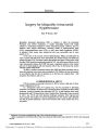
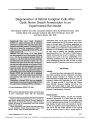
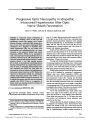
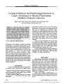
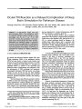
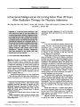
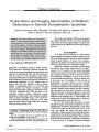
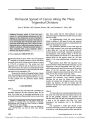
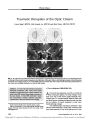
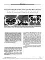
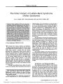
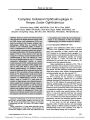
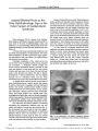
Show ORIGINAL CONTRIBUTION Perineural Spread of Cancer Along the Three Trigeminal Divisions Kara F. Warden, MD, Hemant Parmar, MD, and Jonathan D. Trobe, MD Abstract: Perineural spread of head and neck cancers is a well- documented phenomenon, but the diagnosis is often delayed due to lack of familiarity with clinical manifestations, anatomy of the head and neck, and imaging signs. We present single cases of perineural spread along each of the 3 divisions of the trigeminal nerve in which the perineural spread was initially overlooked. Although perineural spread is often associated with a poor prognosis, earlier detection may improve outcome. ( J Neuro- Ophthalmol 2009; 29: 300- 307) P erineural spread of cancer refers to the noncontiguous antegrade or retrograde extension of tumor cells along the tissues of the nerve. In head and neck cancers, it occurs in 2.5%- 5% of patients ( 1). The second and third trigeminal nerve divisions and the facial nerve are most often involved ( 2,3). The cancers are usually adenoid cystic or squamous cell carcinoma, but any head and neck cancer can spread via this mechanism ( 2,4). We present the clinical and imaging findings of single cases demonstrating perineural spread along the first ( V1), second ( V2) and third ( V3) trigeminal divisions. In none of these patients was the perineural spread initially recognized. CASE REPORTS Case 1 A 74- year- old man was treated with cryoablation for presumed squamous cell skin carcinoma above the left brow. Five weeks later he developed paresthesias in the previously treated area that gradually became a deep aching pain. Nine months after his initial treatment, he noted a droopy left upper lid followed by double vision and a firm left brow nodule. An ophthalmologist found left ocular ductional deficits in addition to left upper lid ptosis and suspected a third cranial nerve palsy. Results of a nondedicated brain MRI were interpreted as normal. Our examination disclosed 4 mm of left upper lid ptosis. Pupils measured 5 mm on the right, reacting briskly to direct light, and 7 mm and very irregular on the left without reaction to direct light. Adduction, supraduction, and infraduction were partially reduced in the left eye. There was hypesthesia over the left V1. A firm nodule was palpated above the left superior orbital rim. All other aspects of the neuro- ophthalmologic examination were normal. High- resolution brain MRI with trigeminal nerve protocol showed a lobular left forehead mass ( Fig. 1A) and enlargement of the left supraorbital nerve ( Fig. 1BC). A subtle enhancing soft tissue lesion was seen in the anterior aspect of the left cavernous sinus ( Fig. 1D). The presumptive diagnosis was perineural spread of skin cancer along V1 into the cavernous sinus ( Fig. 2AB). Biopsy of the solid skin nodule showed invasive squamous cell carcinoma. Local radiation performed elsewhere did not arrest the disease process. Within months, the patients had developed several more scalp nodules and proptosis. He died 20 months after the treatment of his skin tumor and 8 months after diagnosis of perineural spread. Case 2 A 65- year- old woman experienced paresthesias in her left check. One month later she noted reduced vision in the left eye that gradually worsened. Left proptosis soon followed. Brain and orbit MRI was interpreted as showing a mass in the left posterior orbit ( Fig. 3A). Local meningioma and lymphoma were in the differential diagnosis. Our examination 3 weeks later showed a best-corrected visual acuity of 20/ 15 in the right eye and 20/ 40 in the left eye, a left afferent pupillary defect, and mild left Department of Ophthalmology and Visual Sciences ( KFW, JDT), Department of Neurology ( JDT), and Department of Radiology ( Neuroradiology) ( HP), Kellogg Eye Center, University of Michigan Medical Center, Ann Arbor, Michigan. Address correspondence to Jonathan D. Trobe, MD, Kellogg Eye Center, 1000 Wall Street, Ann Arbor, MI 48109; E- mail: jdtrobe@ umich. edu 300 J Neuro- Ophthalmol, Vol. 29, No. 4, 2009 proptosis. A nerve fiber bundle defect was present in the visual field of the left eye. Ophthalmoscopy showed slight temporal optic disc pallor in the left eye. All other aspects of the neuro- ophthalmological examination were normal, including examination of trigeminal function. Review of the outside MRI confirmed the presence of the posterior orbital mass ( Fig. 3A) but also disclosed a mass in the left hard palate ( Fig. 3B) with perineural spread via the greater and lesser palatine nerves into the left pterygopalatine fossa ( PPF) ( Fig. 3C) and anteriorly into the left orbital apex via the left V2 and Vidian nerves ( Fig. 3D). The presumptive diagnosis was a left palatal carcinoma with perineural spread along the greater and lesser palatine nerves into the PPF and then along V2 into the left orbit ( Fig. 4A). Biopsy of the hard palate mass disclosed a large B-cell lymphoma. Results of systemic staging were negative. She was treated with corticosteroids and chemotherapy. Within 6 months, visual symptoms and paresthesias had resolved, but we have not undertaken a follow- up examination. Case 3 A 60- year- old man was noted to have 2 lesions on the left ear, one extending into the external auditory canal. Biopsy disclosed squamous cell carcinoma. Three weeks later he developed a left seventh cranial nerve palsy thought to be due to tumor invasion. MRI showed abnormal soft tissue thickening and enhancement involving the left periauricular region, the extracranial portion of the left seventh cranial nerve, and the area posterior to the mandibular condylar neck ( Fig. 5AB) with suspected involvement of the left auriculotemporal ( AT) nerve, a branch of the V3. The patient underwent total parotidectomy and auriculectomy with facial nerve sacrifice, selective neck dissection, and lateral temporal bone resection. Histopath-ologic examination demonstrated moderately differentiated squamous cell carcinoma with bony and perineural invasion. Two months after completing local radiation and chemotherapy, the patient reported left- sided jaw pain like an ‘‘ electrical sensation,'' attributed to the surgery. FIG. 1. Case 1. Perineural spread of squamous carcinoma along the first trigeminal division ( V1). Postcontrast T1 axial MRI with fat suppression shows a mass on the left forehead ( A, arrows) and enhancement of a thickened anterior orbital segment of the supraorbital nerve ( B, arrows). T1 coronal MRI ( C) shows a mass adjacent to the left superior rectus- levator complex ( arrow). Postcontrast T1 axial image at the level of cavernous sinuses ( D) shows abnormally enhancing soft tissue at the anterior and lateral aspect of the left cavernous sinus ( arrows). Compare with the normal cavernous sinus on the right side. 301 Perineural Spread of Cancer J Neuro- Ophthalmol, Vol. 29, No. 4, 2009 FIG. 2. Case 1. Schematic illustration of Figure 1. A. Axial view shows that forehead squamous cell carcinoma has spread along the left supraorbital branch of V1 to the cavernous sinus. B. Coronal view shows that the tumor has extended from V1 ( black arrow) to the third cranial nerve ( arrowhead) and fourth cranial nerve ( white arrow), probably via nervi nervorum. 302 q 2009 North American Neuro- Ophthalmology Society J Neuro- Ophthalmol, Vol. 29, No. 4, 2009 Warden et al However, a routine surveillance MRI 2 months later disclosed left middle ear cavity recurrence and a mass in the left Meckel's cave ( Fig. 6A) with enhancement along V3 ( Fig. 6B). He underwent another round of radiation and 6 cycles of chemotherapy. Despite these treatments, he developed extensive involvement of the cisternal segment of the left trigeminal nerve ( Fig. 7A) and antegrade involvement of the left inferior alveolar nerve ( Fig. 7B). He died 23 months after the initial diagnosis. The presumptive diagnosis was perineural spread along the AT nerve with spread to Meckel's cave and then to other branches of V3 and the trigeminal root ( Fig. 8A- B). DISCUSSION We have presented the clinical and imaging findings of single cases demonstrating perineural spread of squamous cell carcinoma along V1, V2, and V3. The V1 case was initially believed to be a compressive third cranial nerve palsy. The V2 case was initially diagnosed as a primary intraorbital mass. The V3 case was considered to be a squamous cell carcinoma limited to the region of the left ear. Overlooking perineural spread of head and neck cancer occurs because of incomplete understanding of the clinical manifestations, the anatomy of the sensory and motor nerves of the head and neck, and the often subtle imaging signs. Although some studies have estimated that up to 30%- 45% of patients may be asymptomatic even with extensive perineural spread ( 5), more often the clinical manifestations are subtle and overlooked by the physician. The most common initial symptoms of trigeminal perineural spread are painful dysesthesias and numbness. Hypesthesia is often found on examination ( 6). Trigeminal motor weakness ( of masticator, pterygoid, or temporalis muscles) is usually a much later finding ( 6,7). Familiarity with the innervation of the head and neck will allow the clinician to predict likely areas of perineural spread based on the location of the tumor. V1 innervates the forehead, orbit, eye, frontal sinus, lacrimal gland, and dorsum of the nose ( 8). Tumors in these areas of the face tend to spread along this division. Tumors of the mid- face tend to spread along V2, which serves the skin over the zygomatic arch, anterior temporal region, cheek, lower lid, lateral portion of the nose, upper lip, and maxillary sinus ( 3,8). V3 carries tumors located in the anterior portion of the ear, and the skin and mucosa of the lower portion of the FIG. 3. Case 2. Perineural spread along the second trigeminal division ( V2). Postcontrast T1 axial MRI with fat suppression ( A) shows an avidly enhancing left orbital mass ( arrows). Further review of precontrast T1 non- fat- suppressed coronal ( B) and axial ( C) images revealed a mass in the left palate ( white arrows) and left retromaxillary region ( black arrows). Invasion of the left pterygopalatine fossa is highlighted by replacement of the high signal of fat visible on the uninvolved right side ( black arrowhead). D. There is enhancement and enlargement of the left V2 branch ( small black arrow) and the nerve of the Vidian canal ( large black arrow) compared with normal structures on the right side ( white arrows). 303 Perineural Spread of Cancer J Neuro- Ophthalmol, Vol. 29, No. 4, 2009 FIG. 4. Case 2. Schematic illustration of Figure 3. Coronal ( A) and sagittal ( B) views show lymphoma originating in the left palate ( arrow) which has spread from the hard palate via greater and lesser palatine nerves to the pterygopalatine ganglion ( B, arrowhead) and then via a V2 branch into the orbit. 304 q 2009 North American Neuro- Ophthalmology Society J Neuro- Ophthalmol, Vol. 29, No. 4, 2009 Warden et al FIG. 5. Case 3. Perineural spread of squamous carcinoma along the third trigeminal division ( V3): initial scan. Precontrast T1 axial ( A) and T2 axial ( B) MRI images display a hypointense right parotid mass ( arrows) posterior to the mandibular ramus and along the expected course of the auriculotemporal nerve. Lateral and anterior extension was suspicious for facial nerve involvement as well. FIG. 6. Case 3. Perineural spread of squamous cell carcinoma along the third trigeminal division ( V3): 6 months later. A. Postcontrast T1 axial MRI shows an enhancing mass in the left Meckel's cave ( arrows). B. Postcontrast T1 coronal MRI shows enhancement and enlargement of the left V3 ( arrows). FIG. 7. Case 3. Perineural spread of squamous cell carcinoma along the third trigeminal division ( V3): 12 months later. A. Postcontrast T1 axial MRI shows retrograde extension of tumor along the cisternal segment of the left trigeminal nerve ( arrow). B. There is antegrade extension of tumor into the left inferior alveolar nerve that enhances ( arrows). Compare with the normal unenhancing inferior alveolar nerve in the right mandibular foramen ( arrowhead). 305 Perineural Spread of Cancer J Neuro- Ophthalmol, Vol. 29, No. 4, 2009 FIG. 8. Case 3. Schematic illustration of perineural spread of squamous cell carcinoma along the third trigeminal division ( V3). Sagittal ( A) and coronal ( B) views show tumor spreading from the parotid gland along the auriculotemporal branch of V3 to the trigeminal ganglion ( A) via the foramen ovale ( B). 306 q 2009 North American Neuro- Ophthalmology Society J Neuro- Ophthalmol, Vol. 29, No. 4, 2009 Warden et al cheek, chin, and lower lip. Tumors of the parotid gland travel along the facial nerve or AT branch of V3 ( 7- 9). With high field strength magnets, thin sections, and fat suppression, the changes associated with perineural spread should be evident to careful readers in 95% of patients ( 10). MRI changes include 1) alterations in the normal fat planes surrounding the nerve or within the basal skull foramina, 2) subtle enhancement of the involved nerve, and 3) enlargement of the involved nerve. CT changes include bone erosion and enlargement of the basal skull foramina ( 7). On MRI or CT, cavernous sinus involvement is evident as a convex bulge of its lateral wall ( 11). Collaboration between the clinician and the radiol-ogist plays a critical role in detection of perineural spread. Detailed requisitions including signs and symptoms help the radiologist assign the correct protocol for the study and focus on critical areas that may otherwise be overlooked. However, the entire course of the nerve must be evaluated, given the noncontiguous nature of the spread. Recognizing perineural involvement has important implications for treatment and prognosis. Once perineural spread has been recognized, treatment will usually take the form of radiation or chemotherapy rather than surgery. If surgery is to be performed, it must encompass a much wider region ( 4). In some cases, a multifocal hematopoietic tumor may be the cause ( as in our Case 2), in which case aggressive chemotherapy may be curative. REFERENCES 1. Maroldi R, Farina D, Borghesi A, et al. Perineural tumor spread. Neuroimaging Clin N Am 2008; 18: 413- 429. 2. Ballantyne AJ, McCarten AB, Ibanez ML. The extension of cancer of the head and neck through peripheral nerves. Am J Surg 1963; 106: 651- 667. 3. Ginsberg LE, DeMonte F. Imaging of perineural tumor spread from palatal carcinoma. AJNR Am J Neuroradiol 1998; 19: 1417- 1422. 4. DeMonte F, Hanna E. Transmaxillary exploration of the intracranial portion of the maxillary nerve in malignant perineural disease. J Neurosurg 2007; 107: 672- 677. 5. Mendenhall WM, Parsons JT, Mendenhall NP. et al. Carcinoma of the skin of the head and neck with perineural invasion. Head Neck 1989; 11: 301- 308. 6. Boerman RH, Maassen EM, Joosten H, et al. Trigeminal neuropathy secondary to perineural invasion of head and neck carcinomas. Neurology 1999; 53: 213- 216. 7. Ibrahim M, Parmar H, Gandhi D, et al. Imaging nuances of perineural spread of head and neck malignancies. J Neuroophthalmology 2007; 27: 129- 137. 8. Moore KL, Dalley AF II. Head: Clinically Oriented Anatomy. 4th ed. Philadelphia: Lippincott Williams & Wilkins; 1999: 832- 971. 9. Schmalfuss IM, Tart RP, Mukherji A, et al. Perineural tumor spread along the Auriculotemporal nerve. AJNR Am J Neuroradiol 2002; 23: 303- 311. 10. Nemzek WR, Hecht S, Gandor- Edwards R, et al. Perineural spread of head and neck tumors; how accurate is MR imaging. AJNR Am J Neuroradiol 1998; 19: 701- 706. 11. Caldemeyer KS, Mathews VP, Righi PD, et al. Imaging fea-tures and clinical significance of perineural spread or exten-sion of head and neck tumors. Radiographics 1998; 18: 97- 110. 307 Perineural Spread of Cancer J Neuro- Ophthalmol, Vol. 29, No. 4, 2009 |