| OCR Text |
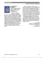
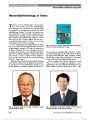
Show Visual and Neurological Outcomes Following Endovascular Stenting for Pseudotumor Cerebri Associated With Transverse Sinus Stenosis Martin G. Radvany, MD, David Solomon, MD, PhD, Satnam Nijjar, MD, Prem S. Subramanian, MD, PhD, Neil R. Miller, MD, Daniele Rigamonti, MD, Ari Blitz, MD, Philippe Gailloud, MD, Abhay Moghekar, MBBS Background: Pseudotumor cerebri (PTC) is characterized by raised intracranial pressure (ICP) without an identifiable mass, evidence of hydrocephalus, or abnormal cerebrospi-nal fluid content. In the past, most cases of PTC appeared to have no identifiable etiology, and thus, they were classified as "idiopathic intracranial hypertension" (IIH). Recently, however, a subset of patients with presumed IIH has been found to have evidence of cerebral dural sinus stenoses, particularly involving one or both transverse si-nuses (TS). The belief that the stenoses are the cause, rather than an effect of the increased ICP, has led investi-gators to recommend stenting of the stenosed sinus for the treatment of the condition. We describe detailed visual and neurological outcomes after stenting for PTC associated with hemodynamically significant dural sinus stenosis. Methods: All patients with PTC had initial neurological, neuro-ophthalmological, and imaging assessments. Regard-less of the findings, all were treated with medical therapy. If medical therapy failed and TS stenosis was detected on contrast-enhanced magnetic resonance or computed tomographic venography, catheter cerebral angiography with venous manometry was performed. If a mean pressure gradient (MPG) of 4 mm Hg or greater was present, unilateral transverse sinus stenting was performed. Results: Twelve patients with PTC and TS stenosis associ-ated with an MPG of .4 mm Hg who failed medical therapy were identified. TS stenting significantly decreased the pres-sure gradient in all cases. Unilateral stenting was sufficient to reduce pressure gradients even when the stenosis was bilateral. At a mean follow-up of 16 months (range, 9-36 months), tinnitus had improved in all patients, and 10 of 12 patients had improvement in visual function. Seven patients had significant improvement in headaches. Conclusion: In this small series of patients with PTC associated with TS stenosis, endovascular stent placement was generally effective in treating visual dysfunction and tinnitus, although not headaches. The optimum gradient and vascular characteristics amenable for selection of patients for stenting needs further research. Journal of Neuro-Ophthalmology 2013;33:117-122 doi: 10.1097/WNO.0b013e31827f18eb © 2013 by North American Neuro-Ophthalmology Society Pseudotumor cerebri (PTC) is defined as raised intracra-nial pressure (ICP) without clinical or imaging evidence of space-occupying intracranial pathology and with normal cerebrospinal fluid (CSF) analysis (1,2). A major morbidity of PTC is visual loss caused by prolonged papilledema with secondary optic nerve atrophy, occurring in 10%-20% of patients (2). Debilitating headache is the usual presenting symptom. Oral acetazolamide is the primary medical treat-ment; however, side effects, such as paresthesias, excessive fatigue, and kidney stones, limit its long-term use (3). Optic nerve sheath fenestration and CSF diversion procedure are potential surgical options (4). Although some cases of PTC are associated with medication use or systemic inflammatory disorders, the majority of cases have no obvious etiology and are referred to as "idiopathic intracranial hypertension" (IIH). Recently, some patients with IIH have been found to have neuro-imaging evidence of stenosis of one or both transverse si-nuses (TS) (5). In some of these cases, the stenosis resolves if ICP is lowered and, thus, is thought to be caused by the elevated ICP. In others, the stenosis remains until the affected sinus is mechanically opened by stenting, Departments of Radiology and Radiological Science (MGR, AB, PG); Neurology (DS, SN, PSS, NRM, AM); Ophthalmology (PSS, NRM), and; Neurosurgery (PSS, NRM, DR), Johns Hopkins University School of Medicine, Baltimore, Maryland; and Departments of Radiology (MGR); and Department of Ophthalmology (PSS), Uni-formed Services University of the Health Sciences, Bethesda, Maryland. The authors report no conflicts of interest. Address correspondence to Abhay Moghekar, MBBS, Department of Neurology, Johns Hopkins Hospital, Phipps 126, 600 North Wolfe Street, Baltimore, MD 21287. E-mail: amogheka@jhmi.edu Radvany et al: J Neuro-Ophthalmol 2013; 33: 117-122 117 Original Contribution Copyright © North American Neuro-Ophthalmology Society. Unauthorized reproduction of this article is prohibited. suggesting a role for venous outflow obstruction as an eti-ologic factor in these cases. It is unsettled if this group of patients should be called "idiopathic" or intracranial hyper-tension secondary to venous sinus stenoses. To date, most reports of intracranial venous sinus stenting for PTC have documented visual and headache outcomes qualitatively rather than quantitatively (6-11). We report results of stenting in 12 patients with PTC associated with TS stenosis, with quantitative assessment of several aspects of visual and neurological function. METHODS Institutional Review Board approval was obtained for this study. Between January 2008 and June 2011, a total of 88 adult patients with IIH were screened. All patients satisfied modified Dandy criteria for IIH, were not pregnant, and were not on medications or had medical conditions associated with intracranial hypertension. None of these patients had under-gone shunt or bariatric surgery. We evaluated 14 of these patients with evidence of unilateral or bilateral TS stenosis who had failed medical therapy; 12 subsequently underwent stent placement. Medical treatment failure was defined as allergy to acetazolamide, intolerance to escalating doses of this medication, or worsening of papilledema, despite treatment of up to 3,000 mg of acetazolamide daily. Detailed neurological and neuro-ophthalmological evaluations were obtained before and after the treatment. Neurological assessment included a detailed headache history and examination to exclude other causes of PTC and lumbar puncture. Opening pressure was measured with the patient in the left lateral decubitus position with hips and knees extended and head neutral. Neuro-ophthalmological assessment included measurement of visual acuity, color vision using Hardy-Rand-Rittler pseudoisochro-matic plates, automated visual field testing, and funduscopic examination with grading of papilledema according to the Frisén classification (12). For the purpose of analysis, visual acuity results were converted to logarithm of the minimal angle of resolution units. Headache intensity was assessed using the Headache Impact Test-6 (HIT-6) score (13). Improvement in headache was defined as a decrease in the HIT-6 score of 1 standard deviation (10 points). After review by a multidisciplinary team, including neurologists, neurosurgeons, neuro-ophthalmologists, and interventional neuroradiologists, these patients were offered optic nerve sheath fenestration, a CSF diversion procedure, or TS stent on an off-label basis. Informed consent was obtained from all patients. All patients had magnetic resonance imaging of the brain and magnetic resonance venography (MRV) with contrast. The neuroradiologist reviewed the raw data in multiple planes and performed 3-dimensional rendering (Webspace; Siemens, Erlangen, Germany). Once the patient chose stent placement, the diagnosis of TS stenosis was confirmed with computed tomographic venography (CTV) using a 320 detector computed tomo-graphic scanner (Aquilion; Toshiba, Tustin, CA). CTV was chosen to confirm the diagnosis because of our experience of overestimating stenosis with MRV (unpublished data, Gailloud 2012). In 6 of the 12 patients, CTV was performed immediately after the reduction of CSF pressure to ,15 cm of water by lumbar puncture. If CTV demonstrated unilateral TS stenosis (Fig. 1), bilateral stenoses, or stenosis of a single dominant TS (contralateral TS, aplastic or absent) that did not resolve with normalization of ICP, the patient was scheduled for cerebral catheter angiography, endovenous pressure meas-urements, and possible stent placement (Fig. 2A). Endovascular stent placement was performed when a pressure gradient of at least 4 mm Hg was documented. In the case of bilateral stenoses, the side with the larger gradient was targeted (Fig. 2B). Endovenous pressure meas-urements were repeated after stenting. Clopidogrel and aspi-rin, which were initiated 4 days before angiography, were continued for 6 months. All patients had follow-up appointments for neurological and neuro-ophthalmological evaluations at 6 weeks, 3 months, FIG. 1. Patient 4. A. Computed tomographic venography showing right transverse sinus stenosis (arrows). B. Following stenting, the right transverse sinus is widely patent (arrows). 118 Radvany et al: J Neuro-Ophthalmol 2013; 33: 117-122 Original Contribution Copyright © North American Neuro-Ophthalmology Society. Unauthorized reproduction of this article is prohibited. 6 months, and then every 6 months. Stent patency was evaluated with CTV at 6 months (Fig. 1B) or earlier if a patient had recurrent symptoms. Acetazolamide was con-tinued for 3-6 months after the surgery in those taking it prior to the surgery, and the dose was tapered gradually after assessing response to stenting. RESULTS There were 11 female patients and 1 male patient ranging in age from 21 to 55 years (mean age, 39 years) with a mean body mass index of 32.6 kg/m2 (range, 27.3-45.7 kg/m2). All patients had headaches and had papilledema. All but 1 had subjective pulsatile tinnitus. A recent lumbar puncture in all patients demonstrated elevated CSF pressures ranging from 29 to 55 cm of water. Stent insertion was followed by a significant reduction in the trans-stenotic gradient in all 12 patients (Table 1). There were no serious complications after stent placement; however, in 1 patient, the procedure was interrupted after stagnation of contrast was observed in the venous sinus wall that raised concerns for dissection. The patient was dis-charged after overnight observation and subsequently trea-ted successfully. Two additional patients who underwent catheter venography had no pressure gradient, despite apparent TS stenosis on CTV, and were not stented. All patients experienced retro-orbital pain ipsilateral to the stent placement immediately after the procedure, which resolved in all cases over a few days. When ex-amined between 6 and 12 weeks following stent place-ment, 11 of the 12 patients showed improvement in visual function or stable visual function and decreased papil-ledema. Visual acuity was stable or improved in 22 of 24 eyes, and color vision was stable or improved in 21 of 24 eyes (Table 2). Visual fields improved or remained stable in 19 of 24 eyes, as measured by mean deviation, and there FIG. 2. Patient 6. A. Venous phase of cerebral angiogram demonstrating bilateral transverse sinus stenosis (arrows). B. Venous phase cerebral angiogram following right transverse sinus stenting. Change in flow dynamics with decreased flow in left transverse sinus was noted. TABLE 1. Patient demographics and treatment summary Patient Age (y)/Sex Body Mass Index (kg/m2) Opening Pressure (cm of Water) Treatment Failure TS Stented Gradient (mm Hg) Gradient After Stent (mm Hg) 1 37/F 28.1 43 Shunt infected · 2 Right 28 0 2 42/F 35.5 35 Intolerant to acetazolamide Right 5 0 3 37/M 35.7 42 Nephrolithiasis Left 22 2 4 36/F 27.3 31 Intolerant to acetazolamide Right 12 2 5 48/F 27.4 44 Intolerant to acetazolamide Right 10 1 6 55/F 32.3 29 Medical treatment failed Right 17 4 7 21/F 29 42 Medical treatment failed Right 8 1 8 33/F 45.7 55 Medical treatment failed Right 19 0 9 44/F 40.2 32 Nephrolithiasis; medical treatment failed Left 8 2 10 51/F 30.3 32 Medical treatment failed Right 6 1 11 34/F 30.9 55 Medical treatment failed Right 8 1 12 25/F 29.2 33 Medical treatment failed Right 6 1 Patients 8 and 11 developed stenosis proximal to the stent. F, female; M, male; TS, transverse sinuses. Radvany et al: J Neuro-Ophthalmol 2013; 33: 117-122 119 Original Contribution Copyright © North American Neuro-Ophthalmology Society. Unauthorized reproduction of this article is prohibited. was resolution of papilledema in 11 of the 12 patients (Table 3). Only patient 12 had worsening of visual parameters and persistent papilledema following stenting. Patients 8 and 11 developed recurrent papilledema and worsening headaches at 6 months. In both cases, CTV demonstrated a widely patent TS when stented without thrombus formation. However, a new TS stenosis was noted proximal to the stent (Fig. 3). Both patients underwent repeat stenting and had initial resolution of symptoms and signs. Patient 11 had an 8-mm Hg gradient across her right transverse sinus ste-nosis, and following stenting, she had improvement in visual acuity, visual field, and papilledema. However, at 6- month follow-up, her color vision and visual fields had worsened, although papilledema had resolved. CTV con-firmed a patent stent but showed TS stenosis proximal to the stent. The patient underwent ventriculoperitoneal shunt surgery that stabilized her visual function and eliminated her headaches. Following stenting, headaches resolved completely in 2 patients and improved in 5 but persisted in the remaining 5 patients. On the HIT-6 assessment, 36 is the lowest score obtainable (Table 4). Between 6 and 12 months after TS stenting, all 5 patients with persistent severe headache underwent a lumbar puncture or continuous spinal fluid pressure monitoring, 4 of whom were found to have normal ICP. The fifth patient (patient 8) had a mean pressure of .20 mm Hg (approximately 26 cm of water) for 68 mi-nutes of the 490 minutes that she was monitored. Tinnitus improved on the stented side in all 11 of the patients in whom it had been present (Table 4), whereas 4 of 7 patients with bilateral tinnitus experienced improve-ment on both sides. DISCUSSION Although Johnston and Paterson (14) first proposed venous hypertension as a potential cause of intracranial hyperten-sion in 1974, it was not until 1995 that King et al (15) documented elevated sagittal and proximal transverse sinus pressures in patients with presumed IIH. In 2003, Higgins et al (7) reported a series of 12 patients with IIH associated with dural sinus stenosis, in whom stenting produced vari-able resolution of papilledema and headache. Although sim-ilar results have been reported (6,8-11), there is a lack of uniformity in methods and outcomes in these studies. We undertook a quantitative analysis of neurological and visual function in patients with PTC associated with venous sinus stenosis and a pressure gradient of .4 mm Hg who under-went stenting following unsuccessful medical therapy. Regarding neurologic findings, headache usually is the presenting symptom in patients with increased ICP. However, all headaches in this setting may not be caused by increased pressure and not resolve despite lowering it to a normal range (16). Our patients who continued to have TABLE 2. Visual outcomes after transverse sinus stenting: visual acuity and color vision Patient VA-Right Eye, logMAR (Snellen) VA-Left Eye, logMAR (Snellen) CV-Right Eye CV-Left Eye Pre Post Pre Post Pre Post Pre Post 1 20.04 (20/1623) 20.1 (20/16) 20.04 (20/1623) 20.1 (20/16) 10 10 10 10 2 0.3 (20/40) 0 (20/20) 0.1 (20/25) 0.1 (20/25) 7.5 10 7.5 10 3 0.44 (20/5023) 20.06 (20/1622) 0 (20/20) 20.06 (20/1622) 9 10 10 10 4 0.02 (20/2021) 20.08 (20/1621) 0.06 (20/2023) 20.08 (20/1621) 9 10 10 10 5 0.16 (20/2523) 0.04 (20/2022) 0.02 (20/2021) 20.1 (20/16) 10 10 10 10 6 0 (20/20) 0 (20/20) 0.1 (20/25) 0.1 (20/25) 10 10 10 10 7 0.04 (20/2023) 0 (20/20) 0.1 (20/25) 0 (20/20) 9 10 9 10 8 0 (20/20) 20.04 (20/16-3) 0 (20/20) 20.1 (20/16) 10 10 10 10 9 0.02 (20/2021) 0 (20/20) 0.2 (20/32) 0.04 (20/2022) 9 10 9.5 10 10 0 (20/20) 20.1 (20/16) 0 (20/20) 20.1 (20/16) 10 10 10 10 11 0.3 (20/40) 0.2 (20/32) 0.2 (20/32) 0.3 (20/40) 10 8 10 0.5 12 0.04 (20/2022) 0.16 (20/2523) 0.06 (20/2023) 0.14 (20/2522) 8.5 7.5 8.5 8.5 CV, color vision; logMAR, logarithm of the minimal angle of resolution; pre, before stenting; post, after stenting; VA, visual acuity. 120 Radvany et al: J Neuro-Ophthalmol 2013; 33: 117-122 Original Contribution Copyright © North American Neuro-Ophthalmology Society. Unauthorized reproduction of this article is prohibited. headaches despite stenting and normalization of ICP had many features associated with migraine. Of 5 patients in this series who continued to have headaches despite normal-ization of CSF pressure after stenting, 4 responded to migraine prophylactic medications. Transient ipsilateral headaches were present immediately after stent placement in all patients. This is not an unexpected finding considering the innervation of the meninges in this location. The intracranial branch of the ophthalmic division of the fifth nerve (tentorial nerve of Arnold) supplies the tentorium, superior surface of the transverse and straight venous sinuses, and the inferior two thirds of the falx cerebri (17). Pulsatile tinnitus is a common symptom in PTC. In patients with transverse sinus stenosis, it is likely secondary to turbulent flow across the stenotic region, located in proximity to the inner ear. Tinnitus resolved in all our patients on the side ipsilateral to TS stenting. Possibly, the reduction of flow across the contralateral stenotic side also resulted in improved tinnitus in 4 of the 7 patients who presented with bilateral TS stenosis. The normal gradual drop in intrasinus pressure from the superior sagittal sinus to the internal jugular bulb is 0-3 mm Hg (15), and a threshold of 4 mm Hg gradient has been proposed for stenting (11). Reduction in the TS gra-dient was achieved in all our patients with a gradient of 4 mm Hg or greater. Two patients developed a new stenosis proximal to the stent, an observation previously described (6,18). We postulate that continued expansion of the sinus by the stent may have caused stenosis adjacent to the treated segment, possibly as a result of the uneven force exerted by the stent on the noncylindrical sinus. Alternatively, with TABLE 3. Visual outcomes after transverse sinus stenting: visual fields and papilledema Patient MD-Right Eye MD-Left Eye Papilledema Grade, Right Eye Papilledema Grade, Left Eye Pre Post Pre Post Pre Post Pre Post 1 22.63 22.19 21.37 21.33 1 0 1 0 2 228.03 0.15 23.66 0.77 3 0 2 0 3 21.75 21.37 20.67 20.45 2 0 1 0 4 22.35 22.29 22.94 23.42 1 0 1 0 5 20.83 20.17 22.82 21.25 1 0 4 0 6 0.01 20.51 22.53 22.04 1 0 2 0 7 27.40 24.33 29.88 27.94 2 0 1 0 8 22.34 21.70 25.57 22.66 2 0 3 0 9 24.67 20.57 27.22 21.12 2 0 2 0 10 23.24 21.18 25.44 21.84 2 0 3 0 11 215.01 213.04 219.09 230.82 2 0 2 0 12 22.48 213.24 24.82 27.74 1 1 1 1 MD, mean deviation; pre, before stenting; post, after stenting; VA, visual acuity. FIG. 3. Patient 8. Computed tomographic venography re-veals area of smooth stenosis (arrowhead) immediately adjacent to stent (arrow). TABLE 4. Outcomes of headache and tinnitus following transverse sinus stenting Pt HIT-6 Score Tinnitus Pre Post Pre Post 1 78 78 Bilateral Absent 2 62 36 Unilateral Absent 3 48 40 Unilateral Absent 4 50 40 Bilateral Improved on stented side 5 40 36 Bilateral Absent 6 78 66 Bilateral Absent 7 78 40 Unilateral Absent 8 69 66 Bilateral Improved on stented side 9 78 58 Absent Absent 10 78 38 Unilateral Absent 11 64 44 Bilateral Absent 12 67 68 Bilateral Improved on stented side HIT-6 = Headache Impact Test-6; pre, before stenting; post, after stenting. Radvany et al: J Neuro-Ophthalmol 2013; 33: 117-122 121 Original Contribution Copyright © North American Neuro-Ophthalmology Society. Unauthorized reproduction of this article is prohibited. increased blood flow through the stented transverse sinus, flow may be diverted from the contralateral side leading to lowered intraluminal pressure within the transverse sinus causing it to collapse in the region not supported by the stent (Bernoulli effect) (19). Rohr et al (20) theorized that there were 2 types of venous sinus stenoses: stenoses caused by elevated ICP nar-rowing the sinus extrinsically and fixed stenoses in which reduction of CSF pressure would not affect the diameter of the sinus. In 6 patients, we normalized ICP before perform-ing CTV and did not observe resolution of stenosis. One of these patients (patient 11) developed a new stenosis proxi-mal to the stent, and her trans-stenotic gradient was 8 mm Hg. Some patients with lower gradients (patients 2 and 10) responded to stenting. The explanation that these venous sinus stenoses were secondary to extrinsic compression from raised ICP appears unlikely. The dispute about extrinsic venous stenoses caused by intracranial hypertension vs intrinsic stenosis has been extensively debated without clear resolution (21). We recognize the limitations of our study. Treatment was not standardized or randomized, and the number of patients was small. In addition, only a carefully screened subset of patients who had failed medical therapy was considered for the procedure. Further studies are needed to evaluate patient selection parameters, the long-term efficacy of this technique, and its effectiveness vs CSF diversion procedures. REFERENCES 1. Ball AK, Clarke CE. Idiopathic intracranial hypertension. Lancet Neurol. 2006;5:433-442. 2. Acheson JF. Idiopathic intracranial hypertension and visual function. Br Med Bull. 2006;79-80:233-244. 3. Ko MW. Idiopathic intracranial hypertension. Curr Treat Options Neurol. 2011;13:101-108. 4. McGirt MJ, Woodworth G, Thomas G, Miller N, Williams M, Rigamonti D. Cerebrospinal fluid shunt placement for pseudotumor cerebri-associated intractable headache: predictors of treatment response and an analysis of long-term outcomes. J Neurosurg. 2004;101:627-632. 5. Farb RI, Scott JN, Willinsky RA, Montanera WJ, Wright GA, terBrugge KG. Idiopathic intracranial hypertension: the prevalence and morphology of sinovenous stenosis. Neurology. 2003;60:1418-1424. 6. Kumpe DA, Bennett JL, Seinfeld J, Pelak VS, Chawla A, Tierney M. Dural sinus stent placement for idiopathic intracranial hypertension. J Neurosurg. 2012;116:538-548. 7. Higgins JNP, Cousins C, Owler BK, Sarkies N, Pickard JD. Idiopathic intracranial hypertension: 12 cases treated by venous sinus stenting. J Neurol Neurosurg Psychiatry. 2003;74:1662-1666. 8. Bussière M, Falero R, Nicolle D, Proulx A, Patel V, Pelz D. Unilateral transverse sinus stenting of patients with idiopathic intracranial hypertension. AJNR Am J Neuroradiol. 2010;31: 645-650. 9. Arac A, Lee M, Steinberg GK, Marcellus M, Marks MP. Efficacy of endovascular stenting in dural venous sinus stenosis for the treatment of idiopathic intracranial hypertension. Neurosurg Focus. 2009;27:E14. 10. Donnet A, Metellus P, Levrier O, Mekkaoui C, Fuentes S, Dufour H, Conrath J, Grisoli F. Endovascular treatment of idiopathic intracranial hypertension: clinical and radiologic outcome of 10 consecutive patients. Neurology. 2008;70: 641-647. 11. Ahmed RM, Wilkinson M, Parker GD, Thurtell MJ, Macdonald J, McCluskey PJ, Allan R, Dunne V, Hanlon M, Owler BK, Halmagyi GM. Transverse sinus stenting for idiopathic intracranial hypertension: a review of 52 patients and of model predictions. AJNR Am J Neuroradiol. 2011;32:1408-1414. 12. Frisén L. Swelling of the optic nerve head: a staging scheme. J Neurol Neurosurg Psychiatry. 1982;45:13-18. 13. Kosinski M, Bayliss MS, Bjorner JB, Ware JE Jr, Garber WH, Batenhorst A, Cady R, Dahlöf CG, Dowson A, Tepper S. A six-item short-form survey for measuring headache impact: the HIT-6. Qual Life Res. 2003;12:963-974. 14. Johnston I, Paterson A. Benign intracranial hypertension. II. CSF pressure and circulation. Brain. 1974;97:301-312. 15. King JO, Mitchell PJ, Thomson KR, Tress BM. Cerebral venography and manometry in idiopathic intracranial hypertension. Neurology. 1995;45:2224-2228. 16. Friedman DI, Rausch EA. Headache diagnoses in patients with treated idiopathic intracranial hypertension. Neurology. 2002;58:1551-1553. 17. Feindel W, Penfield W, McNaughton F. The tentorial nerves and Iocalization of intracranial pain in man. Neurology. 1960;10: 555-563. 18. Owler BK, Parker G, Halmagyi GM, Dunne VG, Grinnell V, McDowell D, Besser M. Pseudotumor cerebri syndrome: venous sinus obstruction and its treatment with stent placement. J Neurosurg. 2003;98:1045-1055. 19. Imbesi SG, Kerber CW. An experimental and angiographic explanation of why ulcerated carotid bulbs embolize. Interv Neuroradiol. 1999;5:11-18. 20. Rohr A, Dörner L, Stingele R, Buhl R, Alfke K, Jansen O. Reversibility of venous sinus obstruction in idiopathic intracranial hypertension. AJNR Am J Neuroradiol. 2007;28: 656-659. 21. Ahmed R, Friedman DI, Halmagyi GM. Stenting of the transverse sinuses in idiopathic intracranial hypertension. J Neuroophthalmol. 2011;31:374-380. 122 Radvany et al: J Neuro-Ophthalmol 2013; 33: 117-122 Original Contribution Copyright © North American Neuro-Ophthalmology Society. Unauthorized reproduction of this article is prohibited. |