| OCR Text |
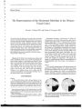
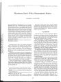
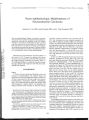
Show Journal of Neuro- Ophthalmology 20( 2): 111- 115, 2000. © 2000 Lippincott Williams & Wilkins, Inc., Philadelphia Vision Loss as the Presenting Sign in Juvenile Neuronal Ceroid Lipofuscinosis Lisa I. Bohra, MD, Jennifer S. Weizer, BA, Andrew G. Lee, MD, and Richard Alan Lewis, MD, MS Objective: To review cases of juvenile neuronal ceroid lipofuscinosis ( JNCL) and highlight salient clinical and diagnostic features, thereby enhancing recognition of this disease among ophthalmologists. Materials and Methods: Twelve cases of JNCL seen from 1982 to 1999 were reviewed. Diagnosis was based on characteristic clinical history, ophthalmoscopic findings, electroretin-ography, neuroimaging, histopathology, and molecular analysis. Results: Vision loss was the first subjective symptom of the disease in all 12 cases. Among these cases, nine of 12 patients ( 75%) developed neurologic deficits an average of 3 years after the onset of visual deterioration. Conclusion: Because visual symptoms usually precede neurologic dysfunction, JNCL should be considered in the differential diagnosis when an apparently healthy child presents with unexplained bilateral vision loss. Key Words: Batten disease- Bull's- eye maculopathy- Spielmeyer- Vogt disease- Lipopigment storage disease. The neuronal ceroid lipofuscinoses are progressive neurodegenerative disorders inherited as autosomal recessive traits and characterized pathologically by the accumulation of autofluorescent lipopigment within lyso-somes. Historically, they are classified as four major subtypes based on age at onset ( infantile, late infantile, juvenile, and adult), clinical course, and, recently, genetic locus. The clinical features include vision loss, alterations in recent and fixed memory, motor dysfunction, and, later, seizures and death. Because of these constitutional signs, patients often devolve to a neurologist. However, the most common type, juvenile neuronal Manuscript received February 2, 2000; accepted March 28, 2000. From the Cullen Eye Institute ( LIB, JSW, RAL), Baylor College of Medicine, Houston, Texas; and the Departments of Ophthalmology, Neurology, and Neurosurgery ( AGL), the University of Iowa Hospitals and Clinics, Iowa City, Iowa. Presented at the 2000 North American Neuro- Ophthalmologic Society Meeting, March 2000, Montreal, Canada. This study was supported in part by Research to Prevent Blindness, New York, New York ( support to RAL and the University of Iowa). Richard Alan Lewis, MD, MS, is a Research to Prevent Blindness Senior Scientific Investigator. Address correspondence and reprint requests to Andrew G. Lee, MD, Department of Ophthalmology, 200 Hawkins Drive, PFP, the University of Iowa Hospitals and Clinics, Iowa City, Iowa 52242. ceroid lipofuscinosis ( JNCL; eponymic terms are Spielmeyer- Vogt or Batten disease), most often presents with vision loss first and with neurological symptoms or signs later, making it relevant for all ophthalmologists, especially those evaluating children and adolescents. From 1982 to 1999, one of the authors ( RAL) has ascertained this diagnosis in 12 children from ages 4 to 15 years old. We review 12 cases of JNCL presenting initially with visual loss and highlight the pertinent characteristics that should prompt consideration of this diagnosis. To our knowledge, this is the largest case series of patients with JNCL in the ophthalmologic literature. METHODS From 1982 to 1999, 28 cases of JNCL, of which 12 cases from ten families met study criteria, were reviewed at Baylor College of Medicine in Houston, Texas. The inclusion criterion for the study was history of substantial progressive visual loss in childhood. Exclusion criteria included intracranial masses, nonretinal intraocular pathology, and/ or alternative etiologies resulting in vision loss. We reviewed medical history, family history, developmental history, age of onset, the course of clinical symptoms and signs, and results of comprehensive ophthalmologic examinations. Results of electroretinog-raphy ( ERG) and/ or neuroimaging obtained in some individuals to aid the exclusion of other diagnoses were also reviewed. Diagnosis was confirmed by electron microscopic examination of either serum lymphocytes or conjunctiva and/ or by DNA analysis. All patients had thorough pediatric neurologic and clinical genetic evaluations. RESULTS The clinical features of our 12 cases are summarized in Table 1. The results of electroretinography, neuroimaging, pathology, and DNA analysis are summarized in Table 2. Patients 1 and 2 and patients 9 and 10 are each a pair of biological siblings. Patient 7 was the product of a consanguineous ( uncle- niece) marriage. In all other cases, family history was unremarkable for the presence of disease and for consanguinity. The mean age of onset of visual loss was 6.6 years 111 112 L. I. BOHRA ET AL. TABLE 1. Clinical features of juvenile neuronal ceroid lipofuscinosis Patient Age of onset of visual loss ( y) Visual acuity at time of presentation Neurologic signs or symptoms Period of time between vision loss and onset of neurologic symptoms or signs ( y) 10 11 12 3.5 5.1 4.2 5.3 5.5 4.0 7.2 8.5 13.5 11.8 2.5 7.5 20/ 400 OU 20/ 80 OU LPOU HMOU LPOU 5/ 200 OU HMOU HMOU 20/ 60 OD 20/ 70 OS HMOU 20/ 40 OU CFat 12ftOD CF at 6 ft OS None None Rotary nystagmus Absence seizures and cognitive impairment Cerebellar dysfunction None Upbeat nystagmus in upgaze Intellectual impairment and absence seizures Intellectual impairment and tremor Intellectual impairment Generalized seizures Intellectual impairment and absence seizures N/ A N/ A 3 2 1 N/ A 2 1 > 3 > 3 9 1 CF, count fingers; HM, hand motion; LP, light perception. ( range, 2.5- 13.5). The mean time from onset of visual loss to presentation at our institution was 3.5 years ( range, 8 months- 9 years). Visual acuity at presentation was uniformly poor; six out of 12 ( 50%) patients had visual function of hand motion ( four patients) or light perception ( two patients) only. Results of ophthalmoscopic examination were abnormal in all patients, revealing characteristic changes of the neuroepithelium and retinal pigment epithelium in the macula. However, severity ranged from a subtle diffuse grainy appearance of the retinal pigment epithelium to a more discrete bull's eye pattern with temporal pallor of the optic disc ( Fig. 1). The foveal reflex and umbo were diminished in all cases. At presentation, mild to moderate diffuse optic pallor was evident in nine out of 12 ( 75%) patients, and vascular attenuation was noted in four out of 12 ( 33%) patients. Neurologic signs, such as nystagmus, tremors, gait disturbance, and cognitive impairment, developed in nine ( 75%) patients during the course of the study. One of these patients died at age 29. Two of the nine patients ( 22%; aged 7.2 and 9.2 years) had abnormal neurologic findings on examination but were otherwise asymptomatic. One of these two patients developed rotary nystagmus 3 years after presentation, while the other patient was found at presentation to have upbeat nystagmus in upgaze only. The mean time from discovery of visual loss to development of neurologic symptoms or signs ranged from 1 to 9 years ( mean, 3; n = 9). During this interval, seven patients were given initial incomplete or erroneous diagnoses, including possible Stargardt disease ( four out of seven patients), retinitis pigmentosa ( two out of seven patients), and rod/ cone dystrophy ( one out of seven patients). No evidence of neurologic deficit was discovered in the remaining three patients ( 25%) after 1, 4, and 9 years from onset of vision loss, respectively. Electroretinography performed in six cases revealed a substantial diminution of a- and b- wave amplitudes in scotopic and photopic situations ( Fig. 2). Neuroimaging studies ( computed tomography [ CT] or magnetic resonance imaging [ MRI]) performed for seven patients at TABLE 2. Patient Electrophysiology, ERG neuroimaging, pathology, and DNA analysis Neuroimaging in juvenile neuronal ceroid lipofuscinosis Pathology DNA analysis 9 10 11 12 Decreased a- and b- wave amplitudes* Normal MRI Decreased a- and b- wave amplitudes Decreased a- and b- wave amplitudes Decreased a- and b- wave amplitudes Decreased a- and b- wave amplitudes Decreased a- and b- wave amplitudes Diffuse cortical atrophy on MRI Diffuse cortical atrophy on CT Mild bilateral optic nerve and tract atrophy on MRI Mild ventricular dilatation on CT Mild cortical atrophy on CT Normal MRI Conjunctival biopsyt Conjunctival biopsyt Conjunctival biopsyt Lymphocyte analysisf Lymphocyte analysisf Lymphocyte analysisf Conjunctival biopsyt Lymphocyte analysisf Conjunctival biopsyt Conjunctival biopsyt 1.02 KB deletion in CLN3 1.02 KB deletion in CLN3 1.02 KB deletion in CLN3 Negative for 1.02 KB deletion in CLN3 * To photopic and scotopic stimuli. t Inclusion bodies seen on electron microscopy, characteristic of JNCL. MRI, magnetic resonance imaging; CT, computed tomography; ERG; electroretinography. J Neuro- Ophthalmol, Vol. 20, No. 2, 2000 VISION LOSS AS THE PRESENTING SIGN IN JNCL 113 FIG. 1. Fundus photographs reveal corrugation in the internal limiting membrane reflexes, the fine granularity in the macular areas, and the lack of a strong foveal light reflex. There is an indistinct hyperpigmented bull's eye zone of atrophy in each eye. presentation revealed three ( 43%) patients with diffuse, mild cortical atrophy. One patient had evidence of bilateral mild optic nerve and optic tract atrophy without cortical involvement on MRI results. One patient had mild ventricular dilatation revealed by CT; the remaining two patients had normal imaging studies at ages 6 and 7.5 years, respectively. Histopathologic examination of peripheral blood lymphocytes and/ or conjunctiva revealed characteristic fingerprint inclusion bodies within lysosomes in all but two patients ( Fig. 3). The diagnosis in patient 2 was based on presentation symmetric to that of her older brother ( patient 1), as well as on DNA analysis. The diagnosis in patient 10 was based on presentation, age of onset, ophthalmoscopic findings, and clinical course symmetric to that of her older sister ( patient 9), for whom pathologic and DNA confirmation of JNCL was obtained previously. Three out of 12 ( 25%) patients had confirmation of the diagnosis by DNA analysis, which demonstrated homozygosity for a common ( North American) mutation, a 1.02 kB deletion in the CLN3 gene of chromosome 16 ( 1). One patient had a negative test result for this particular mutation. Genetic testing was not commercially available at the time of diagnosis of the other nine patients. DISCUSSION The clinical and histologic similarities in presentation among our cohort further illustrate the classic presentation of JNCL, whose incidence is estimated to be up to one out of 25,000 live births worldwide, with an increased relative prevalence in northern Europe ( 2). Juvenile neuronal ceroid lipofuscinosis is not only the most prevalent form of all the neuronal ceroid lipofuscinoses in the United States ( 3), but it is also among the most common progressive childhood encephalopathies. In our series, as in others, visual loss was the most common presenting sign ( 4,5). We reported a mean age of onset of vision loss at 6.6 years, which is comparable with the published range of 6.2 ± 1.8 years ( 6). Parents report changes in a child's visual behavior, but they undoubtedly miss subtle early changes; therefore, it is likely that the onset of vision loss is earlier than has been reported or recognized. Early in its course, there is a reduction of central vision with color deficiency and relative preservation of night vision compared with day vision ( 7). The rate of progression from normal visual acuity to visual impairment varies considerably, ranging from several years to as few as 8 months ( 5,8). For example, patient 1 had a documented decline in visual acuity from 20/ 60 OU to 20/ 400 OU over 3 months. On average, neurologic symptoms and signs in the patients in our series began 3 years after recognition of visual signs. Previous reports describe a typical progression of neurologic symptoms, beginning with cognitive impairment at 7.4 ± 2.0 years of age, seizures and motor disturbances at 9.5 ± 3.5 years, onset of a vegetative state at 18.4 ± 2.8 years, and finally death at 20.2 ± 6.3 years ( 6). Many cases with a more protracted clinical course OD Scotopic Flash ERG " " T 200.00 uV 12 J DIKE [ 00.00 uV 12.5 msec 2 200.00 uV 12.5 mJ « 6 l200. OOuV12.5mMC Tim* Par 3 200.' 7 IZOO. i Tim. P » r nivi. inn 00 uV 12.3 raw: 00 uV 12.5 miec 200.00 uV 12.5 m< « I200. OO uV 12.5 raiec FIG. 2. Flash electroretinogram demonstrates markedly abnormal a and b waves. _, J Neuro- Ophthalmol, Vol. 20, No. 2, 2000 114 L I. BOHRA ET AL. FIG. 3. Transmission electron microscopy of conjunctival biopsy specimen. Note the fingerprint inclusion body. have been described; these patients tend to have different genetic mutations than patients homozygous for the 1.02 kB deletion in the CLN3 gene have, who demonstrate the classic rate of progression ( 6,8,9). Patients 1 and 2 ( aged 7.1 and 5.1 years, respectively) are likely to show neurologic signs within the next several years, given their positive DNA analysis for the homozygous 1.02 kB deletion in CLN3. Patient 6 ( aged 9.3 years) has not exhibited any neurologic signs or symptoms to date and has not had DNA analysis; whether he will follow a protracted course remains to be seen. Ophthalmoscopic examinations in our cohort illustrate the classic physical findings of JNCL. Typically, examination shows an early loss of the foveal light reflex, as well as a bull's- eye or " pickling" of the macular pigment epithelium, often before visual impairment is manifested clinically. Progression of visual loss is accompanied by neuroepithelial atrophy, optic atrophy, and attentuation of the retinal vessels. Subsequently, a more diffuse granular pigmentation of the retinal pigment epithelium develops, without pigment migration into the retina. Results of electroretinography, visual evoked response, and electroencephalography have characteristic but nondefining abnormalities in patients with JNCL ( 10). Even early in the disease, the ERG shows severe abnormalities with reduced rod- mediated activity and subnormal cone responses; b- wave amplitudes are lost disproportionately to a- wave amplitudes. This loss of b-wave amplitude corresponds with the localization of the mutated gene product in the rod or cone photoreceptor inner segment ( 11). Eventually, the ERG shows completely nonrecordable responses for all test conditions. Visual evoked response demonstrates a gradual reduction in amplitude until, finally, no responses can be obtained. Electroencephalography shows a progressive deterioration of background activity during wakefulness, with the occurrence of widespread bursts of 1 to 2.5 Hz discharges, although rapid eye movement and non- rapid eye movement phases may still often be identified ( 10). Typical neuroimaging patterns in JNCL have yet to be established. Boustany and Filipek ( 12) reported a reduction in caudate, lenticular, and cerebellar volume, which may correlate to clinical symptoms of late rigidity, dystonia, and tremor. In our cohort, five out of seven patients demonstrated abnormalities ranging from nonspecific mild ventricular dilatation to diffuse cortical atrophy. Because JNCL is a lipopigment storage disease, the abnormal stored substance is evident in lysosomes, as seen with electron microscopy ( 7), and its presence has been used as a diagnostic criterion. The simplest tissues to biopsy are conjunctival, or blood can be used for lymphocyte analysis. Classic fingerprint inclusion bodies occur in the cytoplasm of vascular endothelial cells or of lymphocytes and were demonstrated in each of the affected families. The accumulations, which histochemi-cally resemble ceroid and lipofuscin, are composed of lipoproteins and other peptides. One such protein has been identified as subunit C of mitochondrial adenosine triphosphatase synthase, but the relationship of this protein to the clinical manifestations of the disease is not yet established ( 11). Until the advent of specific genetic testing, specimen examination by electron microscopy, in the appropriate clinical presentation, was considered the definitive diagnostic criterion for JNCL. Juvenile neuronal ceroid lipofuscinosis is inherited as an autosomal recessive trait. Recently, a gene associated with JNCL has been mapped to chromosome 16 and is termed CLN3 ( 13). This gene contains 15 exons over 15 kB of genomic sequence, encoding a 1689 base pair transcript, which translates into a 438 amino acid protein ( known as CLN3p) ( 14). Approximately 74% of North American patients with JNCL were found to be homozygous for a specific 1.02 kB deletion in CLN3 ( 3), as were three out of four of our patients who have undergone genetic testing. Our remaining patient tested negative for this common mutation, but this patient may have a different genetic defect in CLN3. In fact, 22 novel mutations recently have been identified, including missense, nonsense, small deletions, and small insertions ( 6,15). Patients who were compound heterozygotes for a mis-sense mutation and the common deletion showed an unusually mild phenotype in which the neurodegeneration was delayed substantially after the onset of visual loss ( 3,8,9). Other patients with JNCL without the classic 1.02 kB deletion exhibited amorphous granular osmio-philic deposits on electron microscopy, rather than the usual fingerprint inclusion bodies ( 4). Variant forms of the disease and their respective genetic mutations currently are being investigated ( 15). The specific structure and role of CLN3p in the pathogenesis of JNCL is unknown, although it has been shown that neurons of patients with JNCL containing the mutated CLN3 protein have higher rates of apoptosis than control neurons ( 16). Recent studies have localized CLN3p to the mitochondria of Miiller cells, of inner retinal neurons, and of rod and cone photoreceptor cell inner segments ( 17). In one study, CLN3p was found to be a Golgi integral membrane protein ( 14), while in another study, CLN3p is described as a transmembrane lysosomal protein ( 13). J Neuro- Ophihalmol. Vol. 20, No. 2, 2000 VISION LOSS AS THE PRESENTING SIGN IN JNCL 115 Because patients with JNCL usually present first with visual loss with only subtle objective findings, the diagnosis may be delayed. Patients have been labeled with many other diseases, such as retinitis pigmentosa, Leber congenital amaurosis, and nonorganic visual loss ( 4). In our cohort, seven patients were given other diagnoses before their neurologic symptoms became manifest. At present, JNCL is not treatable. The physician is responsible for diagnosis, genetic counseling, and supportive management. Because visual signs often precede the onset of neurologic dysfunction, the ophthalmologist should consider JNCL when an otherwise healthy child presents with unexplained visual loss. Acknowledgement: The authors thank the families described in this article for their willing and continued cooperation in these investigations, and they thank the attending physicians, who readily shared anamnestic information. The authors also thank Joiner Cartwright, PhD, and Joel Kirkpatrick, MD, of the Department of Pathology, Baylor College of Medicine, for assistance in recovering pathologic materials. REFERENCES 1. Zeman W. Studies in the neuronal ceroid lipofuscinoses. J Neuro-pathol Exp Neurol 1974; 33: 1- 12. 2. International Batten Disease Consortium. Isolation of a novel gene underlying Batten disease ( CLN3). Cell 1995; 82: 949- 57. 3. Wisniewski KE, Zhong N, Kaczmarski W, Kaczmarski A, Sklower- Brooks S, Brown WT. Studies of atypical INCL suggest overlapping with other NCL forms. Pediatr Neurol 1998; 18: 36- 40. 4. Crow YJ, Tolmie JL, Howatson AG, Patrick WJ, Stephenson JB. Batten disease in the west of Scotland 1974- 1995 including five cases of the juvenile form with granular osmiophilic deposits. Neu-ropediatr 1997; 28: 140- 4. 5. Horiguchi M, Miyake Y. Batten disease: deteriorating course of ocular findings. Jpn J Ophthalmol 1992; 36: 91- 6. 6. Munroe PB, Mitchison HM, O'Rawe AM, et al. Spectrum of mutations in the Batten disease gene, CLN3. Am J Hum Genet 1997; 61: 310- 6. 7. Lavery MA. Batten's disease: neuronal ceroid lipofuscinoses. In: Gold DH, Weingeist TA, eds. The eye in systemic disease. Philadelphia: J. B. Lippincott, 1990: 350- 2. 8. Wisniewski KE, Zhong N, Kaczmarski W, et al. Compound heterozygous genotype is associated with protracted juvenile neuronal ceroid lipofuscinosis. Ann Neurol 1998; 43: 106- 10. 9. Lauronen L, Munroe PB, Jarvela I, et al. Delayed classic and protracted phenotypes of compound heterozygous juvenile neuronal ceroid lipofuscinosis. Neurol 1999; 52: 360- 5. 10. Piatella L, Cardinali C, Zamponi N, Papa O. Spielmeyer- Vogt disease: clinical and neurophysiological aspects. Childs Nerv Syst 1991; 7: 226- 30. 11. Weleber RG. The dystrophic retina in multisystem disorders: the electroretinogram in neuronal ceroid lipofuscinoses. Eye 1998; 12: 580- 90. 12. Boustany RM, Filipek P. Seizures, depression and dementia in teenagers with Batten disease. J Inker Metab Dis 1993; 16: 252- 55. 13. Goebel HH. Seventh international congress on neuronal ceroid-lipofuscinoses ( NCL- 98), 13- 16 June 1998, Dallas, USA. Brain Pathol 1998; 8: 809- 10. i4. Kremmidiotis G, Lensink IL, Bilton RL, et al. The Batten disease gene product ( CLN3p) is a Golgi integral membrane protein. Hum Mol Genet 1999; 8: 523- 31. 15. Goebel HH, Sharp JD. The neuronal ceroid- lipofuscinoses: recent advances. Brain Pathol 1998; 8: 151- 62. 16. Lane SC, Jolly RD, Schmechel DE, Alrqy I, Roustany RM. Ap-optosis as the mechanism of neurodegeneration in Batten's disease. J Neurochem 1996; 67: 677- 83. 17. Katz ML, Gao CL, Prabhakaram M, Shibuya H, Liu P, Johnson GS. Immunochemical localization of the Batten disease ( CLN3) protein in the retina. Invest Ophthalmol Vis Sci 1997; 38: 2375- 86. J Neuro- Ophthalmol, Vol. 20, No. 2, 2000 |