| OCR Text |
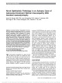
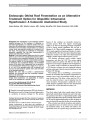
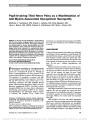
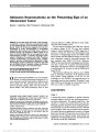
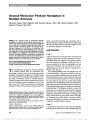
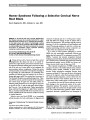
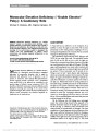
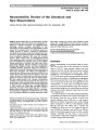
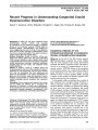
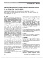
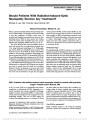
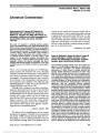
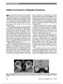
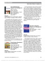
Show Abducens Neuromyotonia as the Presenting Sign of an Intracranial Tumor Daniel J. Salchow, MD, Thomas K. Wermund, MD Abstract: In this case series and review of the literature, we describe 2 cases of abducens neuromyotonia (ANM) as the presenting sign of an intracranial tumor (menin-gioma). Review of the literature suggests that the path-ophysiology of ocular neuromyotonia is incompletely understood. Most patients with ANM have a history of radiation therapy. The diagnosis of ANM is made on the basis of clinical findings and can be supported by elec-trophysiological studies. A complete neurologic exami-nation is mandatory for patients with ANM. Treatment consists of eliminating the underlying cause; carbama-zepine is effective in alleviating the symptoms of ANM. Neuroimaging should be performed if patients with ANM lack the typical history of radiation therapy, as ANM may be the presenting sign of an intracranial mass. Journal of Neuro-Ophthalmology 2011;31:34-37 doi: 10.1097/WNO.0b013e3181f33bc2 2011 by North American Neuro-Ophthalmology Society Ocular neuromyotonia (ONM) is a rare eye movement disorder. It consists of paroxysmal contraction of 1 or more extraocular muscle(s) innervated by an affected ocular motor cranial nerve. The activated muscle does not relax until the paroxysm subsides, which may take seconds to several minutes. The third (oculomotor) nerve is most commonly affected in ONM, followed by fourth (abdu-cens) and sixth (trochlear) nerves. The vast majority of patients with ONM have a history of radiation therapy (1-13). The time from radiation therapy to clinical manifestation of ONM ranges from 2 months to 18 years (7). Some of the reported patients had intracranial or nasopharyngeal masses, but ONM usually did not present until after radiation therapy. In a few cases, nerve compression caused by intracranial aneurysms has been associated with ONM (5,12,14-16). Moreover, ONM has been described after myelography with thorium (13), after a stroke (17), in patients with thyroid-related orbitopathy (18) and with cavernous sinus thrombosis due to mucormycosis (19). In some, no cause for ONM can be found (20-25). Neuromyotonia affecting the abducens nerve (ANM) leads to paroxysmal contraction of the ipsilateral lateral rectus muscle, causing abduction of the eye. To our knowledge, no case of ANM as the presenting sign of an intracranial tumor has been described. We present 2 cases where ANM was the first sign of an intracranial tumor. In both cases, consent to publish was granted. CASE REPORTS Case 1 A 48-year-old woman complained of episodic binocular horizontal diplopia, which she first noted approximately 1 year before presentation. The episodes lasted from a few seconds to 1 minute and occurred several times per day. During the episodes, the patient noted a ‘‘pulling sensation'' around the left eye, she felt nauseated and had a mild left frontal headache, which was not present between episodes. Her medical history was significant for excision of a benign breast cyst at the age of 18 years; there was no history of an intracranial mass or radiation therapy. Social history was significant for smoking (half pack-year). She denied alcohol use and did not take any medications. Augenklinik (TKW), HELIOS Kliniken Schwerin, Germany; and Department of Ophthalmology & Visual Science (DJS), Yale Uni-versity School of Medicine, New Haven, Connecticut. Supported in part by the Departmental Challenge Grant from Re-search to Prevent Blindness, Inc, New York, NYand by National Eye Institute/National Institutes of Health grant number EY000785. Conflict of interest: None. Supplemental digital content is available for this article. Direct URL citations appear in the printed text and are provided in the HTML and PDF versions of this article on the journal's Web site (www.jneuro-ophthalmology.com). Address correspondence to Daniel J. Salchow, MD, Department of Ophthalmology & Visual Science, Yale University School of Medi-cine, 40 Temple Street, 3rd Floor, Suite 3D, New Haven, CT 06510; E-mail: daniel.salchow@yale.edu 34 Salchow and Wermund: J Neuro-Ophthalmol 2011; 31: 34-37 Original Contribution Copyright © North American Neuro-Ophthalmology Society. Unauthorized reproduction of this article is prohibited. Best corrected visual acuity was 20/20 in each eye. External and anterior segment exams were unremarkable except for mild nuclear sclerotic cataracts. Stereopsis and color vision were normal, and visual fields were full to confrontation in both eyes. Pupils reacted equally to light, there was no afferent pupillary defect, and the fundi were normal. The patient was orthophoric in primary gaze at distance and near, and ocular versions were full. There was no strabismus in any gaze position except for esotropia of 14D on left gaze. After maintaining left gaze for 20-30 seconds, the left eye assumed a maximally abducted position and could not be adducted beyond midline. Attempted adduction during the attack resulted in retraction of the left globe (Fig. 1; see also Video, Supplemental Digital Content 1, http://links.lww.com/WNO/A13). On further questioning, the patient reported mild tingling over the left cheek, although sensation to touch in this area was intact and not different from the right cheek. All other cranial nerve functions were intact. MRI of the brain showed a large homogenously en-hancing mass originating from the prepontine cistern, compatible with a meningioma (Fig. 2). Neurosurgical removal of the tumor was undertaken, and pathological evaluation confirmed the diagnosis of meningioma. Post-operatively, the patient had paresis of the third through seventh cranial nerves. ANM did not recur, but the patient was left with a mild left sixth nerve palsy. Case 2 A 49-year-old woman complained of transient diplopia associated with a sensation of pressure around the right eye lasting seconds to 1 minute and occurring 10-15 times per day. Visual acuity was 20/20 in each eye, and ocular exam was normal. Ocular motility testing revealed paroxysmal abduction of the right eye after right gaze, lasting approx-imately 30 seconds (Fig. 3). At other times, ocular align-ment and motility were completely normal. Electromyo-graphy (EMG) of the right lateral rectus muscle during the FIG. 1. Case 1. A. Between episodes of abducens neu-romyotonia, the eyes are aligned. B. The patient directs gaze left for 15-20 seconds. C. With return to primary gaze, the left eye maintains an abducted position. D. On attempted right gaze during the episode, the left eye cannot be adducted beyond the midline. There is re-traction (note narrowing of the left palpebral fissure) and involuntary elevation of the left eye. FIG. 2. Case 1. Contrast-enhanced T1 MRI of the brain. A. Axial scan reveals a mass (3.5 3 3.6 3 3.3 cm) compressing the midbrain and pons, extending into the left cavernous sinus, and causing narrowing of the left internal carotid artery. B. Reformatted T1 sagittal view shows the mass extending over the clivus into the sella turcica. Salchow and Wermund: J Neuro-Ophthalmol 2011; 31: 34-37 35 Original Contribution Copyright © North American Neuro-Ophthalmology Society. Unauthorized reproduction of this article is prohibited. attack showed a high-frequency, repetitive, continuous discharge. Extraocular EMG and electroneurography showed no evidence of a generalized myogenic or neuro-genic disorder. Brain MRI revealed a mass within the right cavernous sinus, compatible with a meningioma (Fig. 4). The patient was started on 600 mg/day carbamazepine, and the paroxysmal ocular deviation disappeared. Stereotactic conformal radiation therapy (total dose: 56 Gy) was given, and carbamazepine was tapered and discontinued. Brain MRI 2.5 years later showed considerable reduction in the size of the tumor, and the patient remained free of diplopia. DISCUSSION The term ONM was introduced by Ricker and Mertens (26) to describe a peculiar disturbance of ocular motility, consisting of paroxysmal contraction of an extraocular muscle, causing the eye to deviate with resultant diplopia. The muscle does not relax until the paroxysm subsides, which may take minutes. A pulling sensation around the involved eye and headaches may be reported. The majority of cases with ANM have a history of previous radiation therapy. Only 1 reported case had resection of an in-tracranial tumor (clivus chordoma) without radiation therapy before developing ANM (27). We could not find any published cases of ANM as the presenting sign of an intracranial mass. Oculomotor neu-romyotonia associated with compressive lesions without radiotherapy has been reported (5,12,14,16). One patient with oculomotor neuromyotonia secondary to a cavernous sinus meningioma also had ‘‘paroxysmal electrical dis-charges in the ophthalmic division of the left trigeminal nerve,'' indicating involvement of the fifth cranial nerve (28). One of our patients (Case 1) complained of par-esthesias in the distribution of the maxillary division of the left trigeminal nerve, emphasizing the importance of a complete cranial nerve exam in patients with ONM. ONM has to be distinguished from oculomotor paresis with cyclic spasm, which is characterized by a cycle of pa-resis and spasm of muscle(s) innervated by the third nerve. FIG. 3. Case 2. A. The eyes are aligned in primary posi-tion. B. Ocular versions are full. C. Occasionally, the right eye persists in abduction after right gaze or spontane-ously deviates to the right for 30-60 seconds. FIG. 4. Case 2. Contrast-enhanced T1 MRI of the brain. Axial (A) and coronal (B) scans show a mass (arrow) (1.9 3 1.3 3 1.6 cm) within the right cavernous sinus extending dorsally toward the tentorium and encircling the right internal carotid artery. 36 Salchow and Wermund: J Neuro-Ophthalmol 2011; 31: 34-37 Original Contribution Copyright © North American Neuro-Ophthalmology Society. Unauthorized reproduction of this article is prohibited. The spasms are neither induced nor altered by eccentric gaze (29). In ONM, the deviation can often be induced by looking in the direction of action of the involved extraocular muscle. Another differential diagnostic consideration is myokymia. Although rare, superior oblique myokymia is more common than trochlear neuromyotonia. Myokymia consists of an ocular microtremor that is not present in neuromyotonia. EMG shows phasic contractions in my-okymia in contrast to tonic contractions in ONM (7). The pathophysiology of ONM is not well understood. Potential mechanisms include 1) ephaptic transmission along the affected nerve, 2) disturbances of potassium channels in the neuronal cell membrane, and 3) central neural reorganization. In most published cases of ONM, an extra-axial lesion is present, but Banks et al (17) indicated that a brainstem lesion may lead to the eye movement disorder. A more detailed discussion on pathogenesis of ONM has been published elsewhere (30). In summary, ANM should prompt a complete neuro-logic exam of the patient, with special attention to the cranial nerves. Most often, it is associated with previous radiation therapy. Neuroimaging should be performed in patients with ANM who lack such a history because it may be the presenting sign of an intracranial mass. REFERENCES 1. Bacskulin A, Guthoff R. Neuromyotonia of the abducens nerve after hypophysectomy and radiation. Strabismus. 1999;7:37-40. 2. Barroso L, Hoyt WF. Episodic exotropia from lateral rectus neuromyotonia-appearance and remission after radiation therapy for a thalamic glioma. J Pediatr Ophthalmol Strabismus. 1993;30:56-57. 3. de Saint Sardos A, Vincent A, Aroichane M, Ospina LH. Ocular neuromyotonia in a 15-year-old girl after radiation therapy. J AAPOS. 2008;12:616-617. 4. Ela-Dalman N, Arnold AC, Chang LK, Velez FG, Lasky JL III. Abducens nerve ocular neuromyotonia following non-sellar or parasellar tumors. Strabismus. 2007;15:149-151. 5. Ezra E, Spalton D, Sanders MD, Graham EM, Plant GT. Ocular neuromyotonia. Br J Ophthalmol. 1996;80: 350-355. 6. Fricke J, Neugebauer A, Kirsch A, Ru¨ssmann W. Ocular neuromyotonia: a case report. Strabismus. 2002;10: 119-124. 7. Haupert CL, Newman NJ. Ocular neuromyotonia 18 years after radiation therapy. Arch Ophthalmol. 1997;115: 1331-1332. 8. Helmchen C, Dieterich M, Straube A, Bu¨ttner U. Abduzensneuromyotonie mit partieller Okulomotoriusparese [‘‘Abducens neuromyotonia'' with partial oculomotor paralysis]. Nervenarzt. 1992;63: 625-629. 9. Koop G, Gra¨f M. Okula¨re Neuromyotonie - Ein Fallbericht [Ocular neuromyotonia]. Klin Monbl Augenheilkd. 2006; 223:247-251. 10. Lessell S, Lessell IM, Rizzo JF III. Ocular neuromyotonia after radiation therapy. Am J Ophthalmol. 1986;102: 766-770. 11. Oohira A, Furuya T. Ocular neuromyotonia with spastic lid closure. J Neuroophthalmol. 2006;26:244-247. 12. Tilikete C, Vial C, Niederlaender M, Bonnier PL, Vighetto A. Idiopathic ocular neuromyotonia: a neurovascular compression syndrome? J Neurol Neurosurg Psychiatry. 2000;69:642-644. 13. Yee RD, Purvin VA. Ocular neuromyotonia: three case reports with eye movement recordings. J Neuroophthalmol. 1998;18:1-8. 14. Abdulla N, Eustace P. A case of ocular neuromyotonia with tonic pupil. J Neuroophthalmol. 1999;19:125-127. 15. Park HY, Hwang JM, Kim JS. Abducens neuromyotonia due to internal carotid artery aneurysm. J Neurol Sci. 2008;270: 205-208. 16. Versino M, Colnaghi S, Todeschini A, Candeloro E, Ravaglia S, Moglia A, Cosi V. Ocular neuromyotonia with both tonic and paroxysmal components due to vascular compression. J Neurol. 2005;252:227-229. 17. Banks MC, Caruso PA, Lessell S. Midbrain-thalamic ocular neuromyotonia. Arch Ophthalmol. 2005;123:118-119. 18. Chung SM, Lee AG, Holds JB, Roper-Hall G, Cruz OA. Ocular neuromyotonia in Graves dysthyroid orbitopathy. Arch Ophthalmol. 1997;115:365-370. 19. Harrison AR, Wirtschafter JD. Ocular neuromyotonia in a patient with cavernous sinus thrombosis secondary to mucormycosis. Am J Ophthalmol. 1997;124:122-123. 20. Choi KD, Hwang JM, Park SH, Kim JS. Primary aberrant regeneration and neuromyotonia of the third cranial nerve. J Neuroophthalmol. 2006;26:248-250. 21. Frohman EM, Zee DS. Ocular neuromyotonia: clinical features, physiological mechanisms, and response to therapy. Ann Neurol. 1995;37:620-626. 22. Papst W. Zur Differentialdiagnose der okula¨ren neuromyotonie [Differential diagnosis of ocular neuromyotonia]. Ophthalmologica. 1972;164:252-263. 23. Safran AB, Magistris M. Terminating attacks of ocular neuromyotonia. J Neuroophthalmol. 1998;18:47-48. 24. Shults WT, Hoyt WF, Behrens M, MacLean J, Saul RF, Corbett JJ. Ocular neuromyotonia. A clinical description of six patients. Arch Ophthalmol. 1986;104:1028-1034. 25. Yu¨ru¨ten B, Ilhan S. Ocular neuromyotonia: a case report. Clin Neurol Neurosurg. 2003;105:140-142. 26. Ricker K, Mertens HG. Okula¨re neuromyotonie [Ocular neuromyotony]. Klin Monbl Augenheilkd. 1970;156: 837-842. 27. Metz HS, Sterns G. Varying esotropia-exotropia. J Pediatr Ophthalmol Strabismus. 1985;22:97-99. 28. Jacob M, Vighetto A, Bernard M, Tilikete C. Ocular neuromyotonia secondary to a cavernous sinus meningioma. Neurology. 2006;66:1598-1599. 29. Miller NR, Lee AG. Adult-onset acquired oculomotor nerve paresis with cyclic spasms: relationship to ocular neuromyotonia. Am J Ophthalmol. 2004;137:70-76. 30. Wermund TK, Salchow D. Ocular neuromyotonia-clinical appearance and thoughts on pathogenesis. Klin Monbl Augenheilkd. 2009;226:881-885. Salchow and Wermund: J Neuro-Ophthalmol 2011; 31: 34-37 37 Original Contribution Copyright © North American Neuro-Ophthalmology Society. Unauthorized reproduction of this article is prohibited. |