| OCR Text |
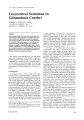
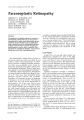
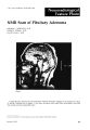
Show J. Clill. Nellrr-ophthalmol. 4: 225-228, 1984. Ophthalmoplegic Migraine THOMAS D. BAILEY, Major, USAF, MC PATRICK S. O'CONNOR, M.D. THOMAS ]. TREDICI, Colonel, USAF, MC DAVID E. SHACKLETT, M.D. Abstract A 5-year-old girl developed severe periocular pain lasting 4 days. As the pain subsided, a right oculomotor palsy developed which slowly resolved without aberrant regeneration over a 21J2-month period. A classic history and normal high-resolution CT scan were essential in making the diagnosis of ophthalmoplegic migraine. This is a rare disorder classically requiring normal cerebral angiography for diagnosis. With the advent of high-resolution CT scanning, angiography may no longer be indicated in the typical case. Although migraine is commonly encountered in ophthalmologic and neurologic practice, ophthalmoplegic migraine (recurrent oculomotor palsy without a demonstrable organic etiology) is rare. Since its first descriptions in the late 1800s, approximately 200 cases have been reported. Recent authors l have suggested that most of these early cases should be excluded because the third nerve palsy was actually caused by aneurysm, infection, or tumor. These authors also believe a negative cerebral angiographic study is a minimum requirement for diagnosis. The advent of high-resolution CT scanning, combined with a typical presentation, may make angiography unnecessary. An illustrative case is presented. Case Report The patient was a 5-year-old female referred to us in March 1980 with a chief complaint of, "My right eye is closed." The present illness began 11 days before with severe pain in and around the right eye, for which the patient stayed indoors, supine, with an ice bag over her eye. Four days later her parents noted a slight ptosis. Upon From the Ophthalmology Branch, USAF Schl'l,ll" Aerospace Medicine. Aeromedical Division (AFSC), Brooks Air Force Base. Texas (TDB, TJT); and the Department of Ophthalmology, University of Texas Health Science Center, San Antonio, Texas (PSOC. DES) December 1984 awakening the following day, her pain was gone, but she now had a complete right oculomotor palsy. Past history was significant for an episode of partial oculomotor palsy with ptosis at age 2 which cleared in 13 days. A CT scan at that time was negative and a diagnosis of partial oculomotor palsy-resolving-etiology unknown was made. At age 31h and 4, the patient had recurrences of headache and mild ptosis, both of which cleared in approximately 1 week, except for a mild residual ptosis noted after the second episode. Her father and maternal grandmother had classic migraine and her maternal grandfather had congenital ptosis. There was no history of allergy. High-resolution CT scanning, with and without contrast, skull and sinus x-rays, a complete blood count, and tensilon testing were normal. A tentative diagnosis of Tolosa-Hunt syndrome was made. The patient was started on 40 mg of Prednisone per day and referred to us. On examination 11 days after onset, the right pupil was 6.0 mm and reacted 1+ to light and near. The left pupil was 4 mm and reacted 4+ to light and near. Accommodation was markedly reduced on the right. No Marcus Gunn pupillary phenomenon was noted. Extraocular muscle examination revealed a complete ptosis with a 95% right superior rectus palsy, an 85% right inferior rectus and complete right medial rectus palsy (FIgs. 1a and 1b). Best-corrected vision was 20/ 25 on the right and 20/25 on the left. Confrontation visual fields, slit lamp, and funduscopic examInatIons were normal. Angiography was not performed. A diagnosis of ophthalmoplegic migraIne was made and Prednisone discontinued. One week later, the extraocular motility was unchanged, but the ptosis was improving. Gradual resolution occurred over the next 10 weeks, although the patient continued having intermittent headaches, unresponsive to Inderal. At 6 months, a1.0-mm ptosis and mild internal ophthalmoplegIa remaIned, but there was no evidence of aberrant regeneration. The patient continued to have recurrent bouts of headache over the next 3 years whIC~ were frequently accompanied by either transIent ptosis, mydriasis, or both, lasting from 225 Ophthalmoplegic Migraine 30 minutes tu 2-3 huurs. In August 1983, fullowing a particularly severe headache, a tutal right oculumutor palsy recurred and is again resulving. Discussion Althl)ugh an isolated oculomotor p~lsv is a relatlvelv commun finding in adults, - ' it is an infrequent neurolugic sign in chJldren.J Ophthalmuplegic migraine is an even rarer syndrome whose apparent incidence markedly decreased with the advent of neuroradiologic procedures in the 1930s. These have led to increasing recognition of other etiulogies of oculomotor paralysis. There still remains, however, a number of patients whose symptoms can be ascribed to no other cause. Miller,; in a review of 3 million admissions to the Johns IIopkins Hospital over a 25-year period, found only 30 cases of isolated third nerve palsy in children. Of these, unly two were diagnosed as ophthalmoplegic migraine. Both (ages 3 and 6 years) had the onset of oculomotor palsy associated with headache and both resolved within 3 weeks. Another review of 5,000 admissions for , Figure la. Tlltal pIllS" 11 davs after onset of llphthalmoplegia. migraine at the Montefiore hospital from 1932 to 1962 revealed only eight cases of ophthalmoplegic migraine" Walsh and Daughertl have established the following diagnostic criteria for oph thalmoplegic migraine: 1. A history of typical migraine headache with a crescendo quality. 2. Ophthalmoplegia, including one or more nerves and pussibly alternating sides with attacks, the paralysis usually appearing subsequent to an established migraine pattern. 3. Exclusion of other causes by arteriography, surgery, or autopsy. Arteriography is considered by them a minimum for diagnosis. In most patients with well-defined cases, the onset of ophthalmoplegia occurs before age 10, the youngest recorded patient presenting with an oculomotor palsy at the age of 8 months.~ In ophthalmoplegic migraine, the third nerve is most frequently involved, with abducens palsy occurring one-tenth as often and fourth nerve palsy occurring even more rarely," In most of these cases, a positive famJly history was not present.' .. Isolated oculomotor palsy in infancy and childhood is rare and most often congenital.; The acquired causes include trauma, intlammatory disease, tumor, aneurysm, and ophthalmoplegi'c migraine. In an utherwise neurolugically normal child who has a sudden onset of oculomotor palsy associated with severe headache, especially if this is the first episode, aneurysm must be ruled out. Does this mean that every child, even with a classic history, requires angiography? We think not. Cerebral aneurysms are exceedingly uncommon in children." In fact, Matson]{l found only three cases that occurred under the age of 5, ail presented with symptoms of subarachnoid hem- 226 Figure lb. Almost complete internal and e:>.ternal ophthalmoplegia, again, 11 days a~roo~t . Journal of Clinical Neuro-ophthalmology orrhage, and none had cranial nerve palsies. Patel and Richardson ll reported 58 patients under the age of 19 years with aneurysm. No cases occurred under the age of 7, and none were accompanied by ophthalmoplegia. The cooperative study12 on intracranial aneurysms included 2,627 patients, only one of which was in the age group of 0-4 years. While clinically latent cerebral aneurysms occur in adults at autopsy with a frequency of 0.6-7.6%,13 no clinically latent cerebral aneurysms have been discovered at angiography or serendipitously at autopsy in infants and young children. H Miller5 described two cases (ages 16 and 17) who presented with a posterior communicating aneurysm and oculomotor palsy, severe headache, and stiff neck. Both had large aneurysms of the posterior communicating artery diagnosed angiographically. To our knowledge, the only case of ophthalmoplegia caused by aneurysm in a child under age 10 was reported by Thompson and Pribram.Q This occurred in a I-month-old girl who developed a bilateral ophthalmoplegia associated with lethargy. The child had, in addition, a quadriparesis and was found on pneumoencephalography to have ventricular dilatation with failure of passage of air into the supratentorial cisterns and cortical subarachnoid spaces. Angiography demonstrated a large berry aneurysm of the right internal carotid. In reviewing other reports of children under the age of 2 with aneurysms, they found that most presented with subarachnoid hemorrhage or hydrocephalus." No others were recorded to have an ophthalmoplegia. Unlike the situation in adults, most aneurysms in children are quite largeH . I' In the most recent series, they ranged from 5.0 mm to 3.5 cm in diameter, with 50% of the aneurysms being larger than 1.0 cm. 14 A review of the three largest reports of aneurysms in children reveals only one arisin~ from the posterior communicating artery. II.11. 14 Ophthalmoplegia caused by aneurysm must be exceedingly rare in children under 10. In fact, we were unable to find any report of aneurysm presenting solely with an isolated oculomotor palsy and no symptoms of subarachnoid hemorrhage in a child under this age. Childhood aneurysms may occur in association with other abnormalities, and Patel and Richardson 11 reported that of 58 children under the age of 19 years with cerebral aneurysms, nine were found to have coarctation of the aorta. In addition, polycystic kidney disease occurred in 5.9% of another large series of congenital intracranial aneurysms. 1h An association between intracranial aneurysm and persistent trigeminal artery has also been reported. 17 The pathophysiology of ophthalmoplegic migraine remains obscure. Although Walsh and December 1984 Bailey et al. O'Doherty I suggested that narrowing of the carotid artery with resultant edema may compress adjacent cranial nerves within the cavernous sinus, the frequent occurrence of negative arteriograms durin?, attacks does not support this theory. 1H Vijayan H believes that most of the clinical evidence in documented cases suggests the ophthalmoplegia is due to a delayed ischemic neuropathy. This, he postulated, results from swelling of the walls of the carotid and/or basilar arteries leading to occlusion of the ostia of smaller vessels which supply the involved cranial nerves. Although this is an interesting possibility, it does not account for the transient episodes of mydriasis and ptosis experienced by our patient. Others have reported a similar unusual case in which attacks of ophthalmoplegia lasted for 45 minutes, recurred weekly, and occurred with the headache rather than following the headache. 19 The question of angiography is an important one. If a child has a headache followed by third nerve palsy, but is otherwise well, with a normal high-resolution CT scan, the decision to perform angiography should be delayed. Conversely, in those patients who have an onset after age 10, the absence of a previous history of migraine, or persistent headache in the face of a total ophthalmoplegia and symptoms and/or signs of subarachnoid hemorrhage, angiography is certainly indicated. Finally, others have also reported their lack of success in preventing recurrences with Inderal, 14.2<1 although some success has been achieved in this syndrome with the prophylactic use of fluophenamic acid, a prostaglandin inhibitor. 21 Summary The case of a 5-year-old girl with multiple attacks of classic ophthalmoplegic migraine who did not respond to Inderal is presented. Cerebral angiography has been recommended previously In all such cases. The large size and rarity of aneurysms in children, especially involving the posterior communicating artery, and presenting With an Isolated painful ophthalmoplegia is stressed. We feel that any child with a classic history of ophthalmoplegic migraine and a normal high-resolution CT scan should have angiography delayed. References 1. Walsh, J.P., and O'Doherty, 0.5.: A possible explanation of the mechanism of ophthalmoplegic migraine. Ncur%gy 10: 1079-1084, 1960. 2. Rucker, C.W.: Paralysis of the third, fourth, and sixth cranial nerves. Alii. J. Ophtha/lllol. 46: 787794, 1958. 227 Ophthalmoplegic Migraine 3. Rucker, CW.: The causes of paralysis of third, fourth, and sixth cranial nerves. Am. f. Ophthalmol. 61: 1293-1298, 1966. 4. Green, W.R., Hackett, E.R., and Schlezinger, N.5.: Neuro-ophthalmologic evaluation of oculomotor paralysis. Arch. Ophthalmol. 72: 154-167, 1964. 5. Miller, N.R.: Solitary oculomotor nerve palsy in childhood. Am. f. Ophthalmol. 83: 106-110, 1977. 6. Friedman, A.P., Harter, D.H., and Merritt, H.H.: Ophthalmoplegic migraine. Arch. Neurol. 7: 320327, 1962. 7. Van Pelt, W.: On the early onset of ophthalmoplegic migraine. Am f. Dis. Child. 107: 628-631, 1964. 8. Walsh, F.B., and Hoyt, W.F.: Clillical Neuro-ophtllall/ lolt)~;'ll (3rd ed.). Williams & Wilkins, Baltimore, 1969, p. 1675. 9. Thompson, R.A, and Pribram, H.F.: Infantile cerebral aneurysm associated with ophthalmoplegia and qUildriparesis. Neurology 19: 785-789, 1969. 10. Matson, D.O.: Intracranial arterial aneurysms in childhood. f. Neurosurg. 23: 578-583,1965. 11. Patel, AN., and Richardson, A.E.: Ruptured intracranial aneurysms in first two decades of life; study of 58 patients. f. Neurtlsurg. 35: 571-576,1971. 12. Locksley, H.B.: Report on the cooperative study of intracranial aneurysms and subarachnoid hemorrhage. f. Neurosurg. 25: 219-239,1966. 13. Stehbens, W.E.: Pathology (If the Cerebral Blood Vessels. CV. Mosby Co., St Louis, Missouri, 1972. 14. Harwood- ash, D.C: Neuroradiology ill Illfallts lllld Childn'll; with the Assistallce of Charles R. Fitz, Vol. 3. C V. Mosby Co, SI Louis, Missouri, 1976, pp. 908-913 228 15. Thompson, J.R., Harwood-Nash, D.C, and Fitz, CR.: Cerebral aneurysms in children. Am. f. Roelltgellol. Radium Ther. Nuc/. Med. 118: 163-175, 1973. 16. Bigelow, N.H.: The association of polycystic kidneys with intracranial aneurysms and other related disorders. Am. f. Med. Sci. 225: 485-494, 1953. 17. George, A.E., Lin, J.P., and Morantz, R.A: Intracranial aneurysms on a persistent primitive trigeminal artery; case report f. Neurosurg. 35: 601-604, 1971b. 18. Vijayan, N.: Ophthalmoplegic migraine: Ischemic or compressive neuropathy? Headache 20: 300304, 1980 19. Cruciger, M.P., and Mazow, M.L.: An unusual case of ophthalmoplegic migraine. Am. J. Ophthalmol. 86: 414-417.1978. 20. Durkan, G.P., Troost, B.T., Slamovits, T.L., Spoor, I.C, and Kennerdell, ].5.: Recurrent painless oculomotor palsy in children. A variant of ophthalmoplegic migraine? Headache 21: 58-62, 1981. 21. Rabey, ].M., Vardi, Y., Van Dyck, D., and Streifler, M.: Ophthalmoplegic migra(ne: Amelioration by flufenamic acid, a prostaglandin inhibitor. Ophthalmologica 175: 148-152, 1977. Acknowledgment This study was supported in part by an unrestricted grant from Research to Prevent Blindness. Special thanks to Dr. Thomas Hedges of Philadelphia for his follow-up care and notes on this patient Write for reprillts to: Patrick S. O'Connor, M.D., Department of Ophthalmology, University of Texas Health Sciences Center, 7703 Floyd Curl Drive, San Antonio, Texas 78284. Journal of Clinical Neuro-ophthalmology |