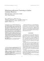
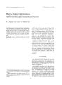
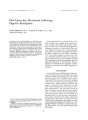
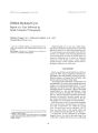
| OCR Text |
Show .~ 1992 Rave" r,...~~. LId . Ne... York Reflex Blink to Visual Threat Grant T. Liu, M. D. and Michael Ronthal, M, B. Beh. The requisite visual modalities for the TlO'OeX blink to visual threat hav.. nol been thoroughly studied. We idenlifit'd five patil'nts with different focal cerebrill lesions documented on computerized tomography scan who had abnormal blink- Ie- threat reflcx(' s, One had a homonymous hemianopia secondary to posterior cerebral arlery occlusion; another had a unilateral pilrictal neglect; and a third had a frontal neglect. They did not blink in response to visual stimuli contralateral to their lesion. A cortically blind palienl and one with Balint's syndrome did not have a blink response. Observation of these and other similar patients and animals previously reported suggests that the blink- to- threat r{" nex is cortically mediated and requires intact primary visual cortex as wt" ll as higher order mechanisms for visual attention mediated in the inferior parietal lobule and frontal eye' fields. Key Words: Blink- Reflex- Threat- Neglect- Attention- Balint's syndrome, From the Department of Neurology ( G. T. L M,~,), Beth Israel Hospital: Division of Neurology ( G, T. L), Bngham and Women's Hospital. Harvard- longwuod Neurology Program, Harvard Medical School. Boston, Massachusetts, U. S, A. Address correspondence and reprint rt'quests to Dr. G. T. Liu, Division of Neurology, Brigham and Women's Hospital. 75 Francis Street, Boston, MA 02115, U. S. A. 47 When an object is brought suddenly toward the open eyes of a normal individual there is a prompt blink response, Obviously the optic nerve serves as the afferent pathway. , , ( and) the cortex almost certainly must be in the reflex arc, but what part it plays is not cle3rly understood. F. B, Walsh " Reflex Blinking and tile Meuace RespmtSt''' ( 1) The blink reflex to Ihreat or menace is a standard bedside method for testing visual field defects ( 24), especially in patients uncooperative with standard visual confrontation ( 5). In response 10 a sud+ den, unexpected gesture directed toward the eyes, a normal person promptly contracts both orbicularis oculi to close the eyelids momentarily. Despite its common use, the pathways and the requisite cortical modalities for this response still remain uncertain. In spite of Walsh's statement ( 1), studies of the role of the cortex in the blink reflex are sparse ( 6). In 1929 Ehlers ( 7) wrote, " The blinking reflex for threatening danger requires involvement of that portion of Ihe cerebral cortex which is the seat of visual perception," implying the need for an occipital cortex. Moses ( 8) felt the reflex was cortically mediated and required an intact occipital lobe and " its connections with the Rolandic area." Ross Russell ( 9) invoked an ocdpitopontine pathway without involvement of the frontal lobe. Hoyt and Loeffler ( 10) and then Miller ( 4) added that parieto- occipital and parietotemporOll areas may be necessary for the blink- to- threat reflex. Hoh et al. ( 11), on the other hand, suggested that the reflex could be mediated entirely in thc brain stem. We identified five patients with varied focal cortical lesions and absent blink- to- threat reflex. Although others have made similar anecdotal observations, to our knowledge no comprehensive review on the reflex blink to visual threat exists. Based on our patients and other similar cases and animal studies reported in the literature, we propose that the lack of a blink- to- threat reflex does 48 G. T. LJU AND M. RONTHAL not always reflect a visual field deficit, bul instead could also indicate a defect in visual attention. METHODS Patients with cortical lesions documented on imaging studies who had absent blink to visual threat were identified from the inpatient services of the Beth Israel and Brigham and Women's Hospitals from February 199010 May 1990. All patients were examined by at least one of us ( GTL). The examiner eliciled the blink- Io- threat reflex by suddenly thrusting his fingertips towards the patient's eyes from the periphery. Menacing from the periphery was preferred because it was difficult to decide which nasal hemifield detected stimuli that was directly in front of the patient. Covering one of the patient's t.' yes to test monocular fields was felt to detract from the surprise of the thrt.' at. Likt.' wist.', auditory cues were not given to direct the patients attention. Jerking the entire hand with the palm or the back of the hand facing the patient was avoided in order to prevent evok · ing a tactile blink reflex by moving air onto the cornea ( 12). The blink reflex was considered intact if there was either partial or complt.' tt.' closure of the eyelids. Visual fields were determined by confrontation ( 5), but in some cases formal Goldmann perimetry was also obtained. Patients with significant ophthalmological dis · ease affecting the cornea, lens, vitreous, choroid, retina, optic nerve, and chiasm, as well as patients with nuclear and peripheral facial nerve lesions. diseases of the neuromuscular junction, and myopathies involving the orbicularis oculi were excluded. Comatose patients wcre also excluded. Five representative patients with focal cerebral lesions and abnormal blink- to- threat reflexes were identified. All patients blinked spontaneously, indicating completeness of the facial nerve motor unit. The ages of the patients ranged from 36 to 81; four were men. Three had acute cerebral infarctions; one had intracerebral hemorrhages; and another had intracerebral cyst formation. CASE REPORTS Patient 1 A 67- year- old man with hypertension and diabetes was admitted with complaints of unsteadiness and difficulty seeing with his left eye. His mental status was normal. but he drew a clock with numbers only on the right side. His corrected visual acuity was 20/ 20 au. Funduscopic examina- I Cli" NnJro- ophtltal" lCl. Vol. J2. No. 1. 1992 tion was normal. Goldmann perimetry demonstrated a dense congruous left homonymous hemianopia with macular sparing, and he did not blink in response to threatening gestures presented in the left hemifield, but he did blink in response to those in the right. His eye movements and optokinetic response were normal. The rest of his neurological examination was significant for mild left facial weakness, a mild left pronator drift, and an increased left biceps reflex. Computerized tomography ( CT) scan of the brain revealed decreased allenuation in the right occipital lobe, consistent with infarction ( Fig. I). The mild left hemiparesis and congruous homonymous hemianopia with macular sparing suggested posterior cerebral artery occlusion ( 13-- 15). COli/ 11Im!. This patient, who developed an oce! p · itallobe infarct, did not blink to threat in his blind hemifield. Patient 2 A 75- year- old man presented with bilateral cystic occipital lobe lesions secondary to resection of a parasagittal occipital meningioma 3 years previously. The cysts were removed initially in 1988, but they recurred. In late 1988, he developed right · sided weakness that was felt to be associated with a lacunar infarction. He required a ventriculoperitoneal shunt for treatment of hydrocephalus in January 1990; he was then readmitted in April FIG. 1. Computed tomography scan of patient 1. demonstrating a right occipital lobe infarction. REFLEX BUNK TO VISUAL THREAT 49 1990 for further surgical drainage of the occipital cysts. On neurological exam he was abulic, but he followed simple commands. Funduscopic examination was normal, and his pupils were briskly reactive to light. The patient appeared not to see anything. He did not blink to visual threat in any field. He was unable to point or direct his gaze to any face or object requested. His eyes roved randomly. On command, he could move his eyes conjugately left and right, but he had some difficulty with upgaze. Optokinetic responses were absent. The rest of his exam was notable for a mild right hemipa · resis and a right extensor plantar response. Computed tomographic scan of the head revealed the occipital cysts, a left frontal subdural fluid collection, and a shunt lying in the frontal horn of the right lateral ventricle ( Fig. 2). Commellt. In this patient bilateral occipital lobe lesions produced cortical blindness ( 16). The preserved pupillary reflexes, mediated in the pretectum ( 17), indicate intact pregeniculatc visual pathways. He did not have optokinetic nystagmus, consistent with the diagnosis ( 16), and he had no blink to threat. Patient 3 A 36- year- old man with a postviral dilated cardiomyopathy underwent a cardiac transplantation on May 2, 1990. After surgery he developed ventricular tachycardia, necessitating cardioversion; FIG. 2. In patient 2, these occipital cysts led to c? rlical blindness. A shunt lies in the right lateral ventricle. right ventricular failure, requiring an intra- aortic balloon pump and a right ventricular assist device; and disseminated intravascular coagulation. His platelet count fell to 45,000. After an episode of hypotension on May 4, 1990, he became unresponsive to voice or deep pinch, and he made no spontaneous limb movements. Head CT demonstrated bilateral parieto- occipital hemorrhages with significant surrounding edema and mass effect. By May 11, 1990, he was alert, attentive, and able to follow simple verbal commands. He did not blink to threat. His pupils were 4 mm bilaterally and reactive. When asked to do so, he was able to direct his gaze in all directions. One week later he was able to recognize bright light and detect movement, but in the left homonymous hemifield only. He was not able to follow visual targets. He was unable to count fingers, but when verbally instructed he held up the correct number of fingers. He wore his eyeglasses and claimed to watch television, although he was unable to describe what he saw. Dilated funduscopic examination at this time revealed normal discs, vessels, maculae, and periphery. Repeat head CT documented the bilateral parieto- occipital hemorrhages ( Fig. 3A). Five weeks postoperatively his vision had recovered significantly, although there were still noticeable deficits. Goldmann perimetry revealed an inferior homonymous altitudinal hemianopia with sparing of the temporal crescent ( 18) OS ( Fig. 3B and C). There was still no blink to threat in any visual field. He was able to look left and right on command. On attempting to shift gaze to an object in the periphery, he had difficulty locating it without ' scanning' for several seconds. When he attempted to reach for objects, he frequently missed his target, especially with his right arm. Finally, he was given a postcard of a lake and asked to identify the scene. His eyes searched over the card for several seconds. Correctly, he said " I see chairs . I see flowers ... I see pines," and then, after 15 seconds of thought, he guessed, " Is this a lake scene?" Commellt. Bilateral parieto- occipital hemorrhages in this patient initially resulted in cortical blindness. He first manifested Anton's syndrome ( 16); when he improved, he displayed the elements of Balint's syndrome ( 19) ( see below). He had no blink to threat. Patient 4 An 80- year- old right- handed woman with a history of hypertension and a stable thoracic aortic aneurysm was admitted after falling at home. I Clin N( w.(>- oplrtfull""". Vol. 12. No. 1. 1992 50 G. T. LlU AND M. RONTHAL • FIG. 3. A: Bilateral parietal- occipital hemorrhages in patient 3 first produced cortical blindness, then Bal · int's syndrome 5 weeks later. Visu~ 1 fi~ ld testin~ reo vealed an inferior homonymous altitudinal hemianopia: Left eye, with sparing of the temporal crescent ( B); right eye ( C). • " e'" "'" , t' "- I .' She was mildly inattentive, but her language was normal. She was initially totally unaware of the left hemispace and denied that her left arm belonged to her. She preferred to look to her right, but she had normal extraocular movements. Her left face, arm, and leg were paretic, and the left plantar response was extensor. The next day she was felt not to have a visual I eliM NrIl..,...,; J: tlvllrroC'l. VIIl. 12. No. 1. 1992 field deficit because, with verbal cues, she ac · knowledged objects and individuals to her left. With double simultaneous stimulation she extinguished visual and tactile stimuli on the left. She did not blink to threat from the left periphery but did from the right. Head CT demonstrated an infarct involving the right parietal lobe but also extending into the pos- c .... REFLEX BUNK TO VISUAL THREAT / FIG. 3. Continued. ....... '"..~. • • '•••• < , •• " , 51 terior portion of the frontal lobe ( Fig. 4). Magnetic resonance imaging of the chest revealed dilatation of the entire thoracic aorta with dissection extending from the descending aorta into the AG. 4. Computed tomography scan of patient 4. who had left- sided neglect caused by this right parietal infarction. abdomen. Thrombus occluded a false lumen. She was thought to have had an embolus from aortic dissection. and she received anticoagulant treatment. Comment. This woman, who had a right parietal infarction, neglected visual stimuli in the left hemispace and did not blink to threat from the left. However, when her attention was directed to the left with auditory cues. it was clear she had no major visual field deficit. Patient 5 A 68- year- old right- handed hypertensive man developed right- sided weakness after coronary artery bypass grafting. He was thought to have had a subendocardial myocardial infarction with hypotension during the surgery, The first day after surgery he did not move his right side as well as his left. He was drowsy but easily arousable. There was a tendency to ignore the right side, but he had no visual field deficit. He easily recognized objects on his right if his attention was directed that way, He did not blink in response to threatening gestures from the right periphery, but he did from the left. Funduscopic examination was normal. Conjugate extraocular f 0", NtutO- Ophrlullrnol. VQj, 12. N". 1. 1992 52 G. T. LlU AND M RONTHAL movements were present, but gaze to his right was incomplete. His face and arm were paretic on the right. and his right brachiuradialis and biceps reflexes were increased. The right plantar response was extensor. Language examin< ltion was limited because he was initially ventilator- dependent. On the fifth postoperative day he was extubated, and his speech was nonlluent and dysarthric. He followed simple verbal commands. but he had difficulty with reading aloud. naming, and repetition. Heat CT demonstrated two hypodense areas in the left frontal lobe: one in the lateral aspect, which also involved the head of the caudate. putamen, and anterior limb of the internal capsule; and the other in the medial and superior portion ( Fig. 5). These were felt to be acute infarcts in the distributions of the left middle cerebral and anterior cerebral arteries, respectively. Carotid noninvasive studies revealed a totally occluded left internal carotid artery. COII/ Illt''' I. The right hemiparesis and mild difficulty with right lateral gaze were consistent with the radiologic involvement of the left motor strip as well as the more anterior frontal eye fields which mediate contralateral eye deviation ( 20). He neglected stimuli on the right and did not blink to threat from the right. •.' FIG. S. In patient 5, these infarctions in the medial ( anterior cerebral artery territory) and lateral ( middle cerebral artery territory) portions of the left Irontal lobe led to right- sided neglect. DISCUSSION An absent blink- to- threat reflex could result from a lesion in the afferent visual pathway by interrupting the presentation of a full array of visual information from eye to brainstem and cortex. Alternatively, a defect in the efferent pathway to orbicularis oculi might abolish the ability to blink. We excluded patients with these mechanisms and will confine our discussion to " central" mechanisms. TeleOlogically, reflex blinks are mechanisms for eye protection in response to external stimuli ( 21). Blink to threat should be distinguished from other reflex blinks that are mostly brainstem mediated, such as the orbicularis oculi reflex ( 4,8,22,23), the corneal blink reflex, and the cochleopalpebral reflex ( 4). The reflex blink to light or dazzle requires an intact optic nerve and connections to pretectum, but not cortex ( 4,24,25). Neonates blink in response to light and corneal stimulation, and the light reflex occurs as early as 25 weeks of gestation ( 26,27). Newborns also have the ability to fix, follow, and be alert to visual stim~ uli, and most have optokinetic responses ( 26,28). However. reflex blink to visual threat is not present in neonates ( 21), and it does not emerge until the infant is 2- 4 months of age ( 12,26). The reflex is believed to be a learned response ( 26). This suggests that the blink to threat is not a primitive reflex, but one that requires higher- order cortical processing of a stimulus more complex than a flash of light or touch on the cornea. Further evidence for the role of the cortex in blink- to- threat reflex is its absence in patients who are effectively decorticate. Keane ( 24) reported a 54- year- old woman who suffered from anoxic neocortical death with isoelectric encephalograms and abSl'nt visual evoked responses. Her corneal and cochleopalpebral reflexes were intact, and her blink- to- light reflex persisted. although she did not blink to visual threat. Pathological examination revealed almost complete cortical neuronal loss but preservation of the brain stem ( 24). Tavy et al. ( 25) described a patient with similar clinical findings who at postmortem was found to have diffuse hypoxic changes of the cerebral and cerebellar cortices because of a cardiac arrest. Hill et al. ( 29) described a l- month- old hydranencephalic girl who " did not respond to thrust movements" but blinked to light. Postmortem examination revealed an intact brain stem, cerebellum, and basal ganglia, and a thin membrane- most likely residual cerebral c~ rtex- s~ rroundinga fluid- filled cavity. Our patients With focal cortical lesions and ab- REFLEX BUNK TO VISUAL THREAT 53 sent blink- to- threat reflex offered an opportunity to examine the modulation of the response by the cortex. We shall discuss the lesions topographically. Occipital Lobe Patients 1, 2, and 3 developed blink- to- threat reflex abnormalities as a result of occipital lobe lesions. Patient I did not blink to threat in his blind hemifield, and cortically blind patients 2 and 3 had no blink to threat. Several reported cases of cortical blindness have also demonstrated absent blink- to- threat reflex. Cases 3 and ~ of Barnet et a1. ( 30) had normally reactive pupils and blinked to light but not " to menace." Silverman et a1. ( 31) and Denny- Brown ( 32) described similar patients. Multiple references ( 16,33,34) list loss of blink- to- threat reflex as one of the principle clinical features of cortical blindness. Experimental studies in monkeys ( 32,35) confirm the above observalions. Marquis and Hilgard ( 35) found that a monkey with one occipital lobe removed did not blink to threat in the contralateral hemifield. After removing both occipital lobes, the researchers demonstrated that the pupillary response was unaffected, but that blinking in response to " threatening gestures" was absent in all visual fields ( 35). Denny- Brown ( 32) ablated are;) 17 bilaterally and produced similar findings. These observations and our own suggest that the blinkto- threat response requires an intact contralateral occipital lobe and primary visual cortex. Patient 3' s initial ability to detect light and motion in his left homonymous hemifield ( 36,37) without a blink- to- threat reflex implies that the response requires more than those visual modalities. Denny- Brawn's cortically blind patient ( 32) similarly improved to appreciate moving objects and light and also did not blink to visual threat. Similar patients with " blindsight" after unilateral or bilateral occipital lesions lose object recognition, but in some instances they may have an ability to locate light sources and detect moving targets in affected fields via retino- tectal- pulvinar pathways ( 38- 43). It would be informative for one of these patients to be tested for blink to threat. Parietal Lobe The findings in patient 4 demonstrate that an individual with unilateral visual inallention from a right parietal lesion ( 44- 47) may have an absent blink to threat in the neglected left hemispace, despite ( presumably) intact visual fields. This observation suggests that attentional mechanisms are important for blink to threat. The posterior parietal lobe, frontal lobe, and cingulate gyrus are instrumental in proposed cortical networks for directed attention ( 47~ 51). Dorsolateral area PC, located in the inferior parietal lobule or area 7, is the pivotal parietal lobe structure ( 52,53). It receives visual information relayed by polymodal sensory cortex from striate, peristriate, and inferotemporal cortices ( 49). An intact area PC is probably necessary for an intact blink- to- threat reflex. In 1881 Munk ( 54) observed that monkeys with pos+ terior parietal- anterior occipital cortex lesions did not blink in response to contralateral threat. Similarly, Heilman et ,11. ( 55) noticed that monkeys with inattention to the contralateral space from ablation of the inferior parietal lobule had a diminished blink- to- threat response in the unattended contralateral hemifield, despite intact visual fields. Shibutani ( · t al. ( 6), recording from monkey PC neurons and electromyography of the orbicularis oculi muscles, demonstrated a population of PC neurons which caused blinking when stimulated directly. Furthermore, when they moved a 10 x 20- cm plate quickly towards the monkey's face, the same neurons fired rapidly, and then, after a delay of 190 msec, the monkey blinked. The results suggest that certain PC neurons are responsive to visual threat and have the capability of coupling the striate cortex's perception of the threat with motor pathways for blinking. This is consistent with PC's proposed role in one of the models for allention ( 49): linking the visual stimulus of motivational significance ( threat of injury to the eye) with motor behavior ( eyelid closure). Thus the absence of a blink- to- threat response in the setting of injury to the inferior parietal lobule may be indicative of a defect in visual attention. In humans, the counterpart to monkey area 7 would correspond either to superior parietal lobule ( human area 7) or inferior parietal lobule ( human areas 39 and 40) or both ( 45). The parietal infarct in patient 4 involves at least areas 39 and 40 on the right ( 56), causing the contralateral neglect and a loss of blink- to- threat response from the left as well. Though motor cortex was likely involved, the frontal eye fields, area 8, were spared ( see below). Balint's Syndrome Balint ( 57) first analyzed this symptom complex [ which was later elaborated by Holmes ( 58) 1, of ocular apraxia ( a deficit in shifting gaze), optic ataxia ( a defect in reaching under visual guidance), and simultanagnosia ( 59). The lesions that cause I elm N~ u'''-( lpllllul/ n101. V( l/. 12. No /. / 992 54 G. T. LllI AND M. RONTHAL Balint's syndrome are typically bilateral and occipitoparietal in areas 7. 19, and sometimes 39 ( 60), zones highlighted above as important for visual attention and foveal rdixation. As in patient 3. the visual field defect is usually inferior ( 61,62). but sometimes the fields art' normal ( 59). Like our patient, five of the six patients Holmes « , ported in ) 918 did not " blink or otherwise respond to any threatening action, as \'\ lhen a hand or other largt' object was suddenly swung towards their faces" ( 58). He believed that " blinking seems to be a forebrain Tl., nex dependent on the functioning of centres which were injured by the lesions presented in these cases" ( 58). Holmes' second patient blinked when his own arm was moved passively towards his face, and Holmes therefore surmised that the absence of the blink renex resulted from defective distance perception. $ e\' eral other patients \ vith Balint's syndrome and absent blink- to- thr~ at renexes have been reported ( 61-- 65), and two of the more rffent authors ( 63,64) impli(.> d that the lack of a blink renected a defect in visual attention. Hausser et aJ. ( 63) included the absence of the blink renex under " disturbance of visual attention." Pierrot- Deseilligny et at ( 64), whose patient sustained bilateral damage to the inferior parietal lobules, used the loss of a menace response to demonstrate a " neglect ( of) some peripheral stimuli." The loss of a blink- tothreat reflex may simply be a manifestation of the " extreme narrowing of attention" ( 66,67), an inability to notice any obje<: t outside foveal vision, seen in Balint's syndrome. As suggested in the previous section, unilateral damage to the inferior parietal lobule causes a contralateral defect in attention to visual stimuli and loss of blink- to- threat renex. By extension, bilateral injury in Balint's syndrome results in a deficit in visual attention for the whole periphery as well as complete absence of a blink- to- threat response. frontal Lobe PatientS had a defect of directed attention to the right hemispace and did not blink to visual stimuli on the right. Frontal lobe neglect is \... ell recognized in humans ( 68.69). can be very dense ( 45), and usually results from a right- sided lesion. Cogan ( 70) used the term " pseudohemianopsia" to refer to patients with this disorder. Kennard's experiments ( 71.72) in monkeys relate unilateral lesions spedfically in area 8. the frontal eye fields, to pseudohemianopsia and blink- to- threat renex abnormalities. In addition to developing ipsilateral conjugate eye deviation without limb weakness, the animals ignored food on the side contralateral to the lesion but noticed the food as soon as it was brought past midline to the ipsilateral side. Blink to visual threat was absent on the contralateral side. These findings were independent of eye position, and lesions to other frontal lobe structures such as primary motor cortex ( areas 4 and 6) and frontal association fields ( areas 9. 10, and 11) did not result in these visual disturbances. After bilateral ablation of area 8, the animals had no blink to threat from either side. and they bumped into large objects as if they had no vision. Nonetheless, they retained an ability to reach and grasp for objects; therefore Kennard believed the monkeys were not totally blind. Instead, she concluded that lesions to the frontal eye fields caused either a deficit in responding to visual stimuli or an inability to recognize them ( 71.72). Subsequent investigators ( 73--- 75) have repeated these experiments with similar findings. One of the models for directed attention ( 49) predicts that unilateral frontal lobe lesions result in contralateral neglect by disrupting the motor programs for responding to new stimuli. An absent blink- to- threat reflex in the setting of frontal lobe injury therefore might be the result of a defect in the motor plan for eyelid closure. On the other hand, the parceling of motor components to frontal cortex and of sensory components to parietal cortex is not necessarilv exclusive ( 49,51,76). Most Hkely, the frontal lobe ~ Iso contains minor mechanisms for sensory integration as well as a motivational map. Thus, the loss of a blink- to- threat response in patients with frontal neglect may also be explained by a defect in visual spatial recognition. The blink- to- threat renex does not require intact motor cortex. Lessell ( 77) reported a patient with presumed amyotrophic lateral sclerosis and evidence of bilateral pyramidal tract involvement, who could not close his eyelids voluntarily but blinked to visualthreal. Ross Russell ( 9) described three individuals with Creutzfeldt- Jakob disease with pyramidal tract degeneration documented on postmortem. who similarly could not blink on command but blinked to threat. In both reports, the patients also continued to blink spontaneously and in response to corneal stimulation and loud noise. Lessell ( n) and Ross Russell ( 9) argued that the defidency in voluntary eyelid closure resulted from bilateral damage to cortical motor neurons and not from a facial dyspraxia. Kennard's results ( 71,72) instead suggest that the blink- to- threat response depends upon an intact contralateral frontal eye field. Electrical stimulation of the posterior bank of the frontal eye field REFLEX BUNK TO VISUAL THREAT 55 in monkeys produces blinking ( 78,79), although this eyelid closure may not be the same as that elicited in a blink to threat. Area 8 may mediate the motor plan ( or the blink in response to threat or contain the actual corticobulbar neurons. As discussed previously. neurons in area PC ( 6) can also cause blinking. It is likely that PC elicits blinking by way of the frontal eye fields ( SOl. but the reverse may be true, because PC makes descending connections directly 10 the brain stem ( 81). On the other hand, PC or the frontal eye fields may require communication first with the various subcortical structures such as cingulate gyrus and putamen that can elicit eyelid closure ( 4). The pathway governing the blink · to- threat reflex from these cortical areas mediating dttention to the facial nerve nuclei in the pons is not certain. CONCLUSION The blink- to- visual- threat reflex thus requires intact striate cortex as well as higher- order visual attentive mechanisms mediated in the inferior parietal lobules and frontal eye fields. Therefore. an absent blink to threat by itself is nonlocalizing. Acknowledgment: The authors wish to thilnk Drs. M.- Marsel Mesulam and Martin A. Samuels for their helpful hints and discussion. REFERENCES l. Walsh FB. Cl"" c< llllc" ro- OI''' lhal''' lIlog~. B.: alltmofe. Williams &. Wilkms. 1947: 2: 28. 2. Adams RD. Victor M. Prlllcl" l~ s ,,/ ' lfuroloSY. 3rd I'd. New York: McCraw- HilI. 1985: 189. 3. Plum F, Posner JB. Th~ dlasrrt> Sis uf "" por lIud coma. 3rd ed, Philadelphia: FA Davis. 198054. . l. Miller NR. WlIlsh IIrrd Hoyt's c/ l." clIl rrrlln"" l'''' hllhrll> l~.¥. ~ th ed. Baltimore: W, lhams &. Wllkms, 198~: 932- 9~. ~. Harnngton DO. Tht (', sllill f'dds. A ' tx" lOl'IilIud 1111115 ",- etmrCll1 ptTlmtlry. 5th ed. St Louis' CV Mosby, 1981:' J.- 12. 6. Shibutanl H, Sakata 11. tlyvarinen J Saccade and bhnkmg evoked by microstimulation of the posterior panetal association cortex of the monkey. Exl' Bnw, R~ 1984; 55: 1- 8. 7. Ehlers H. The bhnklng renex m hemlplegiC5. ActQ PsyehoJ NturolI929; 4: 47- SO. 8 Moses RA. The eyelids. In: Mosn RA, ed. Adlt1' l'/ nf" wl,' Sy of 1M ryt ClrnlCG/ / Ipp1mltlOn. 7th ed. St. Lou. s: CV Mosby. 1981: 1- 11. 9. Ross Russell RW Supranuclear palsy of eyelid closun!'. B." in 1980; 103: 71- 82. 10. Hoyt WF, Loeffkor JO. "' eurology of the orbiculans oculi. Anatomic, physk> logic. . rid clinical aspects of hd closure In: Smith JL ed. Ntllro- oplrlhDlmoiogy Symposium rrf lilt' Unttll'rSlIy of MlIlmr lind Iht Borsrom Plllmtr Eyt Insl1/ u't. St. l. o\ us: CV Mosby, 1965: 202:. II. ltoh K, Tabda M, Yasul Y, Mixuno N. A pretectofanal proj « non in the OIt: a possible hnk III the visua~ ly- tnggerl'd blink renex pathways. BriM RLs 1983,2: 75: 332-:: 1. 12. ra~ RS, Oppf TE. Nrurologlcal exammation of children C1i" Dtv Mtd 1966; 20f21: 99. 13. McAuley OL, R0S5 Russell RW. Correlation of CAT scan and \ · , sual fldd ddl'<: ts m vascular lesIOns of the posterior v" ual pathways I Ntrrro/ NrufOSllpX P". ychlll'" y 197' 9; 42:: 298) 11. l. l. It-!;~ II S, Lt'Ss... 1I 1M. Glaser JS TopiQl diagnosiS' reno · .. h,. tsmal \' I~ u,) 1 pathways oInd higher cortical funchOn. In: Clast'r IS. t'd N. · UroH. lIJhlhalnlt> log.¥ 2nd L- d. Philadelphia. JB upp.... COtl. 1990.213- 38. 15 Hommel M, 8t~ n C. Poiloick r. Kahane P, Le Bas JF, r .. rrel I. H.. mlplo.' l9a m posterior cerroral artery occlUSion N", m> l,' tY IlNO, 4.0.1496- 9 16 lurnl RI. I- Ionch CW. RubIO AJ, Trudell RC. Ocr: ipltallobe ... yndnnnL'S In FredenhJA! o. t. Vloken P), Bru)' nCW, KIa · wans l- ll, NS Illlndfoool '" c// l'l1( lJ1 ",. urology Vol. 1 ( 4St. elm" lIl , ltun'l"' wllollogv Amsterd; lm. Else" ler, I985A9-() 2. 17 R.: anson SW. Magoun HW l'he central path of the pupllloron~ tnctOI r.. f1." m r~ pon! o « ' to light. Arch NtlIroI P$ ye/ tllllry 1933,30.1193- 120- 1 18. Benton S. Levy I, Swash M. VIStOn m the temporal crescent m ocop, t; ll mfarctlOrl Brill" 1980,103.83- 97. 19. lAo RenZI E Dr~". las '" SIlQCt " rpl" rllllOn lind cogn'lIo" ChIchester: lohn Wiley &. Sons. 1982: 57- 137. 20. Godoy J, Luders II, Dinner OS. Moms HH. Wyllie EVerStilI' eye mo" l'ownts eholed by corltC. ll ShmulahOn of the human bram Ntllrdog~ 1990,4.0: 296- 9 21. Hall A Theongm and purp05" of bhnkmg. 8r I OpIt/ lullmoI 19.15,29.. w~ 7 21. Rushwonh C Observations on blink rene" es. I NtlIrd Nruf< 1S1I'.~ P$ y.- Ilrillrv 1962; 25,93- 107. 23 Sh.: lham BT, Young RR Human orbK: ulans oculi refle,,~. N" lm> l,' t~ 1972,22 149- 55 24. Keane JR 8lrnkmg to sudd.. n IllummatlOn. A bra, n stem rl'f1I''' prl'St'nl In ncocorhc" l de. th. Arclr NtlI, oI 1979; 36: 52- 3 25. Ta\')' DLj, un WO\' rkom TCAM. Bots GTAM. Endtz LJ. Persistence of the blink refle~ to sudden illuminatIon 10 a comilloS(' p< ltienl. A climcal and pathological study. Arch N. · uwll984; 41.32)- 4. 26. Mac Kl." ith RC. The eye and VISion in lhe newborn mfant. In CoIrdlnl." r r. M. tc Keith RC. Smith V, eds. ASI'tt"' s of dn'CI" I"" cl'allllld 11ol,~ IIQt'rc OI'I1t1lalmolas~. ( 1m 0,." / 0.1,'< 11% 9; 32: 9- 14 2: 7. Yasuhdra A. Yamada A. Matsumura T. Photo · evoked eve · lid mluovibrallon ( bhnk n · nl'~ .. hClI!." d by t1ash stimuli) in newborns and chlldren Brulll Del' 1983; 5: 47+.. 7. 28. Br, 17.(' lton TB. Sch,) 11 ML. Robey jS. Visual responses in the newborn, l'drlll,,," s 1966; 37: 28+.. 90. 29. HIll K. Cogan DC. Dadg.. PR Ocular SIgns aSSOCiated wilh h) · dranencephaly. Am I Oj~ I" ralllll) l 1% 1; 51: 267- 75. 30. Barnt't AB. Manson JI. Wilner E Acul.. cerebral bhndness in childhood 5" cases studied clinically and electrophysiologlcollI)'. N( lIn> l,~ l( 1970.2< l11. l7- 56. 31 S, lverman SM. Bergman rs. Bender MB The dynamiCs of [ ranSlt'nt cerebral blindness. Report oj nme epIsodes follOWing ' · ~' rtl." bral ; Inglography. An:/ l N. · llI< lll961 .. l: 33>- lS. ) 2 D.: nny. Brown D, Chambers RA Phys, ologICal aspo.' Cts of Visual perceptIon. I. Funcllonal aspects of '' lsual corlex. Arch N,' u,,> 1 19i6.33219- 27 33. Ibrnngton DO n"' · !" S'IllI".- Ids A IcrIA"'" and a/ III" Of (/ rmCG/ jlClI... drv 5th l'd SI Lou, s. CV Mosb)', 1981: 384- 6 J. I M, ller r-.: R 1\' 111:> 11 . md H'' lfl • .- I",,, · al ". · t< r"....' lJhllunlllolll;\:. 1I 4th ed Baillmore Wllhams &: Wllkms. 1985' 142- 4. ) 5 MarqUIS CX;, Ihlgard ER. Conditioned responses to hght m monk., ys after Tl'mo" al oj th... ocaptta1lobes. B."." 1937; 60' 1- 12 J6 RJddoch G DlSSOOiltlOn of " Isual percepnons due 10 ocap- llal , n, ur! t'S....... llh ~ peClal reference 10 appreoation of movement. B.,, 1Il 1911.. K)- I5-- 57. 31 Holml!' S C DIsturbances of VISIon by O'rebrallt'Slons. Br I 0pIt'h11lmolI918,2.35- J- 8.1 38. Poppel E, Held R, Frosl O. RI!' SKlual \' isual function . lfler bram wounds lnvohing the central visual pathways m man. NII/ llrt 1973, N3: 29S- 6. 56 G. T. LlU AND M. RONTHAL 47. 41. 49. 51. ed. I'mroplfS ' Jf belwl'ioralufllrology. Philadelphia: FA Davis. 1985: 259-- 88. 60. Ddmasio AR, Bl'nlOn AL. Impairmenl of h. lnd movements under visuaJ gUidance. Nt" rology 1979; 29: 170- 8. 61. Holmes G, Horrax G. Disturbances of spatial orienlation . lnd visual attention with loss of stereoscopic vision. Arch Nellrul PSlIch, afry 1919; 1: 3850--- 407. 62. Godw, n- Austen RB. A C. lSC of visual disorientation. , Ntl/ rol NellrOSltrX Psydllalry 1965; 28: 453- 8. 63 Hausser CO. Robl'rt F. Glard N. Balint's syndrome. Can I Nt'urol $<, 1980; 7; 157- 61. 64. Pierrot- D(-' seilligny CH, Gray F, Brunet P. InfarClS of bolh inferior j: h1netallobules with ImpalTmeni of visudlly guid(-' d eye movements. peripheTdi visual inathmllon and optic atHia. Bram 1986; 109: 81- 97 65. Watson RT. RapcS- ilk 52. Loss of spontilneous blinking in a patlenl WIth Billinl's syndrome. Arch NtllroI1989; 46: S67- 70. 66. De Renzi E. DIsorders of spatial orientation. In: Frederiks IA~- I. Vinken 1' 1. Bmyn GW. Klaw. lns HL, MS. Handbook of dllllcal " t"", logy Vol I ( 45). Clllllcal nellTopsychology. Amsterdam: EISt'vier. 1985.405- 22. 67. Rino M, Robin DA Slmulldn. lgnosia: a defect of sustained allentlon YleJds insights on visual inform. ltion processing. Nf" roh> g. lI 199OAO;+\ 7_ 55 68. Hellman KM, Valensleln E. Fronlal lobe neglect in man. N,,, rol,, sy 1972.22,660- 4. 69. Stem S. Volpe BT. Classical " parll-' tal" negJect syndrome i1fler sub,: ortlcal righl frontal lobe infarction. Nturolog\( 1983,33] 97- 9. . 70. COgJfl DC Nfllfology <. If Ihe ocular m,,> cl~. 2nd ed. Springtil'ld: ThomdS. 19'; 6.10+- 5. 71. Kennard MA, Eetors L Forced circling in monkl-' ys following lesions of tht' frontal lob<:' s. / N~" toplr\ lsiol 1938; 1: 45- 54. n. Kennard MA. Alterations \ l1 response to ' visu. ll stimuli fol. lOWing leSions of fron! allobes in monke\' s. Arch Nturol Psy-ciuatrv 1939,~ 1: IlB- t6. . 73. Clar'" G. Lashley KS. VIsual disturbances follOWing frontal ablatIons in the monkev. Allal R « 194797: 326 74. Welch K. 5tuteville P. ' Experimental pr'oducti~ n of unilat. (-' ral negleet In monkevs Btam 1% 8: 8L3- 11- 7. 75 [.- alto R. Cuwey A. VIsual field defects afler frontal eye- field l(-' sJons in monkl" ys. Bram Rf> 1971.30: 1_ 24. 76. BJsid( h E. Gemmianl G. Bl-' rti A. Rusconi : l- 1L. PerCl! ptual and premotor factors of unililteral neglt'Ct. llJ<' lIrology 1990; 40: 1~ 78- 81. . 77. Lessdl S. Supr, lnucle.- n paral~' sis oi volunlary lid closure. Arch Ol'lrlha".,,\ 11972; 88.241~. 78. ) dmpel R5. Convergenc(-', dlvergenc.... pupillary reactions and accommodoillon of the eves from faradic stimulation of the maca' 1ue brain. I COnll' , V"". ol 1960;] 15: 371- 97. 79. Bruce CJ, Goldberg ME, Bushnell Me. Stanton GB. Primate trontal e)' l' fields: 11. Physiological and anatomical correlate~ ? f elt'ClnTally e\' oked eye mowments. , Nfljrophvsiol 198.:>..:>- I: 7H_ 34. . 80. Chavis DA. Pandya D:" J. FurtheT observations on corti. cofron! al connt! clions in the rhesus monkey. Bram Rts 1976 · 117: 369-&. ' 81. Kuypers HGlM, Lawrence DC. CorticilJ projt'Ctions 10 lhe red nucleus and the brainstem in the rhesus monkey. Bram R,,~ 1% 7:- 1: 151- 88. 42. 46. Sand.. rs MD. WarringhlO Et<. Mar~ h .. 11 J. Wlt', kranlz L. " Bhndslghf" viSlun in J field <. ide.:!. LlU',-'/ 1': 174; 1: 707- 8. Daln, lsi" AR. Dam", i" II. Ferro 1M. Caslro · C" ldas A. Ri:" ( uvery frum twmMnupid In n1< ln: (' v, den'" fur ( ulheular VI-sion? Lm,"<,! 1974;~, 110 Wels"- r, mlz L, W. lrringlon EK, S< lnd~' rs MD, Marshall I Vi~ uaJ « ' racily in lh.. hemianupic field following d restncted ocCIpital < lr. ld! ion Br, m, 1974; 97.7O'J- 28. BMbur IL. Ruddoc~ KH, Wal~' rfit'ld VA. Human v'sual "" srons..' S in lit... , lbst'nct' 01 tlt( · g, · nj( Ulu- calcann.. prol"<" llon. 8m", 1980: 103: 905- 28. Blyth... 1M, Kennard C. RuJd< xl< KH. R.. sidual vislun in patients w'lh rdrugl'mcul" t.. lesiuns 01 th.. visual palhways_ BnI'" 1987: 110: 887- 905 Allen [ 11.1. Un; lat" rill visual maltl'nlion. N.' l" Z,' rIiomd M~ d I 19~ 8; 47: 60S- 17. Mcsul,' m M- M AUl'ntl( m, confusion, l! slates, and neglcc! In: Mesulam M- M. ed. Pmlcll, I,'> ofh<'/' IIl'l<", l/ 1lclm, log\(. l'h, l-adl'lphla: FA DavIs. 19i1.~: 12S- 68 Weintraub 5, Ml'sulam M- M. RighI cerebral domin. lnCl' In spallal attentIOn. Further eVidence lMSt'd on Ipsilaleral n. · · gll', t. Ar,. h Nellr,, 1 1987.44.621- 5. Ht> ilman KM. Val(' nslein E. Watson RT. Th(-' negl~ t svndrome. ln: fr(-' d(-' nksJAM. Vmken PJ. Bruyn GW, Klawans til. eds. Hm, dlUlk of "',," cal 1!, · urol"~:. I/. Vol I ( 4~ J. Clmlfal N" uml'sl/ flrultJSY' Amswrdam Elsevlcr, 198~: 15J- 8J. ~ 8 Watson RT, Heilman KM. Cauthen Je Kmg FA. Negle<: t after cingulectomy. Nellr" I,,:.:.\( 1973: 231003- 7 Mcsulam M- M. A mrtical n.. twork for dlH~ c( cd atlcntion and umlateral neglect. Am, Nfl",, 1 1981; 10: 309- 25. Daffner KR, Ahern GL, W(-' intraub 5. Mesulam M · M. DIS' sOClated flt'gl~ t behavior followmg sequenhal strokes in the nght hemIsphere. Am, ,' kllr,, 1 1990; 28: 97- 101. Mesulam M · M. Llrg(-'- scale neurocognitive networks and dlstnbuled processmg for atlenhon, I, mguage. and memory Am, N.." rol 1990; 28: 597- 613. EJdelberg D. Galaburda AM. Inferior pane! al lobule DlVergent architectonic asymml-' tries In the humdn brilln. Arcl, Ntl/ rol 19!\ 4; 41: 84J.-~ 2. Bushn(-' lI MC, Goldberg ME. Robmson DL. Bchal'ioral en. hancemerot of visual responses in monkey cerebr. ll COIlt''- I. Modulation m poslenor polrieldl COrll'; related to sel<' Ctive visual altfmhon. I N'l/ rOI" lyWI/ J981; 46]~ 5- 71. Hyv~ nnen I · TI", pandal ( OrIn " I mO'lk<' l( a" d ma" Berlm Springer- Verl" g. 1982: 1> 5. . Heilman i'(\. l. Pandya DN, Geschwind N Trimodal maltention following parielal lobe ablalions. T"" I> Am NfJm, 1 A> 5IX 1970: 95: 259- 61- ~ amas. io ~. A computenud lomogr" phic guid(' to the IdentIficatIOn of cerebral vascular lerri! om's. Arch Nel",, 1 1983; 40: 138-- 12. Bahnl R. Seelenlahmung des " Schauens." oplischl' A! axie, r~ umhche Slorung der AufmerkS- ilmkel! M"''' lt;> cl" PS\ I<" IIi' atr Ntlmn 1909; 25; 51--. 81. . Holmes G. Dislurbances of VJsudl orientation. Br I O" lll! mlmol 1918; 2: 449----- 86. 506- 518. Damasio AR. Disorders of comple\ visual proceSSing. ago ~ oslas . . lchro. m" topSi. l, Balint's syndrome. dnd rcla! ed dlf. ficulhes of orientation and construction In: : lksulam M- M, 53. 55 52. 54. so. 57. 59. |