| OCR Text |
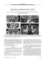
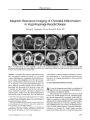
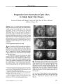
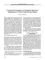
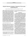
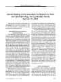
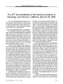
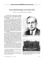
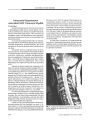
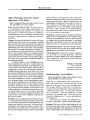
Show VIEWPOINT Magnetic Resonance Imaging of Suspected Cervicocranial Arterial Dissections Gaurang V. Shah, MD, Douglas J. Quint, MD, and Jonathan D. Trobe, MD Abstract: The authors propose that the optimal screening protocol for evaluation of suspected cervicocranial arterial dissections is magnetic resonance imaging ( MRI) that includes three components: 1) contrast- enhanced three- dimensional time- of- flight magnetic resonance angiography ( MRA) through the superior mediastinum, neck, and skull base; 2) three- dimensional multiple overlapping thin-section acquisition MRA of the skull base and Circle of Willis region; and 3) axial non- contrast, non- fat- suppressed Tl-weighted, fat- suppressed Tl- weighted, and T2- weighted spin- echo MRI from the level of the aortic arch through the level of the circle of Willis. MRA permits visualization of vascular luminal narrowing or obliteration, which can suggest vascular dissection but can also be caused by congenital variation, dysplasia, intraluminal thrombus, vasospasm, or extramural compression by tumor. By directly visualizing the blood vessel wall, axial Tl- weighted and T2- weighted spin- echo MRI can identify the intramural hemorrhage of vascular dissection. This protocol is designed to maximize the sensitivity of a noninvasive technique and may eliminate the need for conventional endovascular angiography. ( JNeuro- Ophthalmol 2004; 24: 315- 318) Cervicocranial ( carotid and vertebral) arterial dissection is among the most common causes of ischemic stroke in patients aged younger than 50 years ( 1). There is a premium on early diagnosis because short- term antiplatelet or anticoagulant therapy may be useful in preventing stroke ( 2). Dissections of the carotid and vertebral arteries can be detected by many imaging methods, but there is yet no consensus on the most efficient and effective choice. We propose that a combination of spin- echo magnetic resonance imaging ( MRI) and magnetic resonance angiography ( MRA) is an accurate screening tool and may eliminate the need for more invasive, expensive endovascular angiogra- Departments of Radiology ( Neuroradiology) ( GVS, DJQ), Ophthalmology ( JDT), and Neurology ( JDT), University of Michigan Medical Center, Ann Arbor, Michigan. Address correspondence to Jonathan D. Trobe, MD, Kellog Eye Center, 1000 Wall Street, Ann Arbor, MI 48105; E- mail: jdtrobe@ umich. edu phy. Based on recent clinical experience, we have the impression that clinicians are not optimizing the use of this modality. CASE REPORT Headache and falling to his right suddenly developed in a 42- year- old man. Clinical findings included a right Horner syndrome; nystagmus; skew deviation; left palatal deviation; poor swallowing, speech, and right extremity ataxia; as well as right facial and left extremity pain and temperature loss. These findings were considered typical of a right dorsolateral medullary and possibly cerebellar infarct in the distribution of the right posterior inferior cerebellar artery; in other words, a Wallenberg syndrome. Four hours after onset, computed tomographic ( CT) ( Fig. 1A) scanning revealed a low attenuation area in the right inferior cerebellum with minimal associated mass effect. Intracranial MRI obtained the next day ( Fig. IB) showed high T2 and fluid- attenuated inversion recovery signal, as well as restricted diffusion ( Fig. IB) in the right inferior cerebellum and the right dorsolateral medulla consistent with subacute infarction. Skull base and intracranial MRA with three-dimensional ( 3D) multislice overlapping thin section acquisition performed on the day of admission demonstrated absence of the intracranial ( V4) segment of the right vertebral artery ( Fig. 1C). The contrast- enhanced 3D time- of- flight MRA of the superior mediastinum and neck ( Fig. ID) showed narrowing and tapering of the right vertebral artery involving the V2 segment ( where the vessel enters the foramen transversarium at the C6 level) with complete absence of vessel signal above the lower C3 level. Although these studies disclosed a right vertebral vascular abnormality, they did not distinguish between vascular dissection and other potential causes of luminal compromise such as congenital variation or dysplasia, intraluminal thrombosis, vasospasm, or extramural compression by tumor. Therefore, the patient was recalled to the MR scanner the next day for axial fat- suppressed Tl- weighted imaging ( Fig. 2A), which showed a high signal area indicating methemoglobin- containing thrombus within the wall of the V2 segment of the right vertebral artery. The subacute nature of the thrombus was confirmed on the axial T2 MRI J Neuro- Ophthalmol, Vol. 24, No. 4, 2004 315 JNeuro- Ophthalmol, Vol. 24, No. 4, 2004 Shah et al FIG. 1. A. Axial non- contrast CT imaging in a patient with symptoms consistent with an acute right dorsolateral medullary infarct shows a hypodense area in the inferior portion of right cerebellar hemisphere suggestive of infarction in the right posterior inferior cerebellar artery territory. B. Axial diffusion- weighted magnetic resonance imaging ( MRI) demonstrates restricted diffusion of the inferior portion of right cerebellar hemisphere and also the dorsolateral right medulla ( arrow) consistent with acute/ subacute infarction. The medullary component of the infarct was not visualized on computed tomography ( CT) because of beam-hardening (" streak") artifacts and the intrinsic lower sensitivity of CT when compared with MRI in evaluating the posterior fossa. C. Maximum intensity projection magnetic resonance angiogram ( MRA) of brain vessels with three-dimensional ( 3D) multislice overlapping thin-section acquisition demonstrates absent signal in the expected location of the intracranial ( V4) segment of the right vertebral artery { arrowheads). D. Contrast- enhanced 3D time- of-flight MRA through the neck demonstrates narrowing and tapering of the right vertebral artery at the V2 segment { arrowheads). ( Fig. 2B) because increased intramural T2 signal was seen in the same region. Absence of right vertebral arterial blood flow was also confirmed on these images ( Figs. 2A and B) because the normal " flow void" was absent in the expected location of the vertebral artery. The diagnosis of vertebral artery dissection prompted immediate anticoagulation with heparin and later warfarin. Within 3 months, the patient's clinical manifestations had largely resolved. Repeat MRI showed normal luminal morphology, so the anticoagulation was discontinued. Our patient presented with classic symptoms of dorsolateral medullary and cerebellar infarction ( 3). Initial intracranial imaging demonstrated the changes of subacute ischemic infarction. MRA showed absence of the distal ( intracranial) right vertebral artery ( Fig. IC). However, initial neck evaluation included only MRA { without MRI), which revealed tapering and complete occlusion of the cervical portion of the right vertebral artery ( Fig. ID). This finding was not specific for a cause of the observed vascular occlusion. Only after recalling the patient to the scanner and obtaining axial spin- echo MRI was an intramural hematoma identified. This finding confirmed dissection as the cause of the vertebral arterial occlusion. PROPOSED IMAGING PROTOCOL In the evaluation of suspected cervicocranial dissection, the proper combination of MRI and MRA offers the best diagnostic screening tool currently available, even though endovascular angiography has long been considered the gold standard ( 4,5). As with endovascular angiography, MRA can sometimes identify dissection of cervical arteries by demonstrating specific luminal abnormalities such as aneurysmal dilatation or an intimal flap ( 6), which may be better visualized on the " source images" ( the images from which MRA reformatted images are generated) than on the reformatted maximum intensity projection, the MRA images most familiar to clinicians ( 4). Earlier reports had suggested that MRA was not as sensitive to vertebral artery dissection as to carotid artery dissection caused by the smaller lumen and physiological variations in the caliber of vertebral artery ( 7), but subsequent studies have disputed that notion ( 4,8). 316 © 2004 Lippincott Williams & Wilkins MRI of Cervicocranial Arterial Dissections JNeuro- Ophthalmol, Vol. 24, No. 4, 2004 FIG. 2. A. Axial non- contrast, fat- suppressed, T1- weighted MRI obtained 2 days after the MRI demonstrates high signal within the lumen of foramen transversarial ( V3) segment of the right vertebral artery, compatible with the subacute ( methemoglobin phase) intramural thrombus { arrow). Note the normal appearance of the left vertebral artery { arrowhead). B. Axial non- contrast T2- weighted MRI shows that the expected normal " flow void" has been replaced with high signal within the lumen of the right vertebral artery at the same anatomic level as in A { arrow). Note the normal appearance of the left vertebral artery { arrowhead). C. Axial non- contrast, T1 - weighted MRI obtained after a few months shows persistent lack of normal " flow- void" signal in the right vertebral artery with a concentric high signal around the lumen of the artery { arrow). Compare with normal flow void of the left vertebral artery { arrowhead). However, neither endovascular angiography nor MRA can directly identify a dissection unless a double-lumen, intimal flap, or dissecting aneurysm is found. Narrowing or even obliteration of the vascular lumen can have many causes ( 8), including congenital variation, dysplasia, intraluminal thrombosis, vasospasm, or extramural mass. To differentiate dissection from these other causes, one must be able to directly visualize the vessel wall, which is possible with MRA but not usually possible with endovascular angiography. The typical MRI appearance of a dissected blood vessel in cross- section is increased diameter of the artery with eccentric narrowing of the arterial lumen caused by the presence of an intramural hematoma ( 9- 11). The shape of the intramural hematoma varies with the relationship of the imaging plane to the axis of the dissected vessel ( 4). The hematoma may be crescentic, oval, or circumferential ( 8,12,13). The signal intensity of an intramural hematoma on Tl- weighted images depends on the age of the hemorrhage ( 9,14). The Tl signal appears essentially isointense for the first few days after bleeding occurs (" deoxyhemoglobin phase") and then becomes hyperintense in the subacute stage (" methemoglobin phase"). The abnormal signal persists for a few months ( 15), but disappears as the intramural blood is absorbed ( 15,16). A follow- up MR examination with the proposed protocol showed concentric intraluminal high Tl signal in the right vertebral artery ( Fig. 2C). Because the high Tl signal of intramural thrombus in the subacute ( methemoglobin) phase can blend with surrounding fat on conventional spin- echo Tl- weighted images, fat-suppressed Tl- weighted scan techniques ( 17,18) are used to better delineate subacute hematomas ( 19). However, sometimes these images can be degraded because of artefacts. T2- weighted imaging is performed to confirm the intramural hematoma in the acute phase ( isointense Tl signal with a decreased T2 signal ( 8)), and also to better demonstrate either a residual patent but narrowed vascular " flow void" or complete lack of intraluminal blood flow. Therefore, we believe that the ideal imaging evaluation of a patient clinically suspected of dissection of the cervicocranial arteries consists of a combination of axial noncontrast nonfat- suppressed Tl- weighted and fat- suppressed Tl- weighted images, together with T2- weighted spin- echo MRI and contrast- enhanced 3D time- of- flight MRA through the superior mediastinum, neck, and skull base, and 3D multislice overlapping thin section acquisition MRA of the skull base and circle of Willis region. These imaging principles apply equally to diagnosis of carotid artery dissection. We recently encountered a patient in whom high signal in carotid artery wall on axial MRI disclosed the diagnosis of carotid artery dissection in the presence of a minor MRA luminal abnormality that had been dismissed as a normal variant. A 53- year- old man with the acute onset of left- sided headache and a left- sided Horner syndrome underwent a contrast- enhanced time-of- flight MRA ( Fig. 3A), which showed only subtle asymmetry of the internal carotid arteries. The study was interpreted as normal. His clinical findings were therefore attributed to cluster headache syndrome. However, a subsequent Tl axial neck MRI revealed the high signal of intramural hemorrhage expanding the vessel wall and consistent with dissection ( Fig. 3B). 317 JNeuro- Ophthalmol, Vol. 24, No. 4, 2004 Shah et al FIG. 3. A. 3D time- of- flight MRA of neck in another patient with acute left headache and left Horner syndrome demonstrates minimal asymmetry of the cervical internal carotid arteries with the left internal carotid artery { arrow) appearing somewhat smaller than the right { arrowhead). This appearance could be overlooked or attributed to normal variation. B. Axial non- contrast, T1 - weighted MRI at the base of the skull demonstrates circumferential high signal surrounding the left internal carotid artery { arrow). Compare with the normal flowvoid in the right internal carotid artery { arrowhead). This finding is consistent with subacute intramural thrombus and confirms a recent cervical internal carotid artery dissection even though only minimal luminal compromise is present on the MRA ( A). Ultrasound has been advocated for the evaluation of suspected cervicocranial dissection. A study of suspected cervical carotid dissection 15 years ago reported a sensitivity of 76% ( 20). The diagnosis is suggested by an intense systolic low- frequency Doppler signal of alternating flow direction along the extent of a tapered lumen or proximal to a severe obstruction at the skull base. ( 20). In another study, lack of diastolic flow in a vertebral artery suggested dissection with a sensitivity of 79%> ( 8). However, evaluation of vertebral arteries with ultrasound is hampered by the fact that sound waves cannot penetrate the osseous structures surrounding the foramina transversaria between C6 and C2 ( V2 portion of the vertebral arteries). Moreover, ultrasound is unreliable for luminal compromise of less than 50%>, cannot reliably visualize intramural abnormalities, and cannot evaluate any vessels in the superior mediastinum, behind the mandible, within the skull base, or within the intracranial compartment. With the advent of multidetector helical CT scanners, CT angiography has emerged as a potential rapid alternative in the screening of craniocerebral vascular disease ( 21,22). It can reveal a narrowed arterial lumen with enlargement of the overall vessel diameter caused by intramural hematoma. However, subtle intimal flaps can escape detection with CT angiography ( 22). Its role in this setting will be defined with further experience. REFERENCES 1. Schievink WI. Spontaneous dissection of the carotid and vertebral arteries. N Engl J Med 2001 ; 344; 898- 906. 2. Brandt T, Caplan L. Spontaneous arterial dissection. Curr Treatment Options Neurol 2001 ; 3: 463- 9. 3. Sacco RL, Freddo L, Bello JA, et al. Wallenberg's lateral medullary syndrome. Clinical- magnetic resonance imaging correlations. Arch Neurol 1993; 50: 609- 14. 4. Shin JH, Suh DC, Choi CG, et al. Vertebral Artery Dissection: Spectrum of imaging findings with emphasis on angiography and correlation with clinical presentation. RadioGraphics 2000; 20: 1687- 96. 5. Pelkonen O, Tikkakoski T, Leinonen S, et al. Extracranial internal carotid and vertebral artery dissections: angiographic spectrum, course and prognosis. Neuroradiology 2003; 45: 71- 7. 6. Levy C, Laissy JP, Raveau V, et al. Carotid and vertebral artery dissections: three- dimensional time- of- flight MR angiography and MR imaging versus conventional angiography. Radiology 1994; 190: 97- 103. 7. Levy C, Laissey JP, Raveau V, et al. Carotid and vertebral artery dissections: three- dimensional time- of- flight MR angiography and MR imaging versus conventional angiography. Radiology 1994; 190: 97- 103. 8. Auer A, Felber S, Schmidauer C, et al. Magnetic resonance angiographic and clinical features of extracranial vertebral artery dissection. JNeurol Neurosurg Psychiatry 1998; 64: 474- 81. 9. Kitanaka C, Tanaka J, Kuwahara M, et al. Magnetic resonance imaging study of intracranial vertebrobasilar artery dissections. Stroke 1994; 25: 571- 5. 10. Gelbert F, Assouline E, Hodes JE, et al. MRI in spontaneous dissection of vertebral and carotid arteries: 15 cases studied at 0.5 tesla. Neuroradiology 1991; 33: 111- 113. 11. Zuber M, Meary E, Meder JF, et al. Magnetic resonance imaging and dynamic CT scan in cervical artery dissections. Stroke 1994; 25: 576- 81. 12. Iwama T, Andoh T, Sakai N, et al. Dissecting and fusiform aneurysms of vertebro- basilar systems: MR imaging. Neuroradiology 1990; 32: 272- 9. 13. Goldberg HI, Grossman RI, Gomori JM, et al. Cervical internal carotid artery dissecting hemorrhage: diagnosis using MR. Radiology 1986: 158; 157- 61. 14. Quint DJ, Spickler EM. Magnetic resonance demonstration of vertebral artery dissection: report of two cases. J Neurosurg 1990; 72: 964- 7. 15. Gomori JM, Grossman RI, Goldberg HI, et al. Intracranial hematomas: imaging by high- field MR. Radiology 1985; 157: 87- 93. 16. Yoshimoto Y, Wakai S. Unruptured intracranial vertebral artery dissection: clinical course and serial radiographic imaging. Stroke 1997; 28: 370- 4 17. Rother J, Schwartz A, Rautenberg W, et al. Magnetic resonance angiography of spontaneous vertebral artery dissection suspected on Doppler ultrasonography. J Neurol 1995; 242: 430- 6. 18. Pacini R, Simon J, Ketonen L, et al. Chemical shift imaging of a spontaneous internal carotid artery dissection: case report. AJNR Am JNeuroradiol 1991; 12: 360- 2. 19. Bloem BR, Lammers GJ, Van Buchem MA. Magnetic resonance imaging and vertebral artery dissection. J Neurol Neurosurg Psychiatry 1999; 67: 691- 2. 20. Hennerici M, Steinke W, Rautenberg W. High- resistance Doppler-flow pattern in extracranial carotid dissection. Arch Neurol 1989; 46: 670- 2. 21. Bub L, Hollingworth W, Hallam D, et al. Screening for blunt cerebrovascular injury: Evaluating the quality and accuracy of multidetector computed tomographic angiography ( CTA). ( Abstract). Radiology 2003; 229P: 595. 22. Nunez DB Jr., Torres- Leon M, Munera F. Vascular injuries of the neck and thoracic inlet: Helical CT- angiographic Correlation. RadioGraphics 2004; 24: 1087- 100. 318 © 2004 Lippincott Williams & Wilkins |