| OCR Text |
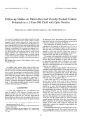
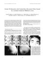
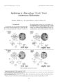
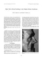
Show Journal of' Neuro- Ophthalmology 18( 1): 53- 55, 1998. © 1998 Lippincotl- Raven Publishers, Philadelphia Pseudotumor Cerebri Sine Papilledema with Unilateral Sixth Nerve Palsy Rohit Krishna, M. D., Gregory S. Kosmorsky, D. o., and Kenneth W. Wright, M. D., A 17- year- old woman presented with a history of 1- week of headache and 3 days of horizontal diplopia. Examination revealed 20/ 20 vision in both eyes, no papilledema, and an abduction deficit in her left eye. Lumbar puncture revealed an opening pressure of 440 mm HzO. After treatment with acet-azolamide, the headache and abduction deficit resolved. Papilledema never developed. This is a unique case of pseudotumor cerebri sine papilledema with a unilateral abduction deficit. We suggest that young women with headache and unilateral abduction deficits may be unrecognized cases of pseudotumor cerebri. Key Words: Pseudotumor cerebri- Papilledema- Sixth nerve palsy. Pseudotumor cerebri is characterized by an elevated opening pressure on lumbar puncture examination with normal cerebrospinal fluid ( CSF) composition, normal brain scan, and papilledema. Our report is a unique case of pseudotumor cerebri sine papilledema with a unilateral sixth nerve palsy. CASE REPORT A 17- year- old woman, weighing 114 kg, presented with 1 week of throbbing holocranial headache radiating from her neck to her forehead and 3 days of horizontal diplopia. The patient complained of nausea, but denied recent viral syndrome, transient visual obscurations, tinnitus, visual aura, trauma, vomiting, numbness, seizures, or use of medication. There was no family history of migraine headaches. Attempts to treat the headache with antibiotics, oral nonsteroidal antiinflammatory agents, and prochlorperazine were unsuccessful. On examination, visual acuity was 20/ 20 in both eyes, with normal color vision, no anisocoria or afferent pupillary defect, unremarkable anterior segment, normal intraocular pressures, and no papilledema ( Figs. 1 and 2). There was an abduction deficit of the left eye ( Fig. 3) and Manuscript received February 5, 1997; accepted April 24, 1997. From the Division of Ophthalmology, Cleveland Clinic Foundation, Cleveland, Ohio. Address correspondence and reprint requests to Dr. Gregory S. Kosmorsky, Division of Ophthalmology, Cleveland Clinic Foundation, 9500 Euclid Ave., Cleveland, OH 44195, U. S. A. full motility of the right eye. Forced duction testing revealed absence of restriction. Evaluation included normal magnetic resonance ( MR) imaging, normal MR venogram, and lumbar puncture ( which revealed an opening pressure of 440 mm H20), glucose 60 mg/ dl, protein 11 mg%, and one white blood cell ( WBC) per high power field ( HPF). The patient had some relief of headache with lumbar puncture. Treatment with acet-azolamide 500 mg p. o. b. i. d. was initiated. Ten days later, the headache had decreased, the abduction deficit improved by 50%, and there was no papilledema. At 2- month follow- up, the headache was gone, the abduction deficit had resolved ( Fig. 4), and there was no papilledema. Papilledema is a common, but not necessary, finding for a diagnosis of pseudotumor cerebri. Pseudotumor cerebri with unilateral or asymmetric papilledema has been described in the literature.( 1- 6) Lipton ( 7) was first to describe pseudotumor cerebri without papilledema. Marcelis ( 8) and Mathew ( 9) reported pseudotumor cerebri without papilledema in patients with chronic daily headache syndrome. Spence ( 10) reported on nine patients with pseudotumor cerebri without papilledema, seven of whom had a history of closed head trauma. In a case reported by Chari ( 11), papilledema developed after the diagnosis of pseudotumor cerebri had been secured. The probable cause of asymmetric or unilateral papilledema, or its absence, is best delineated by Hayreh ( 12) in his landmark work with monkeys. Inflatable balloons placed in different compartments of the brain resulted in increased intracranial pressure, with pressure ultimately conveyed to the optic nerves. Varying communication of the subarachnoid space and optic nerve through the optic canal created variable transmission of the intracranial pressure to the optic nerve head. A block of CSF pressure could occur at the level of the optic canal due to an almost nonexistent subarachnoid space, thereby functionally dividing the intracranial and intraorbital compartments. There have been other purported causes of asymmetric papilledema, such as inflammatory gliosis or congenital anomalies of the optic nerve sheath ( 13), obliteration of the subarachnoid space by subarachnoid hemorrhage ( 10), and intermittently increased intracranial pressure allowing axoplasmic flow to equilibrate 53 54 R. KRISHNA ETAL. FIG. 1. Right optic nerve examination reveals no papilledema FIG. 2. Left optic nerve examination reveals no papilledema FIG. 3. On presentation, there is an abduction deficit of the left eye FIG. 4. At 2- month follow- up, there is complete resolution of the abduction deficit ( 1,10). It is possible that our patient did not have increased intracranial pressure long enough or severe enough to produce papilledema, but this suggestion is unlikely because of the duration of her symptoms and the fact that a sixth nerve palsy developed. Sixth nerve palsies associated with pseudotumor cerebri are commonly bilateral and are believed to be due to intracranial pressure transmitted to the sixth nerves by an undetermined mechanism. Inferior displacement of the pons, with traction on the abducens nerves, is one possible explanation. The cause of marked asymmetry of abduction deficit in our case is not clear. It is possible that the anatomy of our patient's sixth nerves differed from one side to the other or that downward brainstem forces were asymmetrically distributed. In a young woman presenting with a unilateral or bilateral abduction deficit, headache, and no papilledema, a diagnosis of postviral abducens palsy should be entertained. However, abducens palsy is seen more commonly in younger children. It is possible that our patient had an aseptic meningitis, with temporary obstruction of CSF egress mimicking the pseudotumor cerebri syndrome. However, lack of inflammatory cells, and normal protein and glucose levels in the CSF militates against such a possibility. Our patient responded in a typical fashion to acetazolamide, as would be expected with pseudotumor cerebri. It is possible that other cases of postviral sixth nerve palsies could be cases of unrecognized pseudotumor cerebri without papilledema. We bring this case to attention to provide an alternative explanation for a young woman complaining of headache with unilateral sixth nerve palsy and no papilledema. REFERENCES 1. Sedwick LA, Burde RM. Unilateral and asymmetric optic disk swelling with intracranial abnormalities. Am J Ophthalmol 1983; 96: 484- 7. 2. To KW, Warren FA. Unilateral papilledema in pseudotumor cerebri. Arch Ophthalmol 1990; 108: 644- 5. 3. Sher NA, Wirtschaffer J, Shapiro SK, See C, Shapiro I. Unilateral J Neuro- Ophlhahnol, Vol. IS, No. I, 1998 PSEUDOTUMOR CEREBRI SINE PAPILLEDEMA 55 papilledema in benign intracranial hypertension. JAMA 1983; 250: 2346- 7. 4. Lepore RE. Unilateral and highly asymmetric papilledema in pseudotumor cerebri. Neurology 1992; 42: 676- 8. 5. Kirkham TH, Sander MD, Sapp GA. Unilateral papilledema in benign intracranial hypertension. Can J Ophthalmol 1973; 8: 533- 8. 6. Strominger MB, Weiss GB, Mehler MF. Asymptomatic unilateral papilledema in pseudotumor cerebri. / Clin Neuro- Ophthalmol 1992; 12: 238- 41. 7. Lipton HL, Michelson PE. Pseudotumor cerebri syndrome without papilledema. JAMA 1072; 220: 1591- 2. 8. Marcelis J, Silberstein SD. Idiopathic intracranial hypertension without papilledema. Arch Neurol 1991; 48: 392- 9. 9. Mathew NT, Ravishanker K, Sanin LC. Coexistence of migraine and idiopathic intracranial hypertension without papilledema. Neurology 1996; 46: 1226- 30. 10. Spence JD, Amacher AZ, Willis NR. Benign intracranial hypertension without papilledema: role of 24- hour cerebrospinal fluid pressure monitoring in diagnosis and management. Neurosurgery 1980; 7: 326- 36. 11. Chari C, Rao NS. Benign intracranial hypertension- its unusual manifestations. Headache 1991; 31: 599- 600. 12. Hayreh SS. Pathogenesis of oedema of the optic disc. Br J Ophthalmol 1964; 48: 522- 43. 13. Maxner CE, Freedman MI, Coibett JJ. Asymmetric papilledema and visual loss in pseudotumour cerebri. Can J Neurol Sci 1987; 14: 593- 6. J Neiira- Ophlhalmol, Vol. IS, No. I, 1998 |