| Title |
Functional Magnetic Resonance Imaging of Visual Function in Postpapilledema Optic Atrophy |
| Creator |
Miki, A; Nakajima, T; Hasebe, H; Abe, H |
| Affiliation |
Department of Ophthalmology, Niigata University School of Medicine, Japan. |
| Abstract |
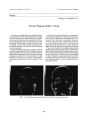
We studied a girl with intraventricular brain tumor who developed postpapilledema optic atrophy and severe concentric visual field constriction in both eyes. The patient had mild symptoms despite severe visual field loss. We performed functional magnetic resonance imaging using two kinds of visual stimulation to assess her residual visual function. The functional magnetic resonance imaging demonstrated that the activity in her association visual cortex was largely intact bilaterally, although the activation of the calcarine cortex was decreased in the left calcarine cortex. Her activity in the visual cortex seemed to correspond well to her visual symptom. Functional magnetic resonance imaging may be useful in objectively documenting residual visual function in patients with severe visual loss. |
| Subject |
Adolescent; Brain Neoplasms/complications; Cerebral Ventricles; Female; Humans; Magnetic Resonance Imaging; Neurocytoma/complications; Optic Atrophy/diagnosis; Optic Atrophy/etiology; Optic Atrophy/physiopathology; Papilledema/complications; Visual Fields/physiology |
| Format |
application/pdf |
| Publication Type |
Journal Article |
| Collection |
Neuro-Ophthalmology Virtual Education Library: Journal of Neuro-Ophthalmology Archives: https://novel.utah.edu/jno/ |
| Publisher |
Lippincott, Williams & Wilkins |
| Holding Institution |
Spencer S. Eccles Health Sciences Library, University of Utah |
| Rights Management |
© North American Neuro-Ophthalmology Society |
| Setname |
ehsl_novel_jno |
| ID |
224885 |
| Reference URL |
https://collections.lib.utah.edu/ark:/87278/s6wx0pr1/224885 |