OCR Text |
Show Journal of Clinical Neuro- ophfhalmology 11( 3): 139- 143, 1991. © 1991 Raven Press, Ltd., New York Meningovascular Syphilis with a Gumma of the Midbrain David G. Standaert, M. D., Ph. D., Steven L. Galetta, M. D., and Scott W. Atlas, M. D. 
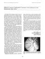
We report a patient with meningovascular syphilis who had a dorsal midbrain syndrome, cognitive dysfunction, and a left peripheral seventh nerve palsy. Magnetic resonance imaging ( MRI) disclosed a large lesion of the midbrain and thalamus with intense enhancement of the interpeduncular cistern, both of which resolved after treatment with intravenous penicillin. The clinical features, radiographic appearance, and response to therapy suggest that this lesion was a focal syphilitic inflammatory process, or gumma. We conclude that MRI with intravenous contrast may reveal the full spectrum of pathologic involvement in neurosyphilis and, in certain situations, may obviate the need for biopsy of an associated mass lesion. Key Words: Neurosyphilis- Magnetic resonance imaging- Dorsal midbrain syndrome-- Gumma. From the Department of Neurology ~ D. G: S., S. L. G.) and D~ partment of Radiology ( S. W. A.), UniversIty of Pennsylvania School of Medicine, Philadelphia, Pennsylvania, U. S. A. Address correspondence and reprint requests to Dr. Steven L. Galetta at Department of Neurology, Hospita~ of the University of Pennsylvania, 3400 Spruce Street, PhJladelphla, PA 19104, U. S. A. 139 A gumma is a circumscribed mass of granulation tissue resulting from syphilitic infection of the brain ( 1). Distinguishing gummas from tumors or other focal cerebral lesions is often quite difficult. Serologic methods may be misleading, as a patient with a tumor of the brain and a positive serologic test for syphilis is more likely to have asymptomatic syphilis and an unrelated neoplasm than a gumma ( 2). Furthermore, patients with proven gummas may have nonreactive serology ( 3). Relatively few gummas have been studied with modern neuroimaging techniques, and the radiographic features that have been described are nonspecific ( 4,5). Even when it is possible to make a diagnosis of a gumma without direct biopsy, there are no clear guidelines for treatment of these lesions. We report a patient with meningovascular syphilis in whom magnetic resonance imaging ( MRI) documented a large mass lesion of the midbrain that resolved following antibiotic treatment. CASE REPORT A 27- year- old left- handed man was evaluated for headache, diplopia, and facial weakness. The patient was well until 2 months previously, when he developed throbbing bifrontal and occipital headaches. Two weeks later, he noted the sudden onset of left facial weakness and slurred speech. Two days before admission he developed vertical diplopia, and his family noticed confusion, somnolence, and gait difficulty. He acknowledged smoking cocaine about twice a week but denied intravenous drug use or homosexual activity. The patient had gonorrhea in the past, but no history of syphilis. He had a temperature of lOO. l° F. Neurologic examination showed that he was drowsy and confused, with prominent impairment of memory. 140 D. G. STANDAERT ET AL. DISCUSSION TABLE 1. Results of cerebrospinal fluid examinations When seen in follow- up 10 months later, the patient had minimal evidence of cognitive impairment. His abnormalities of ocular motility were markedly improved, although he was still troubled by intermittent diplopia. His left facial palsy had largely resolved, but aberrant regeneration was evident. Magnetic resonance imaging showed a small, focal area of cavitation in the right thalamus and superior midbrain, with no pathologic enhancement ( Fig. 3). A serum assay for HIV was again negative, and the CSF was essentially normal ( Table 1). 5 88 41 57 negative 113 88 136 60 488 78 189 53 303 88 251 42 1: 2 Day 1 Day 3 Day 15 10 Months Syphilis remains an important cause of neurologic disease. Over 48,000 cases of primary and secondary syphilis were reported in the United States in 1990 ( 6). It is estimated that at least 10% of patients with untreated primary syphilis will develop neurologic disease ( 1). Syphilitic disease of the nervous system may be classified into three clinical forms: asymptomatic, meningovascular, and parenchymal ( 1,7). Meningovascular syphilis is characterized by a prodromal phase of fatigue, malaise, meningismus, and mild encephalopathy. There may be cranial nerve palsies related to the meningeal inflammation and the stroke- like onset of focal neurologic deficits ( 1,8). Pathologic studies reveal inflammatory infiltration of the pia and arachnoid with variable degrees of vascular involvement ( 1,9). The proportion of meningovascular disease among patients with symptomatic neurosyphilis is about one- third in contemporary series ( 10,11). Focal inflammation with formation of a gumma is an uncommon complication of meningovascular syphilis and is thought to represent extension of the meningeal inflammatory process into the brain parenchyma ( 1,9). The histology of gummatous lesions is variable. Often there is infiltration by lymphocytes and plasma cells as well as giant cell formation, while in some instances fibrosis and caseative necrosis are prominent ( 9). The response to therapy is also variable, with some re- Leukocytes ( N = x 106 / L) Lymphocytes (%) Protein ( mg/ dL) Glucose ( mg/ dL) VDRL His speech was dysarthric but his language was fluent. Upgaze paresis, convergence- retraction nystagmus, and a left peripheral seventh nerve palsy were evident ( Fig. 1). An esotropia and right hypertropia were induced by attempted upgaze. Neither light- near dissociation of the pupils nor lid retraction were detected. The remainder of the examination was significant for a mild left hemiparesis and a wide- based unsteady gait. Cerebrospinal fluid ( CSF) analysis revealed a lymphocytic pleocytosis ( Table 1). An MRl of the head disclosed an area of high signal abnormality on long TRITE images centered in the left cerebral peduncle, and extending rostrally into the thalamus and the hypothalamus and caudally into the pons ( Fig. 2A and B). After intravenous contrast administration, the intra- axial lesion was not enhanced but there was intense enhancement along the left side of the interpeduncular fossa and along the margin of the left cerebral peduncle ( Fig. 2C). The serum RPR was positive at a titer of 1: 16, and a serum microhemagglutination test for Treponema pallidum was also positive. The spinal fluid VORL was positive at 1: 2. A serum assay for antibody to human immunodeficiency virus ( HIV) was negative, and a C04 lymphocyte count was normal. The patient was treated with intravenous penicillin G, 21 million units a day for 3 weeks. There was gradual improvement in his alertness and memory as well as in his ocular motility, although his left facial weakness persisted. A repeat MRl 2 weeks after admission showed a significant decrease in the size of the signal abnormality in the midbrain, while repeat lumbar puncture showed gradual resolution of the CSF abnormalities ( Table 1). FIG. 1. External photograph demonstrating the upgaze paresis and esotropia of the dorsal midbrain syndrome. Note the decreased eyebrow elevation and frontalis contraction consistent with a left peripheral seventh nerve palsy. GUMMA OF THE MIDBRAIN 141 A c FIG. 2. MRI at time of initial evaluation. A: T2weighted image at level of midbrain. B: T2- weighted image at level of caudal thalamus. C: Tl- weighted image at level of the midbrain after intravenous gadolinium- DPTA. High- signal lesion ( closed arrows, A and B) involves left midbrain white matter and thalami bilaterally. Note focal enhancement in the interpeduncular fossa and superficial aspect of the left cerebral peduncle ( open arrow, C) after intravenous contrast. ports demonstrating resolution of the lesions with antibiotic therapy, while others require surgical intervention ( 4,5,12,13). These observations suggest a pathologic continuum in the formation of gummas, ranging from a focal inflammatory infiltrate to a chronic fibrotic lesion. Cerebral gummas have been studied with modem neuroimaging techniques only rarely. On computed tomography, the most common finding is a circumscribed area of decreased attenuation. Enhancement of these areas after intravenous contrast is often observed; however, several biopsyproven gummas have failed to show enhancement (:>- 5,12,13). The most frequently reported MRI abnormality in patients with meningovascular syph-ilis is multiple foci of increased signal on T2weighted images which persist despite therapy and are believed to correspond to focal areas of infarction ( 14,15). Ito et aI. ( 13) have recently described a patient with a gumma of the left parietal lobe diagnosed by biopsy; this lesion also showed nonspecific increased signal on T2 weighted images. Our patient presented with laboratory findings and a history typical of meningovascular neurosyphilis. His examination revealed a dorsal midbrain syndrome with impaired upgaze and convergence- retraction nystagmus. The MRI findings were incompatible with simple ischemia, in that the midbrain lesion involved only white matter , Clin Neuro- ophthalmol. Vol. II, No. 3. 1991 142 D. G. STANDAERT ET AL. A IB c FIG. 3. Follow- up MRI 10 months after therapy. A: T2weighted image at level of midbrain. B: T2- weighted image at level of mid- thalamus. C: T1- weighted image at the level of the midbrain after intravenous gadolinium- DPTA. T2- weighted images ( A and B) show complete resolution of the midbrain lesion, with a small residual area of infarction as focal high intensity in the right thalamus ( arrows, B). Note slight dilation of the third ventricle toward the lesion, indicating its malacic nature. Abnormal enhancement is no longer present ( C). and did not respect a vascular territory ( Fig. 2). The complete resolution of the lesion after therapy also argues strongly that it was primarily inflammatory rather than ischemic or neoplastic in nature ( 14). In addition, our patient sustained a focal thalamic infarction as a result of the meningeal and vascular inflammation, the residua of which is dearly seen on the follow- up MRI ( Fig. 3). The observations in the present case illustrate that in patients with meningovascular syphilis and findings of parenchymal dysfunction, MRI may be able to distinguish between ischemic lesions and the focal inflammation of a gumma. In contrast to infarction, gummatous lesions which are treated early appear to have an excellent prognosis for re-covery without a need for surgical intervention. In addition, serial MRI examinations are an important marker of the activity of the meningeal as well as the parenchymal components of the disease, and are helpful in guiding the type and duration of therapy. As this technique is applied to additional cases of meningovascular syphilis, it seems possible that gummatous disease will be found to be more common than previously appreciated. REFERENCES 1. Merritt HH, Adams RA, Solomon He. Neurosyphilis. New York: Oxford University Press, 1946: 443. 2. Adams RD, Victor M. Principles of neurology. 2nd ed. New York: McGraw- Hill, 1981: 456. GUMMA OF THE MIDBRAIN 143 3. Currie JN, Coppeto JR, LesseIl S. Chronic syphilitic meningitis resulting in superior orbital fissure syndrome and posterior fossa gumma. / Clin Neuro- ophthalmoI1988; 8: 14555. 4. Godt P, Stoeppler L, Wischer U, Schroeder HH. The value of computed tomography in cerebral syphilis. Neuroradiology 1979; 18: 197- 200. 5. Kaplan JG, Sterrman AB, Horoupian 0, Leeds NE, Zimmerman RD, Gade R. Leutic meningitis with gumma: clinical, radiographic and neuropathologic features. Neurology 1981; 31: 464- 7. 6. Centers for Disease Control. Cases of speCified notifiable diseases, United States. MMWR 1991; 39: 994. 7. Rowland LP. Spirochete infections: neurosyphilis. In: Rowland LP, ed. Merritt's textbook of neurology. 8th ed. Philadelphia: Lea & Febiger, 1989: 152~ 2. 8. Simon RP. Neurosyphilis. Arch NeuroI1985; 42: 606- 13. 9. Harriman DGF. Bacterial infections of the central nervous system. In: Blackwood W, Corsellis JAN, eds. Greenfield's neuropathology. 3rd ed. London: Edward Arnold, Ltd., 1976: 23~. 10. Burke JM, Schaberg DR. Neurosyphilis in the antibiotic era. Neurology 1985; 35: 1368- 71. 11. Hotson JR. Modern neurosyphiliS: a partially treated chronic meningitis. West / Med 1981; 135: 191- 200. 12. Punt J. Multiple cerebral gummata./ Neurosurg 1983; 58: 95961. 13. Ito M, Sato K, Tada H, Kuru Y. Neurosyphilis manifesting as a focal mass lesion: computed tomographic and magnetic resonance imaging features. Neurol Med Chir ( Tokyo) 1990; 30: 194- 7. 14. Holland BA, Perrett LV, Mills CM. Meningovascular syphilis: CT and MR findings. Radiology 1986; 158: 439- 42. 15. Johns DR, Tierney M, Parker SW. Pure motor hemiplegia due to meningovascular syphilis. Arch Neurol 1987; 44: 1062- 5. 1Clin Neuro- ophthalmol, Vol. 11, No. 3, 1991 [VBintracranialinfections] |