OCR Text |
Show Acute Retrobulbar Optic Neuritis and Macular Detachment Associated With Morning Glory Optic Disc Anomaly Marjorie A. Murphy, MD, Robert H. Janigian, MD, Theodoros Filippopoulos, MD, Glenn A. Tung, MD, FACR Abstract: 
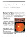
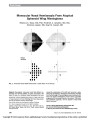
A 31-year-old woman with morning glory optic disc anomaly (MGDA) developed acute retrobulbar optic neuritis and a bullous macular detachment. MRI demon-strated truncation of the perineural space of the affected optic nerve as well as focal optic nerve enhancement. Optical coherence tomography (OCT) showed reti-noschisis associated with the macular detachment. The MRI and OCT findings support the vitreous as the source of the subretinal fluid. This is the first reported case of optic neuritis in MGDA. Journal of Neuro-Ophthalmology 2010;30:123-125 doi: 10.1097/WNO.0b013e3181ce199e 2010 by North American Neuro-Ophthalmology Society Morning glory optic disc anomaly (MGDA) is a con-genital disorder characterized by a funnel-shaped excavation of the optic disc and the surrounding posterior pole of the retina with an annulus of pigmented, elevated chorioretinal tissue (1). Usually sporadic and unilateral, it has been associated with posterior pole retinal detachments in up to 37% of patients (2). Other congenital cavitary optic nerve anomalies associated with serous macular detach-ments include optic pits and optic disc colobomas. To explain the origin of the subretinal fluid and the patho-genesis of the macular detachment associated with these optic disc anomalies, several mechanisms have been pro-posed: 1) an abnormal connection between the subretinal space and the perineural cerebrospinal fluid (CSF) (2-4); 2) a small peripapillary retinal defect creating an abnormal connection between the subretinal space and the vitreous (5-9); and 3) variable interconnections between the sub-retinal, vitreous, and subarachnoid spaces (10,11). We describe a patient with MGDA who developed acute retrobulbar optic neuritis and an associated macular detachment, a combination of findings not previously reported. CASE REPORT A 31-year-old woman with known MGDA in the left eye presented with a 5-day history of ipsilateral retrobulbar pain exacerbated by eye movement. The pain was not relieved by nonsteroidal anti-inflammatory medications. Her past medical history was notable for hypertension, hypercho-lesterolemia, and asthma. She denied any current or prior neurologic or constitutional symptoms. Best-corrected visual acuity was 20/20 in the right eye and hand motions in the left eye, both unchanged from her previously documented baseline. Extraocular motility was full, with a large-angle left exotropia dating back to childhood. A long-standing left afferent pupillary defect was noted. Slit lamp examination of the anterior segment was normal in both eyes. Dilated ophthalmoscopy of the left eye showed a MGDA and a large serous retinal detachment involving the macula, with no visible peripapillary retinal break (Fig. 1). No macular abnormality had been noted in the left eye on a routine examination 1 year earlier. Oph-thalmoscopy of the right eye was normal. MRI of the orbits (Fig. 2) showed focal truncation of the left optic nerve perineural space 4 mm posterior to the sclera and focal enhancement of the corresponding short segment of the retrobulbar left optic nerve. Results of MRI and MRA of the brain were normal. Optical coherence tomograms (Stratus OCT3) of the macula and optic disc showed conical excavation of the optic disc and a bullous macular detachment (Fig. 3). Departments of Ophthalmology (MAM, RJH, TF) and Diagnostic Imaging (GAT), The Warren Alpert Medical School of Brown University, Rhode Island Hospital, Providence, Rhode Island. Address correspondence to Marjorie A. Murphy, MD, Department of Ophthalmology, APC 7, Rhode Island Hospital, 593 Eddy Street, Providence, RI 02903; E-mail: margiemurphy@cox.net Murphy et al: J Neuro-Ophthalmol 2010; 30: 123-125 123 Original Contribution Copyright © North American Neuro-ophthalmology Society.Unauthorized reproduction of this article is prohibited. Retinoschisis was noted to extend temporally from the optic disc. A definitive connection of the schisis cavity to the subretinal space or the vitreous cavity could not be iden-tified by optical coherence tomography (OCT). Based on a diagnosis of left retrobulbar optic neuritis, the patient was treated with 1 g intravenous methylpredniso-lone daily for 3 days, followed by an oral prednisone taper. Periocular pain resolved completely after the 3rd day of treatment. During a follow-up of 1 year, she had no recur-rence of symptoms. Visual function remained unchanged and the macular detachment persisted. Eighteen months after presentation, repeat MRI of the brain and orbits showed no change. At 24 months after presentation, the macular detachment had completely resolved. DISCUSSION This is the first report of MGDA associated with MRI evidence of truncation of the perineural space and retro-bulbar optic nerve. We suggest that this observation indi-cates that the vitreous rather than the cerebrospinal fluid (CSF) is the source of subretinal fluid in our patient's retinal detachment. With cavitary optic nerve anomalies, dysplastic tissue herniates through a defect in the lamina cribrosa and may extend slightly posteriorly into the subarachnoid space of the distal portion of optic nerve sheath (11). However, it is unlikely that such a potential herniation would extend more than 4 mm to reach beyond the point of truncation of the nerve sheath and into the subarachnoid space. In addition, retinoschisis was noted in our patient, similar to that seen in some patients with optic pit maculopathy, in whom a connection between the schisis cavity and vitreous has been demonstrated (12,13). Although a retinal break could not be definitively identified by OCT in our patient, the radial lines protocol uses a 30 arc between scans and could have missed a small FIG. 1. Fundus photograph of the left eye shows a morning glory disc anomaly and associated macular detachment. FIG. 2. Focal absence of the retrobulbar optic perineural space and acute inflammation in the left optic nerve near the globe. Axial T2 (A), FLAIR (B), and postcontrast T1 (C) MRI show focal absence of the left perineural space (arrow, A) corresponding to a short segment of the enhancing retrobulbar left optic nerve (arrow, C). Coronal short-tau inversion recovery images show hyperintense signal within the affected left optic nerve (arrow, D) and around the nerve (arrow, E) compared with the normal signal intensity within and around the unaffected right optic nerve. Original Contribution 124 Murphy et al: J Neuro-Ophthalmol 2010; 30: 123-125 Copyright © North American Neuro-ophthalmology Society.Unauthorized reproduction of this article is prohibited. break. Johnson and Johnson (10) have emphasized that the anatomy of cavitary optic disc anomalies varies from one eye to another, with a communication to the vitreous in some eyes and to the subarachnoid space or both spaces in others. Hence, no single unifying mechanism necessarily accounts for the variability and behavior of macular detachments associated with congenital optic disc anomalies. We believe this to be the first report of acute retrobulbar optic neuritis in association with MGDA. Perkins et al (11) have postulated that undetermined systemic factors may play a role in the development of macular changes in patients with cavitary optic nerve anomalies, noting that one of the patients in their series developed bilateral simulta-neous macular detachments associated with bilateral atyp-ical optic nerve colobomas. Systemic as well as local factors may play a role and the inflammatory process in our patient probably contributed to the concurrent development of a macular detachment. This detachment was a new finding from an examination 1 year earlier and had resolved spontaneously within 2 years of follow-up. This case highlights the fact that patients with profound visual loss from congenital optic disc anomalies may develop superimposed acquired optic neuropathies and emphasizes that those who present with a clinical picture consistent with optic neuritis should undergo neuroimaging. REFERENCES 1. Kindler P. Morning glory syndrome: unusual congenital optic disk anomaly. Am J Ophthalmol. 1970;69:376-384. 2. Haik BG, Greenstein SH, Smith ME, Abramson DH, Ellsworth RM. Retinal detachment in the morning glory anomaly. Ophthalmology. 1984;91:1638-1647. 3. Chang S, Haik BG, Ellsworth RM, St. Louis L, Berrocal JA. Treatment of total retinal detachment in morning glory syndrome. Am J Ophthalmol. 1984;97:596-600. 4. Irvine AR, Crawford JB, Sullivan JH. The pathogenesis of detachment with morning glory disc and optic pit. Retina. 1986;6:146-150. 5. Harris MJ, deBustros S, Michels RG, Joondeph HC. Treatment of combined traction-rhegmatogenous retinal detachment in the morning glory syndrome. Retina. 1984;4: 249-252. 6. Coll GE, Chang S, Flynn TE, Brown GC. Communication between the subretinal space and the vitreous cavity in the morning glory syndrome. Graefes Arch Clin Exp Ophthalmol. 1995;233:441-443. 7. Bartz-Schmidt KU, Heimann K. Pathogenesis of retinal detachment associated with morning glory disc anomaly. Int Ophthalmol. 1995;19:35-38. 8. Ho CL, Wei LC. Rhegmatogenous retinal detachment in morning glory syndrome pathogenesis and treatment. Int Ophthalmol. 2001;24:21-24. 9. Ho TC, Tsai PC, Chen MS, Lin LL. Optical coherence tomography in the detection of retinal break and management of retinal detachment in morning glory syndrome. Acta Ophthalmol Scand. 2006;86:225-227. 10. Johnson TM, Johnson MW. Pathogenic implications of subretinal gas migration through pits and atypical colobomas of the optic nerve. Arch Ophthalmol. 2004;122: 1793-1800. 11. Perkins SL, Han DP, Gonder PE, JR, Colev G. Dynamic atypical optic nerve colobomas associated with transient macular detachment. Arch Ophthalmol. 2005;123: 1750-1754. 12. Lincoff H, Schiff W, Krikov D, Ritch R. Optic coherence tomography of optic disk maculopathy. Am J Ophthalmol. 1996;122:264-266. 13. Rutledge BK, Puliafito CA, Duker JS Hee MR, Cox MS. Optical coherence tomography of macular lesions associated with optic nerve pits. Ophthalmology. 1996; 103:1047-1053. FIG. 3. Optical coherence tomography of the left eye. Radial superotemporal cut of the peripapillary region demonstrates retinoschisis and neurosensory retinal de-tachment. Original Contribution Murphy et al: J Neuro-Ophthalmol 2010; 30: 123-125 125 Copyright © North American Neuro-ophthalmology Society.Unauthorized reproduction of this article is prohibited. |