Contents | 13 of 13
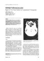
Neuro-anatomical feature photo.


| Title | Journal of Neuro-Ophthalmology, September 1982, Volume 2, Issue 3 |
| Date | 1982-09 |
| Language | eng |
| Format | application/pdf |
| Type | Text |
| Publication Type | Journal Article |
| Collection | Neuro-Ophthalmology Virtual Education Library: Journal of Neuro-Ophthalmology Archives: https://novel.utah.edu/jno/ |
| Publisher | Lippincott, Williams & Wilkins |
| Holding Institution | Spencer S. Eccles Health Sciences Library, University of Utah |
| Rights Management | © North American Neuro-Ophthalmology Society |
| ARK | ark:/87278/s6h73mwt |
| Setname | ehsl_novel_jno |
| ID | 226915 |
| Reference URL | https://collections.lib.utah.edu/ark:/87278/s6h73mwt |
Page Metadata
| Title | Neuro-anatomical feature photo. |
| Creator | N. B. Barton, R. G. Clark |
| Subject | Humans; Visual Cortex |
| Format | application/pdf |
| Publication Type | Journal Article |
| Collection | Neuro-Ophthalmology Virtual Education Library: Journal of Neuro-Ophthalmology Archives: https://novel.utah.edu/jno/ |
| Publisher | Lippincott, Williams & Wilkins |
| Holding Institution | Spencer S. Eccles Health Sciences Library, University of Utah |
| Rights Management | © North American Neuro-Ophthalmology Society |
| Setname | ehsl_novel_jno |
| ID | 226914 |
| Reference URL | https://collections.lib.utah.edu/ark:/87278/s6h73mwt/226914 |