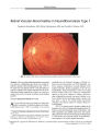
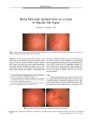
OCR Text |
Show ORIGINAL CONTRIBUTION Interferon- a- Associated Bilateral Simultaneous Ischemic Optic Neuropathy Yoav Vardizer, MD, Yifat Linhart, MD, Anat Loewenstein, MD, Hanna Garzozi, MD, Nail Mazawi, MD, and Anat Kesler, MD Abstract: 
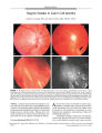
The authors describe one patient with essential thrombocytosis and one with chronic hepatitis C infection who developed bilateral simultaneous anterior ischemic optic neuropathy within 3 months of starting treatment with interferon- a. One patient had several typical risk factors for conventional AION; the other did not. These cases are the fourth and fifth reported examples of this phenomenon. Interferon- a treatment may cause or aggravate the risk of developing anterior ischemic optic neuropathy. Vulnerable patients should be advised of this potential complication, assisted in reducing risk factors, and monitored for optic nerve and retinal vascular complications. ( JNeuro- Ophthalmol 2003; 23: 256- 259) A nterior ischemic optic neuropathy ( AION), a common cause of acute optic neuropathy in the elderly ( 1- 3), is presumed to be a small infarct within the optic nerve head. Many patients have arteriosclerotic risk factors ( 3). We report two patients in whom bilateral AION occurred shortly after onset of treatment with interferon- a for essential thrombocytosis and hepatitis C. Interferon- a is a natural glycoprotein produced and secreted by immune cells in response to viral infections, exerting its effects by binding to a membrane receptor and initiating intracellular signaling that ultimately leads to expression of specific genes. This leads to the enhancement and induction of target cell killing by lymphocytes and inhibition of virus replication in infected cells. Interferon- a has anticytokine, antiviral, immunomodulatory, and antiproliferative activities. It is used to treat cancer, chronic hepatitis C and B, and essential thrombocytosis ( 4). Ophthalmologic complications are rare and include retinal vascular occlusions, retinal hemorrhages, and AION Department of Ophthalmology ( YL, AL, AK), Tel Aviv Medical Center, Sackler School of Medicine, Tel Aviv University, Tel Aviv, Israel; and Department of Ophthalmology ( YV, HG, NM), Haemek Medical Center, Technion School of Medicine, Haifa, Israel. Address correspondence to Anat Kesler, MD, Neuro- ophfhalmology Unit, Department of Ophthalmology, Tel Aviv Medical Center, 6 Weiz-mann Street, Tel Aviv 64239, Israel; E- mail: kesler@ netvision. net. il ( 5- 7). Most reported cases of interferon- a retinopathy are asymptomatic. When treatment with interferon- a is discontinued, the retinopathy usually resolves. Diabetes, hypertension, and anemia are risk factors for the development of interferon- a retinopathy ( 3). Anterior ischemic optic neuropathy is an uncommon complication of interferon- a treatment. Purvin ( 6) reported two cases of sequential AION and Lohmann ( 7) a single case. We describe two cases of AION occurring simultaneously in two eyes shortly after onset of treatment with interferon- a. CASE REPORTS Case 1 A 61- year- old man was admitted due to painless visual loss in the OD. Medical history included insulin-dependent diabetes mellitus, hypertension, ischemic heart disease, and hypercholesterolemia. He also had essential thrombocytosis treated for the past 2 years with anagrelide HC1 ( Agrylin) and hydroxyurea ( Hydrea). Three months before the patient's hospitalization, treatment was changed to 3,000,000 IU of interferon- a four times weekly. Visual acuity was 20/ 100 OD and 20/ 200 OS. An afferent pupillary defect was present in the OS. Fundus examination of the OS revealed a swollen disc and splinter hemorrhages temporal and superior to the optic disc. The right fundus demonstrated flame- shaped hemorrhages temporal to the optic disc ( Fig. 1). Visual field examination disclosed inferonasal nerve fiber bundle defects in both eyes ( Fig. 2). The platelet count was 394,000/ uL. Other blood examination findings were within normal values. Treatment with aspirin 300 mg/ day was initiated. Two months later, the patient developed a sudden further decline in vision in the OD. His best- corrected visual acuities were unchanged, but visual field examination showed progression of the nerve fiber bundle defects in both eyes ( Fig. 3). The erythrocyte sedimentation rate was 20 mm/ h. Orbital ultrasonography and brain computed tomography Copyright © Lippincott Williams & Wilkins. Unauthorized reproduction of this article is prohibited. 256 J Neuro- Ophthalmol, Vol. 23, No. 4, 2003 Interferon- Associated AION JNeuro- Ophthalmol, Vol. 23, No. 4, 2003 FIG. 1. Case 1: Fundi at presentation, showing a swollen optic disc OS and bilateral peripapillary splinter hemorrhages. FIG. 2. Case 1: Visual fields at presentation, showing inferior nerve fiber bundle defects. FIG. 3. Case 1: Visual fields 2 months after presentation, showing progression of nerve fiber bundle defects. findings were within normal limits. Interferon- a treatment was discontinued, as it was presumed to be the cause of these defects. Seven months after the first examination, optic disc pallor supervened and visual fields remained unchanged. Case 2 A 44- year- old man underwent an ophthalmic examination after waking up the same morning with a painless left inferior visual field defect. The patient was a hepatitis C virus ( HCV) carrier, probably as a result of a childhood appendectomy during which he received a blood transfusion. Two and a half months prior to the ophthalmologic examination, he had started a clinical trial of interferon- a polyethylene glycol. The addition of polyethylene glycol to the interferon enables prolongation of the substance's half- life, reduces the number of injections needed per treatment, and thereby improves compliance. Visual acuity was 20/ 30 OD, 20/ 80 OS. There was left relative afferent pupillary defect. Fundus examination of the OD revealed pallid edema of the optic nerve head with splinter hemorrhages and cotton wool spots. In the OS disc, there was optic disc edema, more elevated than in the right, with splinter hemorrhages on the nasal side, and a few cotton wool spots at the edge of the edema ( Fig. 4). Visual field examination demonstrated defects suggestive of inferior nerve fiber bundle defects in both eyes ( Fig. 5), worse in the OS. Neurologic assessment was otherwise normal. Standard blood study results were normal, including erythrocyte sedimentation rate ( 29), white blood cell count ( 3300 with relative lymphopenia [ 16%]), hemoglobin ( 13.5 mg%), prothrombin time, partial thromboplastin time, protein S, protein C, antithrombin III, antiphospholipid Ab, methyltetrahydrofolate reductase, hyperviscosity indices, antinuclear antibody, venereal disease research laboratory titer, and cryoglobulins. Thyroid- stimulating hormone was on the upper edge of normal values; other thyroid function test results were normal. Lumbar puncture had an opening pressure of 80 mm water. Cranial and orbital computed tomography findings were normal. Fluorescein angiography demonstrated late optic disc leakage with no retinal pathology. Interferon- a polyethylene glycol was discontinued. Two days later, the patient noted a black shadow obscuring his right visual field. Visual acuity was 20/ 60 OD, 20/ 80 OS. The relative afferent pupil defect had changed from the left to the OD. Fundus examination in the OD showed enlargement of the optic nerve edema. Visual field demonstrated progression of the field defects in the OD ( Fig. 6). The patient was treated with intravenous methylpred-nisolone 1 g/ d for 3 days, with tapering of prednisone over the next 14 days with no apparent improvement. Over the next 2 months, visual acuities improved to 20/ 30 in both FIG. 4. Case 2: Fundi at presentation, showing swollen discs with splinter hemorrhages. Copyright © Lippincott Williams & Wilkins. Unauthorized reproduction of this article is prohibited. 257 JNeuro- Ophthalmol, Vol. 23, No. 4, 2003 Vardizer et al. FIG. 5. Case 2: Visual fields at presentation, showing inferior nerve fiber bundle defects. eyes without any change in the visual field defects. Three months later, fundus examination demonstrated optic disc pallor in both eyes. DISCUSSION We describe two patients undergoing treatment with interferon- a who developed bilateral simultaneous optic neuropathy within 3 months of starting this medication. Case 1 had established risk factors for conventional AION, including diabetes, hypertension, ischemic heart disease, and hypercholesterolemia. However, we believe that interferon- a treatment may have been a contributing factor, given that conventional AION very rarely develops simultaneously in both eyes ( 8). Furthermore, interferon- a has been implicated in vascular retinopathy ( 5) in patients with diabetes and hypertension. The contribution of the patient's essential thrombocytosis, known to predispose to retinal vascular occlusions, is unknown ( 9- 11). Case 2 had none of the systemic risk factors for conventional AION. We doubt that the HCV alone precipitated AION. In patients with chronic HCV infection, optic neuritis has been reported in only one patient who was not previously treated with interferon- a ( 12). This patient had several signs related to systemic vasculitis, including purpura and cryoglobulinemia. The authors attributed these features to HCV infection. Because our patient did not have systemic symptoms of vasculitis, we had ruled out essential mixed cryoglobulinemia, and because corticosteroids did not reverse the optic neuropathy, it is unlikely that the HCV caused the AION. Alternately, there is evidence that ties AION to interferon- a treatment. Three reported cases of AION ( 6,7) have occurred within 1 week to 3 months of starting interferon- a treatment in patients who lacked typical risk factors for AION. Purvin ( 6) suggested involvement of the posterior ciliary arteries rather than the retinal vessels as a possible cause for AION. Lohmann ( 7) postulated that interferon- a is able to produce autoantibodies and subsequently cause deposition of immune complexes in the small retinal or optic nerve arteries. Interferon- a is also able Copyright © Lippincott Williams & Wilkins. Unai 258 " FIG. 6. Case 2: Visual fields 2 days after presentation, showing progression of nerve fiber bundle defects. to stimulate other cytokines that can cause an inflammatory reaction of the blood vessels that might subsequently lead to ischemia ( 6,7,13). Although Case 2 suffered continued deterioration of vision after discontinuation of interferon- a therapy, this could have been due to the long half- life of the polyethylene glycol variant. The continued deterioration stands in contrast to other patients described, who stabilized or even improved ( 14) after discontinuation of interferon-a treatment. In view of the possible poor visual outcome associated with interferon- a treatment, we suggest that patients who are candidates for this treatment be assessed beforehand for crowded discs and systemic risk factors for conventional AION. It is also important to examine patients during treatment, because some patients who have developed interferon- a- associated retinopathy are asymptomatic, and retinal changes in the reversible phase can be detected. We recommend ophthalmic screening that includes baseline and follow- up examinations every 3 to 4 months ( 15). REFERENCES 1. Hayreh SS, Baines JA. Occlusion of the posterior ciliary artery III: effects on the optic nerve head. Br J Ophthalmol 1972; 56: 754- 64. 2. Hayreh SS. Anterior ischemic optic neuropathy I: terminology and pathogenesis. Br J Ophthalmol 1974; 58: 955- 63. 3. Jacobson DM, Vierkant RA, Belongia EA. Nonarteritic anterior ischemic optic neuropathy: a case control study of potential risk factors. Arch Ophthalmol 1997; 115: 1403- 7. 4. Baron S, Tyring SK, Fleischmann WR Jr, et al. The interferon: mechanism of action and clinical application. JAMA 1991; 266: 1375- 83. 5. Guyer DR, Tiedeman J, Yannuzzi LA, et al. Interferon associated retinopathy. Arch Ophthalmol 1993; 111: 350- 6. 6. Purvin VA. Anterior ischemic optic neuropathy secondary to interferon alfa. Arch Ophthalmol 1995; 113: 1041- 4. 7. Lohmann CP, Kroher G, Bogenrieder T, et al. Severe loss of vision during adjuvant interferon alfa 2b treatment for malignant melanoma. Lancet 1999; 353: 1326. 8. Morrissey SP, Borraut FX, Miller DH, et al. Bilateral simultaneous optic neuropathy in adults: clinical, imaging, serological, and genetic studies. J Neurol Neurosurg Psychiatry 1995; 58: 70- 4. 9. Jabaily J, Hand HJ, Laszlo J, et al. Neurologic manifestations of essential fhrombocyfhemia. Ann Intern Med 1983; 99: 513- 8. orized reproduction of this article is prohibited. © 2003 Lippincott Williams & Wilkins Interferon- Associated AION JNeuro- Ophthalmol, Vol. 23, No. 4, 2003 10. Fenaux P, Simon M, Caulier MT, et al. Clinical course of essential thrombocythemia in 147 cases. Cancer 1990; 66: 549- 56. 11. Yonshizumi MO, To wnesend- Pico W. Essential thrombocythemia and central retinal vein occlusion with neovascular glaucoma. Am J Ophthalmol 1996; 121: 728- 30. 12. Tembl JI, Ferrer JM, Sevilla MT, et al. Neurologic complications associated with hepatitis C virus infection. Neurology 1999; 53: 861- 4. 13. Taylor JL, Grossberg SE. The effects of interferon alpha on the production and action of other cytokines. Semin Oncol 1998; 25( suppl): 23- 9. 14. Gupta R, Singh S. Anterior ischemic optic neuropathy caused by interferon alpha therapy. Am JMed 2002; 112: 683- 4. 15. Neubauer AS, Hoops JP. Interferon- induced retinopathy in asymptomatic cancer patients. Ophthalmol 2002; 109: 821- 2. Copyright © Lippincott Williams & Wilkins. Unauthorized reproduction of this article is prohibited. 259 |