Best known for his world-renowned neuro-ophthalmology unit based at the University of California, San Francisco, William Hoyt, MD collected here more than 850 of his best images covering a wide range of disorders.
William F. Hoyt, MD, Professor Emeritus of Ophthalmology, Neurology and Neurosurgery, Department of Ophthalmology, University of California, San Francisco.
NOVEL: https://novel.utah.edu/
TO
Filters: Collection: ehsl_novel_wfh
1 - 25 of 4
| Title | Description | Type | ||
|---|---|---|---|---|
| 1 |
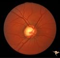 |
Sturge Weber Syndrome (Encephalotrigeminal Angiomatosis) | Sturge Weber Syndrome (Encephalotrigeminal angiomatosis); Color of the retina is deep red (sometimes called tomato catsup) due to a four fold thickening of the choroidal vascular bed. Optic disc is cupped due to elevated intraocular pressure. (Secondary glaucoma) Patient had a major ""port wine"" m... | Image |
| 2 |
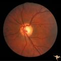 |
Sturge Weber Syndrome (Encephalotrigeminal Angiomatosis) | Left eye is normal, without the deep red from thickened Choroid. Pair with R1_B1a. Anatomy: Optic disc. Pathology: Diffuse choroidal hemangioma; Glaucoma. Disease/Diagnosis: Sturge Weber Syndrome. Clinical: Port wine hemangioma of the face. | Image |
| 3 |
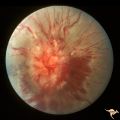 |
Sturge Weber Syndrome (Encephalotrigeminal Angiomatosis) | Sturge Weber Syndrome (Encephalotrigeminal angiomatosis) with retinal evidence of central retinal vein occlusion. Anatomy: Retina. Pathology: Diffuse choroidal hemangioma; Glaucoma. Disease/Diagnosis: Sturge Weber Syndrome. Clinical: Port wine hemangioma of the face. | Image |
| 4 |
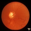 |
Sturge Weber Syndrome (Encephalotrigeminal Angiomatosis) | Sturge Weber Syndrome (Encephalotrigeminal angiomatosis); Color of the retina is deep red (sometimes called tomato catsup) due to a four fold thickening of the choroidal vascular bed. Glaucomatous cupping of the optic nerve. Striking retinal venous vascular anomalies on the disc and in the retina. ... | Image |
1 - 25 of 4