Best known for his world-renowned neuro-ophthalmology unit based at the University of California, San Francisco, William Hoyt, MD collected here more than 850 of his best images covering a wide range of disorders.
William F. Hoyt, MD, Professor Emeritus of Ophthalmology, Neurology and Neurosurgery, Department of Ophthalmology, University of California, San Francisco.
NOVEL: https://novel.utah.edu/
TO
Filters: Collection: ehsl_novel_wfh
1 - 25 of 9
| Title | Description | Type | ||
|---|---|---|---|---|
| 1 |
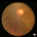 |
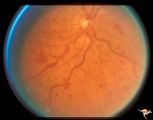
Slow Flow (Chronic Hypoxic) Retinopathy | Examples of Slow flow (chronic hypoxic) retinopathy showing produced by a carotid-cavernous sinus fistula. Arteriole pressure was low in the retina and venous pressure was elevated. Note the characteristic dot and blot hemorrhages in the black and white photo (R3B2b). Anatomy: Retina. Pathology: Car... | Image |
| 2 |
 |
Slow Flow (Chronic Hypoxic) Retinopathy | Examples of Slow flow (chronic hypoxic) retinopathy produced by a carotid-cavernous sinus fistula. Arteriole pressure was low in the retina and venous pressure was elevated. Note the characteristic dot and blot hemorrhages in this black and white photo. Anatomy: Retina. Pathology: carotid-cavernous ... | Image |
| 3 |
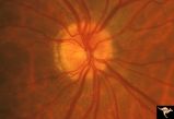 |
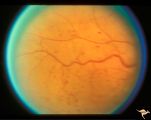
Slow Flow (Chronic Hypoxic) Retinopathy | Slow flow (chronic hypoxic) retinopathy. Optic disc change in left eye (b) secondary to reduced carotid artery perfusion. Patient was an elderly man with a innominant artery occlusion. Note the reduced arteriole caliber in the left disc (b) compared to the right (a). Central retinal artery pressure ... | Image |
| 4 |
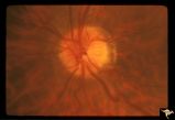 |
Slow Flow (Chronic Hypoxic) Retinopathy | Slow flow (chronic hypoxic) retinopathy. Optic disc change in left eye (b) secondary to reduced carotid artery perfusion. Patient was an elderly man with a innominant artery occlusion. Note the reduced arteriole caliber in the left disc (b) compared to the right (a). Central retinal artery pressure ... | Image |
| 5 |
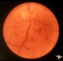 |
Slow Flow (Chronic Hypoxic) Retinopathy | Slow flow (chronic hypoxic) retinopathy from macroglobulanemia. Note the dot and blot hemorrhages. Anatomy: Retina. Pathology: Macroglobulanemia. Disease/Diagnosis: Slow flow (chronic hypoxic) retinopathy from macroglobulanemia. | Image |
| 6 |
 |
Slow Flow (Chronic Hypoxic) Retinopathy | Slow flow (chronic hypoxic) retinopathy (right eye) in a man with polycythaemia rubra vera. Hematological disease. Anatomy: Retina. Pathology: Hematological disease. Disease/Diagnosis: Slow flow (chronic hypoxic) retinopathy secondary to polycythaemia. Clinical: No visual symptoms. | Image |
| 7 |
 |
Slow Flow (Chronic Hypoxic) Retinopathy | Slow flow (chronic hypoxic) retinopathy (right eye) in a man with polycythaemia rubra vera. Hematological disease. Anatomy: Retina. Pathology: Hematological disease. Disease/Diagnosis: Slow flow (chronic hypoxic) retinopathy secondary to polycythaemia. Clinical: No visual symptoms. | Image |
| 8 |
 |
Slow Flow (Chronic Hypoxic) Retinopathy | Examples of Slow flow (chronic hypoxic) retinopathy showing dilated and tortuous retinal veins and multiple capillary hemorrhages. This kind of retinopathy is produced by impaired arteriole circulation to the retina from various causes. Anatomy: Retina. Pathology: Ophthalmic artery venous malformati... | Image |
| 9 |
 |
Slow Flow (Chronic Hypoxic) Retinopathy | Examples of Slow flow (chronic hypoxic) retinopathy showing dilated and tortuous retinal veins and multiple capillary hemorrhages. This kind of retinopathy is produced by impaired arteriole circulation to the retina from various causes. Anatomy: Retina. Pathology: Ophthalmic artery venous malformati... | Image |
1 - 25 of 9