Best known for his world-renowned neuro-ophthalmology unit based at the University of California, San Francisco, William Hoyt, MD collected here more than 850 of his best images covering a wide range of disorders.
William F. Hoyt, MD, Professor Emeritus of Ophthalmology, Neurology and Neurosurgery, Department of Ophthalmology, University of California, San Francisco.
NOVEL: https://novel.utah.edu/
TO
Filters: Collection: ehsl_novel_wfh
1 - 25 of 16
| Title | Description | Type | ||
|---|---|---|---|---|
| 1 |
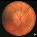 |
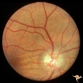
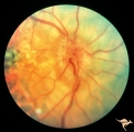
D101 Disc Edema with Systemic Lupus | Unilateral disc swelling with narrowed arterioles. No decrease in visual acuity or field. 19 year old woman. Patient died of cerebral lupus within two months. Optociliary veins dumping into disc edge at 4:00, 9:00, and 11:00. Anatomy: Optic disc. Pathology: Axoplasmic stasis due to vasculitis (Lupu... | Image |
| 2 |
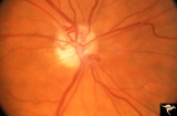 |
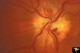
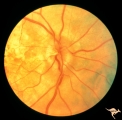
IA01 Atrophy with Optociliary Veins | 1994, perioptic nerve sheath meningioma, right eye, Optociliary vein dumping into disc edge at 4:00. Anatomy: Optic disc. Pathology: Optociliary vein. Disease/ Diagnosis: Perioptic nerve sheath meningioma. Clinical: Progressive visual loss | Image |
| 3 |
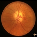 |
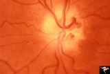
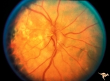
IA02 Atrophy with Optociliary Veins | 1971, left eye, perioptic nerve sheath meningioma, notice how vein dumps into adjacent choroid at 3:00. The darker venous blood can be seen at the disc edge. Anatomy: Optic disc. Pathology: Optociliary vein. Disease/ Diagnosis: Perioptic nerve sheath meningioma. Clinical: Progressive visual loss. | Image |
| 4 |
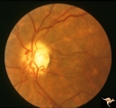 |
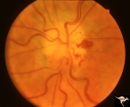
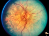
IA03 Atrophy with Optociliary Veins | 1974, left eye, perioptic nerve sheath meningioma, blind eye. Optociliary bypass veins in the nasal disc tissue. Anatomy: Optic disc. Pathology: Optociliary vein. DIsease/ Diagnosis: Perioptic nerve sheath meningioma. Clinical: Blind eye. | Image |
| 5 |
 |
IA04 Atrophy with Optociliary Veins | 1981, right eye, perioptic nerve sheath meningioma with optociliary bypass vein. Notice horizontal choroidal folds in the retina from posterior tumor pressure. Anatomy: Optic disc. Pathology: Optociliary vein. Disease/ Diagnosis: Perioptic nerve sheath meningioma. Clinical: Blind eye. | Image |
| 6 |
 |
IA05 Atrophy with Optociliary Veins | 1971, right eye, perioptic nerve sheath meningioma with optociliary bypass veins on the upper half of the disc. Anatomy: Optic disc. Pathology: Optociliary vein. Disease/ Diagnosis: Perioptic nerve sheath meningioma. Clinical: Blind eye. | Image |
| 7 |
 |
IA06 Atrophy with Optociliary Veins | 1979, left eye, perioptic nerve sheath meningioma with optociliary bypass veins. Anatomy: Optic disc. Pathology: Optociliary vein. Disease/ Diagnosis: Perioptic nerve sheath meningioma. Clinical: Blind eye. | Image |
| 8 |
 |
IA07 Atrophy with Optociliary Veins | Left eye, perioptic nerve sheath meningioma. Anatomy: Optic disc. Pathology: Optociliary vein. Disease/ Diagnosis: Perioptic nerve sheath meningioma. Clinical: Visual loss. | Image |
| 9 |
 |
IA08 Atrophy with Optociliary Veins | 1996, left eye. Chronic pale optic nerve swelling with optociliary bypass veins produced by perioptic nerve sheath meningioma. Anatomy: Optic disc. Pathology: Optociliary vein. Disease/ Diagnosis: Perioptic nerve sheath meningioma evolution. Clinical: Visual loss. | Image |
| 10 |
 |
IA09a Evolution of Optociliary Veins with Perioptic Nerve Sheath Meningioma | April 1975, Normal eye, macular degeneration. Anatomy: Optic disc. Pathology: Optociliary vein. Disease/ Diagnosis: Perioptic nerve sheath meningioma evolution. Clinical: Visual loss. | Image |
| 11 |
 |
IA09b Evolution of Optociliary Veins with Perioptic Nerve Sheath Meningioma | January 1977, macular degeneration, disc swelling begins. Anatomy: Optic disc. Pathology: Optociliary vein. Disease/ Diagnosis: Perioptic nerve sheath meningioma evolution. Clinical: Visual loss. | Image |
| 12 |
 |
IA09c Evolution of Optociliary Veins with Perioptic Nerve Sheath Meningioma | June 1977, continued disc swelling. Anatomy: Optic disc. Pathology: Optociliary vein. Disease/ Diagnosis: Perioptic nerve sheath meningioma evolution. Clinical: Visual loss. | Image |
| 13 |
 |
IA09d Evolution of Optociliary Veins with Perioptic Nerve Sheath Meningioma | October 1977, continued disc swelling. Anatomy: Optic disc. Pathology: Optociliary vein. Disease/ Diagnosis: Perioptic nerve sheath meningioma evolution. Clinical: Visual loss. | Image |
| 14 |
 |
IA09e Evolution of Optociliary Veins with Perioptic Nerve Sheath Meningioma | February 1979, development of optociliary veins at 7:00, 1:00. Anatomy: Optic disc. Pathology: Optociliary vein. Disease/ Diagnosis: Perioptic nerve sheath meningioma evolution. Clinical: Visual loss. | Image |
| 15 |
 |
IA09f Evolution of Optociliary Veins with Perioptic Nerve Sheath Meningioma | August 1979, less disc swelling and development of atrophy with more prominent optociliary veins at 7:00 and 1:00. Anatomy: Optic disc. Pathology: Optociliary vein. Disease/ Diagnosis: Perioptic nerve sheath meningioma evolution. Clinical: Visual loss. | Image |
| 16 |
 |
IA09g Evolution of Optociliary Veins with Perioptic Nerve Sheath Meningioma | April 1980, prominent atrophy and increased numbers of optociliary veins. Anatomy: Optic disc. Pathology: Optociliary vein. Disease/ Diagnosis: Perioptic nerve sheath meningioma evolution. Clinical: Visual loss. | Image |
1 - 25 of 16