Best known for his world-renowned neuro-ophthalmology unit based at the University of California, San Francisco, William Hoyt, MD collected here more than 850 of his best images covering a wide range of disorders.
William F. Hoyt, MD, Professor Emeritus of Ophthalmology, Neurology and Neurosurgery, Department of Ophthalmology, University of California, San Francisco.
NOVEL: https://novel.utah.edu/
TO
Filters: Collection: ehsl_novel_wfh
1 - 25 of 7
| Title | Description | Type | ||
|---|---|---|---|---|
| 1 |
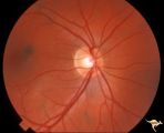 |
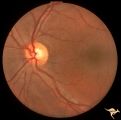
Neurofibromatosis-1 | Normal appearing optic disc with dark pigmented choroidal nevi. The patient had NF-1 and had a subclinical optic glioma on the left eye. This is the right eye. Anatomy: Optic disc. Pathology: Choroidal nevus. Disease/Diagnosis: Neurofibromatosis type 1. Clinical: No visual symptoms. | Image |
| 2 |
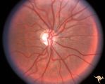 |
Neurofibromatosis-1 | Optic atrophy and hypoplasia of the optic disc associated with chiasmal glioma in a patient with NF-1. Anatomy: Optic disc. Pathology: Chiasmal glioma; Optic atrophy; Hypoplasia. Disease/Diagnosis: Neurofibromatosis type 1. Clinical: Proptosis; Blindness. | Image |
| 3 |
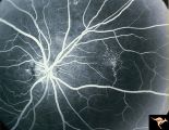 |
Neurofibromatosis-1 | Fluorescein angiogram defines the extent of the microvascular malformation. Pair with R1_E5b. (Ref: BJO 2002:86, p282-284). Anatomy: Retina. Pathology: Retinal microvascular malformations. Disease/Diagnosis: Neurofibromatosis type 1. Clinical: No visual symptoms. Imaging: Fluorescein angiogram. | Image |
| 4 |
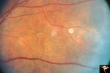 |
Neurofibromatosis-1 | Retinal microvascular malformations in NF-1. Fundus picture shows a somewhat larger vertically running corkscrew malformation between two temporal retinal veins. Pair with R1_E5a. Anatomy: Retina. Pathology: Retinal microvascular malformations. Disease/Diagnosis: Neurofibromatosis type 1. Clinical: ... | Image |
| 5 |
 |
Neurofibromatosis-1 | Retinal microvascular malformations in NF-1 located between the disc and the macula. Anatomy: Retina. Pathology: Retinal microvascular malformations. Disease/Diagnosis: Neurofibromatosis type 1. Clinical: No visual symptoms. | Image |
| 6 |
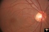 |
Neurofibromatosis-1 | Retinal microvascular malformations between optic disc and macula in NF-1. Anatomy: Retina. Pathology: Retinal microvascular malformations. Disease/Diagnosis: Neurofibromatosis type 1. Clinical: No visual symptoms. | Image |
| 7 |
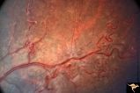 |
Neurofibromatosis-1 | Extensive retinal microvascular malformation involving both small and large retinal vessels. (Ref: BJO 2002:86, p282-284). Anatomy: Retina. Pathology: Retinal microvascular malformations. Disease/Diagnosis: Neurofibromatosis type 1. Clinical: No visual symptoms. | Image |
1 - 25 of 7