Best known for his world-renowned neuro-ophthalmology unit based at the University of California, San Francisco, William Hoyt, MD collected here more than 850 of his best images covering a wide range of disorders.
William F. Hoyt, MD, Professor Emeritus of Ophthalmology, Neurology and Neurosurgery, Department of Ophthalmology, University of California, San Francisco.
NOVEL: https://novel.utah.edu/
TO
Filters: Collection: ehsl_novel_wfh
1 - 25 of 8
| Title | Description | Type | ||
|---|---|---|---|---|
| 1 |
 |
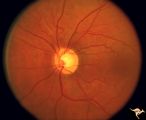
Cerebroretinal Microangiopathy (Susac Syndrome) | Retinal signs of Susac's Syndrome in acute phase consist of areas of retinal artery infarction from branch retinal artery occlusions. These patients are usually women, many of whom are demented and have hearing loss. Refs: 1) Susac, Hardiman, Sellhorst. Neurology. 1979. 29:313-316 2) Susac ""Susa... | Image |
| 2 |
 |
Cerebroretinal Microangiopathy (Susac Syndrome) | This fundus picture from a patient with Susac Syndrome shows a focal shiny plaque in the inferior retinal arteriole. This plaque is not the result of embolism, but is the result of the microangioplastic process underlying the syndrome. (NANOS 2001 by Egan, RA). Anatomy: Retina. Pathology: Microangi... | Image |
| 3 |
 |
Cerebroretinal Microangiopathy (Susac Syndrome) | Retinal signs of Susac's Syndrome in acute phase consist of areas of retinal artery infarction from branch retinal artery occlusions. Shows clearing retinal branch artery occlusion. Pathology: Retina. Pathology: Microangiopathy involving brain, auditory nerve and retina. Disease/Diagnosis: Cerebro... | Image |
| 4 |
 |
Cerebroretinal Microangiopathy (Susac Syndrome) | Retinal signs of Susac's Syndrome in acute phase consist of areas of retinal artery infarction from branch retinal artery occlusions. Branch artery occlusion beginning to clear. Note the occluded arteriole lying on top of the infarcted zone. Anatomy: Retina. Pathology: Microangiopathy involving br... | Image |
| 5 |
 |
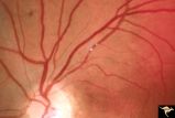
Cerebroretinal Microangiopathy (Susac Syndrome) | There is a plaque superior to the disc at 12:00. This plaque is not the result of embolism, but is the result of the microangioplastic process underlying the syndrome. There is a ghost vessel inferiorly at 5:00 off the disc. (NANOS 2001 by Egan, RA). Anatomy: Retina. Pathology: Microangiopathy invo... | Image |
| 6 |
 |
Cerebroretinal Microangiopathy (Susac Syndrome) | There is an occlusion of the superior nasal retinal arteriole visible as a white ghost vessel at 11:00. Note: Collateral filling of the distal branches in two places. (NANOS 2001 by Egan, RA). Anatomy: Retina. Pathology: Microangiopathy involving brain, auditory nerve and retina. Disease/Diagnosis:... | Image |
| 7 |
 |
Cerebroretinal Microangiopathy (Susac Syndrome) | Two plaques which have been called Psuedo-emboli. This plaque is not the result of embolism, but is the result of the microangioplastic process underlying the syndrome. (NANOS 2001 by Egan, RA). Anatomy: Retina. Pathology: Microangiopathy involving brain, auditory nerve and retina. Disease/Diagnosi... | Image |
| 8 |
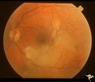 |
Cerebroretinal Microangiopathy (Susac Syndrome) | Retinal signs of Susac's Syndrome in acute phase consist of areas of retinal artery infarction from branch retinal artery occlusions. This fundus shows two area of retinal infarction from occlusion of both superior and inferior branch retinal arterioles. Anatomy: Retina. Pathology: Microangiopathy... | Image |
1 - 25 of 8