Best known for his world-renowned neuro-ophthalmology unit based at the University of California, San Francisco, William Hoyt, MD collected here more than 850 of his best images covering a wide range of disorders.
William F. Hoyt, MD, Professor Emeritus of Ophthalmology, Neurology and Neurosurgery, Department of Ophthalmology, University of California, San Francisco.
NOVEL: https://novel.utah.edu/
Filters: Collection: ehsl_novel_wfh
1 - 25 of 8
| Title | Description | Type | ||
|---|---|---|---|---|
| 1 |
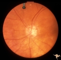 |
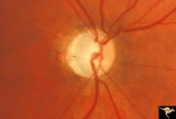
IC102a Central Retinal Artery Occlusion with Cilioretinal Collaterals | Left eye, 1988, Central retinal artery with cilioretinal collaterals due to calcific embolic behind the lamina cribrosa due to calcific valvular heart disease. Collaterals have been called "Nettleship Collaterals", recognizing the British physician who first described them in 1892. Anatomy: Optic di... | Image |
| 2 |
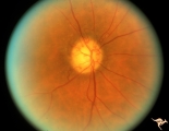 |
IC102b Central Retinal Artery Occlusion with Cilioretinal Collaterals | Right eye, 1991, Central retinal artery occlusion with cilioretinal collateral occlusions due to calcific embolic occlusion behind the lamina cribrosa due to calcific valvular heart disease. Collaterals have been called "Nettleship Collaterals", recognizing the British physician who first described ... | Image |
| 3 |
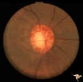 |
IC102c Central Retinal Artery Occlusion with Cilioretinal Collaterals | Right eye, 1982, Central retinal artery occlusion with cilioretinal collateral occlusions due to calcific embolic occlusion behind the lamina cribrosa due to calcific valvular heart disease. Collaterals have been called "Nettleship Collaterals", recognizing the British physician who first described ... | Image |
| 4 |
 |
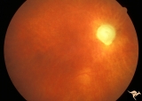
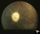
IC103a Central Retinal Artery Occlusion with Choroidal Arteriolar Occlusion | Central retinal artery occlusion and choroidal vascular occlusion due to pressure on the eyeball during craniotomy. Note total loss of vascularity of the optic disc and surrounding choroid. Anatomy: Optic disc. Pathology: Combined central retinal and choroidal arteriolar occlusion. Disease/ Diagnos... | Image |
| 5 |
 |
IC103b Central Retinal Artery Occlusion with Choroidal Arteriole Occlusion | 1980, Evidence of choroidal vascular ischemia. Central retinal artery occlusion and choroidal vascular occlusion from amputation of the optic nerve for meningioma. Anatomy: Optic disc. Pathology: Optic glioma and optic nerve was amputated during excision of the tumor. Disease/ Diagnosis: Optic nerve... | Image |
| 6 |
 |
IC103c Central Retinal Artery Occlusion with Choroidal Arteriole Occlusion | 1988, Central retinal artery occlusion and choroidal vascular occlusion, 70 year old woman with history of central retinal artery occlusion 30 years prior. Anatomy: Optic disc. Pathology: Combined central retinal and choroidal arteriolar occlusion. Disease/ Diagnosis: Combined central retinal and ch... | Image |
| 7 |
 |
IC103d Central Retinal Artery Occlusion with Choroidal Arteriole Occlusion | 1969, Complete loss of blood supply to retina and choroid. Cause unknown. Boy. Anatomy: Optic disc. Pathology: Toxic ischemic retinal damage 22a. Clinical: Blindness. | Image |
| 8 |
 |
IC101 Old Central Retinal Artery Occlusion | Old central retinal artery occlusion without additional retinovascular signs, 1969. Anatomy: Optic disc. Pathology: Central retinal artery occlusion. Disease/ Diagnosis: Central retinal artery occlusion. Clinical: Sudden blindness. | Image |
1 - 25 of 8