Collection of materials relating to neuro-ophthalmology as part of the Neuro-Ophthalmology Virtual Education Library.
NOVEL: https://novel.utah.edu/
TO
- NOVEL720
| Title | Creator | Description | Subject | ||
|---|---|---|---|---|---|
| 201 |
 |
Histiocytosis (PowerPoint) | AAO/NANOS - American Academy of Ophthalmology / North American Neuro-Ophthalmology Society | This 1-year-old child with familial erythrophagocytic lymphohistiocytosis was readmitted with a fever and was noted to have bilateral blindness. The spinal tap showed a protein of 148, with 178 WBC with 98% ""lymphocytes."" This image demonstrates the optic nerve infiltration. He was treated with ra... | Optic Nerve Histiocytosis; Histiocytosis |
| 202 |
 |
Orbital Tumors - Choroidal Folds From Orbital Mass (PowerPoint) | AAO/NANOS - American Academy of Ophthalmology / North American Neuro-Ophthalmology Society | This 30-year-old man had a retrobulbar intraconal mass OS. The CT scans showed a heterogeneous lobulated enhancing mass, 2.2 x 1.9 x 1.8 cm. The case beautifully exhibits chorodial folds. The ultrasound showed internal reflectivity. The patient refused surgery. | Choroidal Folds from Orbital Mass |
| 203 |
 |
Moyamoya Disease (PowerPoint) | AAO/NANOS - American Academy of Ophthalmology / North American Neuro-Ophthalmology Society | This 32-year-old woman was referred with a history of 4 days of loss of vision OD. She had a history of manic depressive illness and IV drug abuse; she had been HIV tested 4 weeks before and was negative. She said she last injected cocaine 5 days before being seen, the night before she awoke with th... | Saturday Night Retinopathy; Moyamoya Disease |
| 204 |
 |
Saturday Night Retinopathy (PowerPoint) | AAO/NANOS - American Academy of Ophthalmology / North American Neuro-Ophthalmology Society | This 32-year-old woman was referred with a history of 4 days of loss of vision OD. She had a history of manic depressive illness and IV drug abuse; she had been HIV tested 4 weeks before and was negative. She said she last injected cocaine 5 days before being seen, the night before she awoke with th... | Saturday Night Retinopathy |
| 205 |
 |
Moyamoya Syndrome (PowerPoint) | AAO/NANOS - American Academy of Ophthalmology / North American Neuro-Ophthalmology Society | A 9-year-old boy had recurrent ischemic episodes that had begun 2 years prior to evaluation. A significant right hemiparesis and a significant speech, learning, and memory disorder were present. His noncontrast axial view CT scan demonstrated multiple cerebral infarcts. Cerebral angiography revealed... | Moyamoya Disease; Moyamoya Syndrome |
| 206 |
 |
Periphlebitis in Optic Neuritis (PowerPoint) | AAO/NANOS - American Academy of Ophthalmology / North American Neuro-Ophthalmology Society | This 35-year-old otherwise-healthy woman developed typical optic neuritis OD with excellent recovery. She had no clinical evidence of multiple sclerosis at that time. She presented in August of 1991, at which time perivenous sheathing was seen in the retinal periphery OU. A limited workup was negati... | Periphlebitis in Optic Neuritis |
| 207 |
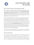 |
Hereditary Optic Neuropathy (Leber's Hereditary Optic Neuropathy) | NANOS | Hereditary Optic Neuropathy - A hereditary optic neuropathy is caused by a genetic variant (or mutation) that causes dysfunction of the neurons (nerve cells) which form the optic nerve. The optic nerve sends information from the back of the eye to the vision center in the brain.The two most common t... | Hereditary Optic Neuropathy; Patient Brochure |
| 208 |
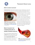 |
Transient Vision Loss | NANOS | Vision loss that is temporary (transient) is a common problem and has many potential causes.Patients with temporary vision loss often do not have any abnormalities on their eye examination, especially once the vision has returned to normal. | Transient Vision Loss; Patient Brochure |
| 209 |
 |
NExT Introduction | Sachin Kedar, MD, Editor-in-Chief | Transcript of video introduction to the NExT curriculum collection. | NANOS Examination Techniques |
| 210 |
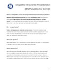 |
Idiopathic Intracranial Hypertension | NANOS | Idiopathic intracranial hypertension (IIH), also called pseudotumor cerebri, is a condition in which there is high pressure in the fluid surrounding your brain, spinal cord, and optic nerves. This can cause headaches and problems with vision. | Idiopathic Intracranial Hypertension; Patient Brochure |
| 211 |
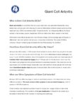 |
Giant Cell Arteritis | NANOS | Giant cell arteritis is a condition that can cause vision loss, new persistent headaches, scalp tenderness, and jaw pain with chewing. It is due to inflammation of blood vessels primarily of the head and neck. | Giant Cell Arteritis; Patient Brochure |
| 212 |
 |
Thyroid Eye Disease | NANOS | Thyroid eye disease, also called dysthyroid orbitopathy, is an autoimmune condition in whichyour body's immune system triggers inflammation in the eye socket (also called the orbit),affecting the muscles that move the eye and the fatty tissue behind the eye. | Thyroid Eye Disease; Thyroid Orbitopathy; Patient Brochure |
| 213 |
 |
Metastatic Glioblastoma to Intracranial Optic Nerves, Optic Chiasm and Optic Tracts | Bashaer Aldhahwani, MD; Mariam S. Vilá-Delgado, MD | The patient with pathology confirmed glioblastoma after resectioning the superior frontal lobe tumor followed by 6 weeks of radiation therapy with concurrent temozolomide. The patient started bevacizumab to treat steroid-refractory vasogenic cerebral edema/radiation necrosis. 8 months after radiatio... | Metastatic Glioblastoma; Infiltrative Chiasmal Lesion |
| 214 |
 |
Lessons From Bench Bedside | Shirley H. Wray, MD, PhD, FRCP | See also: http://content.lib.utah.edu/cdm/ref/collection/ehsl-shw/id/69, http://content.lib.utah.edu/cdm/ref/collection/ehsl-shw/id/282, http://content.lib.utah.edu/cdm/ref/collection/ehsl-shw/id/94, and http://content.lib.utah.edu/cdm/ref/collection/ehsl-shw/id/103 | Bilateral Internuclear Ophthalmoplegia; Pendular Horizontal Oscillations; Lid Nystagmus; Upbeat Nystagmus; Botulinum Toxin Therapy; Multiple Sclerosis; Horizontal Pendular Nystagmus; Gaze Evoked Upbeat Nystagmus; Abducting Nystagmus; Normal Convergence; Gaze Evoked Downbeat Nystagmus; Sac... |
| 215 |
 |
Optochiasmal Tuberculoma | Jeanie Paik, MD; Rudrani Banik, MD | PowerPoint of case of chiasmal tuberculoma causing bitemporal defect in patient with tuberculosis on RIPE treatment; case history, differential diagnosis and treatment discussed. | Chiasmal Disorder; Chiasmal Tuberculoma; Bitemporal Visual Field Defect; Ethambutol Optic Neuropathy |
| 216 |
 |
Unilateral Oculomotor Nerve Palsy Secondary to Internal Carotid Artery Aneurysm Without Pupil involvement: A Case Report | Danilo Andriatti Paulo; Richard J Blanch | Acquired oculomotor palsies (OMP) can result from numerous factors. The most common causes are presumed microvascular, trauma, compressive neoplasm, postneurosurgery and compression from aneurysm.1,2 ONP caused by internal carotid artery (ICA) aneurysm is a common clinical manifestation suggesting i... | Unilateral Oculomotor Nerve Palsy; Internal Carotid Artery Aneurysm; Pupil Involvement; Oculomotor Nerve Palsy; Secondary Oculomotor Nerve Palsy |
| 217 |
 |
Acute Multifocal Pigment Epithelium Epitheliopathy (AMPEE) | Gregory P. Van Stavern, MD | Images providing example of Acute Multifocal Pigment Epithelium Epitheliopathy (AMPEE) | Acute Multifocal Pigment Epithelium Epitheliopathy (AMPEE) |
| 218 |
 |
Acquired Hyperopia | AAO/NANOS - American Academy of Ophthalmology / North American Neuro-Ophthalmology Society | Choroidal folds may result from choroidal tumors, compression on the eye wall from thyroid ophthalmopathy, orbital pseudotumor, orbital tumor, posterior scleritis, hypotony, scleral laceration, retinal detachment, marked hyperopia, or secondary to papilledema. Intraocular pressure measurements, refr... | Acquired Hyperopia |
| 219 |
 |
Amyotrophic Lateral Sclerosis (Guest Lecture) | John Q. Trojanowski, MD | The patient is a 68 year old right handed retired air conditioner repair man who presented with impaired balance and slow walking. For about one year he had noted difficulty lifting his feet high enough when climbing the stairs. From that time on, his movements slowed and worsened so that he had dif... | Saccadic Initiation Deficit of Unilateral Horizontal Gaze; Complete Paralysis of Voluntary Horizontal Saccades on Command to Look Left; Inability to Make a Refixation Saccade on Command to a Target Held on the Left; Normal Voluntary Horizontal Saccadic Eye Movements to the Right; Impaired Pursuit; F... |
| 220 |
 |
Emianopsia Omonima (Italian) | North American Neuro-Ophthalmology Society | This refers to an absence of vision towards one side of the visual world in each eye. The damage that caused this problem is in the brain and not in the eyes. | Homonymous Hemianopsia; Patient Brochure |
| 221 |
 |
Fat Emboli | Kathleen B. Digre, MD; James J. Corbett, MD | Slideshow describing condition. | Emboli |
| 222 |
 |
Fluorescein Angiography | Kathleen B. Digre, MD; James J. Corbett, MD | Fluorescein angiography in neuro-ophthalmology. | Fluorescein Angiography; History |
| 223 |
 |
Fibrin-Platelet Emboli | Kathleen B. Digre, MD; James J. Corbett, MD | Slideshow describing condition. | Emboli; Platelet Emboli |
| 224 |
 |
Giant Cell Arteritis: Diagnostic Prediction Models, Temporal Artery Biopsy and Epidemiology | Edsel Ing MD, PhD FRCSC MPH CPH MIAD MEd MBA, | Giant cell arteritis (GCA) is the most common primary vasculitis in the elderly and can cause irreversible blindness, aortitis, and stroke. Diagnostic confirmation of GCA usually entails temporal artery biopsy (TABx) - a time-consuming and invasive test, or ultrasound. The primary treatment of GCA i... | Giant Cell Arteritis; Diagnostic Prediction Model; Epidemiology; Temporal Artery Biopsy; Differential Diagnosis |
| 225 |
 |
Histoplasmosis | Gregory P. Van Stavern, MD | Histoplasmosis, a fungus, can present acutely as a systemic condition. This image shows signs of Histoplasmosis. | Histoplasmosis |