Collection of materials relating to neuro-ophthalmology as part of the Neuro-Ophthalmology Virtual Education Library.
NOVEL: https://novel.utah.edu/
TO
- NOVEL966
Filters: Collection: "ehsl_novel_novel"
| Title | Creator | Description | Subject | ||
|---|---|---|---|---|---|
| 201 |
 |
Convergence Insufficiency | Shirley H. Wray, MD, PhD, FRCP | The patient is a 73 year old man with a ten year history of idiopathic Parkinson's disease characterized by difficulty in walking, generalized rigidity and a mild tremor of his hands at rest with deterioration in his handwriting. He denied any memory impairment or loss of cognitive function. He was ... | Basal Ganglia; Blepharoclonus; Convergence Insufficiency; Slow Hypometric Saccades; Saccadic Breakdown of Smooth Pursuit; Parkinson's Disease- Dopamine deficiency; Slow Hypometric Horizontal Saccades; Convergence |
| 202 |
 |
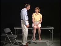
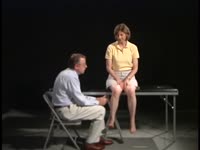
Convergence-Retraction Nystagmus in Dorsal-Midbrain Syndrome | Paul Freund, MD, FRCSC; Edward Margolin, MD, FRCSC | A man in his early twenties was referred by optometrist for abnormal eye motility findings. He had a remote history of an excised pinealoma. On exam he had almost complete upgaze palsy, convergence-retraction nystagmus on attempted upgaze, and light-near dissociation of pupillary reaction, the class... | Dorsal Midbrain Syndrome; Parinaud Syndrome; Convergence-Retraction Nystagmus; Light-Near Dissociation |
| 203 |
 |
Coordination Exam: Abnormal Examples: Finger-to-nose (x2) (includes Spanish audio & captions) | Paul D. Larsen, MD | The patient places her heel on the opposite knee then runs the heel down the shin to the ankle and back to the knee in a smooth coordinated fashion. NeuroLogic Exam has been supported by a grant from the Slice of Life Development Fund at the University of Utah, the Department of Pediatrics and the O... | Coordination Examination; Finger-to-nose Test |
| 204 |
 |
Coordination Exam: Abnormal Examples: Heel-to-shin (x2) (includes Spanish audio & captions) | Paul D. Larsen, MD | The patient with ataxia of the lower extremity will have difficulty placing the heel on the knee with a side-to-side irregular over- and undershooting as the heel is advanced down the shin. Dysmetria on heel-to-shin can be seen in midline ataxia syndromes as well as cerebellar hemisphere disease so ... | Coordination Examination; Heel-shin Test |
| 205 |
 |
Coordination Exam: Normal Exam: Finger-to-nose (includes Spanish audio & captions) | Paul D. Larsen, MD | The patient moves her pointer finger from her nose to the examiner's finger as the examiner moves his finger to new positions and tests accuracy at the furthest outreach of the arm. NeuroLogic Exam has been supported by a grant from the Slice of Life Development Fund at the University of Utah, the D... | Coordination Examination; Finger-to-nose Test |
| 206 |
 |
Coordination Exam: Normal Exam: Heel-to-shin (includes Spanish audio & captions) | Paul D. Larsen, MD | The patient places her heel on the opposite knee then runs the heel down the shin to the ankle and back to the knee in a smooth coordinated fashion. NeuroLogic Exam has been supported by a grant from the Slice of Life Development Fund at the University of Utah, the Department of Pediatrics and the O... | Coordination Examination; Heel-shin Test |
| 207 |
 |
Corectopia | Meagan Seay, DO | These are photos of a patient with unilateral corectopia. This patient's corectopia is of unclear etiology and possibly related to birth trauma. | Corectopia; Unilateral; Photos |
| 208 |
 |
Corneal Staining | Sovik De Sirkar, MSIII; Ore-ofe Adesina, MD | Description of the corneal staining technique. | Corneal Staining |
| 209 |
 |
Cortical Localization: Neuroanatomy Video Lab - Brain Dissections | Suzanne S. Stensaas, PhD | The lobes of the brain are defined together with their major functions. The visual field representation in the occipital lobe is explained with a diagram. Speech areas and the major types of aphasia are discussed in the dominant hemisphere and parietal lesions of neglect and spatial orientation are ... | Cortical Localization; Brain; Dissections |
| 210 |
 |
Cotton Wool Spots: The Basics | Arnav Gupta, BHSc; Rahul Sharma, MD, MPH | A presentation describing cotton wool spots, an abnormal finding on funduscopic exam of the retina of the eye. | Cotton Wool Spots; Retina |
| 211 |
 |
Crafting a Grant Proposal for Research | Silvia Sörensen, PhD | Lecture describing the process of writing a grant for research. | Grant Writing |
| 212 |
 |
Cranial Nerves: Neuroanatomy Video Lab - Brain Dissections | Suzanne S. Stensaas, PhD | The approach is to learn to associate the cranial nerves with their brainstem level and blood supply. Emphasis is given to the midbrain (3, 4), pons (5, 6, 7, 8), medulla (9, 10, 11, 12) and their most important functions. | Cranial Nerves; Brain; Dissection |
| 213 |
 |
Craniopharyngioma and Optic Atrophy | Kathleen B. Digre, MD; James J. Corbett, MD | Slideshow describing condition. | Craniopharayngioma; Otpic Atrophy |
| 214 |
 |
Crowded Disc - Family | William F. Hoyt, PhD | Left eye. PP3 a & b: sister; PP4 a&b: brother; Congenital disc margin blurring with crowded discs. Excellent example of pseudo papilledema. Pathology: Normal variation of the optic disc. Disease/Diagnosis: Normal variation of the optic disc. Crowded disc. Clinical notes: Appearance due to too many f... | Pseudopapilledema; Congenital Blurred Disc |
| 215 |
 |
Curtain Sign (Enhanced Ptosis) | Bashaer Aldhahwani, MD; Hong Jiang, MD, PhD | This is a 78-year-old male patient who presented with diplopia, right eyelid ptosis, and ophthalmoplegia. He had severe ptosis OD and pseudo-proptosis (lid retraction) OS at baseline, but when the right eyelid was manually elevated, there was marked enhanced ptosis of the left eyelid (Video). He was... | Myasthenia GravIs; Clinical Signs |
| 216 |
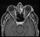 |
Curtain Sign (Enhanced Ptosis) - Associated Image 1 | Bashaer Aldhahwani, MD; Hong Jiang, MD, PhD | This is a 78-year-old male patient who presented with diplopia, right eyelid ptosis, and ophthalmoplegia. He had severe ptosis OD and pseudo-proptosis (lid retraction) OS at baseline, but when the right eyelid was manually elevated, there was marked enhanced ptosis of the left eyelid (Video). He was... | Myasthenia GravIs; Clinical Signs |
| 217 |
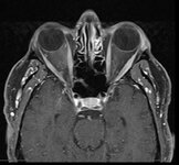 |
Curtain Sign (Enhanced Ptosis) - Associated Image 2 | Bashaer Aldhahwani, MD; Hong Jiang, MD, PhD | This is a 78-year-old male patient who presented with diplopia, right eyelid ptosis, and ophthalmoplegia. He had severe ptosis OD and pseudo-proptosis (lid retraction) OS at baseline, but when the right eyelid was manually elevated, there was marked enhanced ptosis of the left eyelid (Video). He was... | Myasthenia GravIs; Clinical Signs |
| 218 |
 |
Decompensated Phoria | Alex Christoff, MD | An overview of decompensated phoria and its treatment. | Decompensated Phoria |
| 219 |
 |
Dementia with Lewy Bodies: Overview and Neuro-ophthalmologic features | Pavan Vaswani, MD, PhD; Ali G. Hamedani, MD, MHS | Objectives: Recognize the difference between Dementia with Lewy Bodies and Parkinson disease dementia; Recognize the clinical presentation of DLB and differentiating features from Alzheimer disease dementia; Understand the symptomatic therapies and prognosis | Dementia; Lewy Bodies |
| 220 |
 |
Dementia: Overview and Classification | Molly Cincotta, MD; Whitley Aamodt, MD; Ali G. Hamedani, MD, MHS | PowerPoint providing a broad overview of dementia, including definition, clinical findings, work up, diagnosis, classification, and management. | Dementia |
| 221 |
 |
Diagnosis and Evaluation of Stroke in the Pons | Padmaja Sudhakar; Fatai Momodu | This short power point describes the anatomy of the pons, followed by description of stroke in the pons along with clinical presentation and work up. | Crossed Signs; One and Half Syndrome; Pontine Stroke |
| 222 |
 |
Diagnosis and Evaluation of Stroke: Cerebral Hypoxia | Danny Alevy; James Brian Davis; Amanda Dean Henderson | Neurons are particularly vulnerable to oxygen deprivation. Cerebral hypoxia can be caused by arterial thrombosis, embolism, hypoperfusion, cervical artery dissection, or cryptogenic causes. About 1/3 of ischemic strokes are from cryptogenic causes. Embolic strokes are the next most common (20-30%), ... | Atherosclerosis; Cerebral Hypoxia; Cervical Artery Dissection; Cryptogenic Ischemia; Embolism; Hypoperfusion; Ischemia; Stroke; Thrombosis |
| 223 |
 |
Diagnosis and Evaluation of Stroke: Mechanism - Vasculitis | Danny Alevy; Amanda D. Henderson, MD | In this video, we discuss the diagnosis and evaluation of several vasculitides linked to an increase in risk for cerebral ischemic stroke. We focus on ANCA-associated vasculitides, polyarteritis nodosa, Takayasu arteritis, lupus vasculitis, and Susac syndrome, while highlighting their key symptoms a... | Ischemic Stroke; Vasculitis; ANCA-associated Vasculitis; Polyarteritis Nodosa; Takayasu Arteritis; Systemic Lupus Erythematosus; Susac Syndrome |
| 224 |
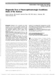 |
Diagnostic Error of Neuro-ophthalmologic Conditions: State of the Science | Leanne Stunkel, MD; David E. Newman-Toker, MD, PhD; Nancy J. Newman, MD; Valérie Biousse, MD | Diagnostic error is prevalent and costly, occurring in up to 15% of US medical encounters and affecting up to 5% of the US population. One-third of malpractice payments are related to diagnostic error. A complex and specialized diagnostic process makes neuro-ophthalmologic conditions particularly vu... | Diagnostic Errors |
| 225 |
 |
Diffusion Tensor Imaging (DTI) | Devin D. Mackay, MD | Explanation of using diffusion tensor imaging (DTI) in examinations. | Diffusion Tensor Imaging (DTI) |