AAO-NANOS Neuro-Ophthalmology Clinical Collection: Derived from the AAO-NANOS Clinical Neuro-Ophthalmology collection produced on CD. The images are of selected cases from the NANOS teaching slide exchange, and the CD was produced under the direction of Larry Frohman, MD and Andrew Lee, MD.
The American Academy of Ophthalmology (AAO); The North American Neuro-Ophthalmology Association (NANOS).
NOVEL: https://novel.utah.edu/
TO
Filters: Collection: "ehsl_novel_aao_nanos"
| Title | Creator | Description | ||
|---|---|---|---|---|
| 201 |
 |
Systemic Disorders With Optic Nerve and Retinal Findings | Larry P. Frohman, MD | This 74-year-old asthmatic male had acute visual loss OS while watching the Super Bowl in 1994. He was seen the next day by a retina specialist, who noted that his optic disc was normal and referred the patient to a neuro-ophthalmologist, who evaluated him about 40 hours after his visual loss. He wa... |
| 202 |
 |
Optic Tract Syndrome Due to Carotid Artery Dolichoectasia | Larry P. Frohman, MD | This 43-year-old man was referred for evaluation of 6 months of visual loss OU. In retrospect, he had noticed increasing difficulty with his tennis game dating back over 3 years, as balls would pass him unexpectedly when hit to his backhand (left) side. The patient did not think this was progressive... |
| 203 |
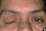 |
Chiasmal Syndromes | Shlomo A. Dotan, MD | A 52-year-old, morbidly obese man with a past medical history that included ischemic cardiac disease with a history of angioplasty, COPD, hypertension, and NIDDM, presented with a severe headache. The next day he had a frozen OD, complete right ptosis, and an unreactive middilated right pupil with V... |
| 204 |
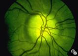 |
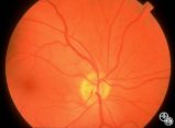
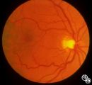
Isolated Congenital Optic Disc Anomalies | Thomas R. Wolf, MD | This optic disc displays multiple drusen. Note the pseudopapilledema here. One can differentiate this from true papilledema in that there is no obscuration of the vessel by the peripapillary nerve fiber layer as they cross the disc margin. This photograph was taken with barrier filters in place, but... |
| 205 |
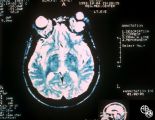 |
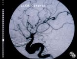
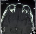
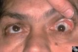
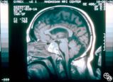
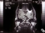
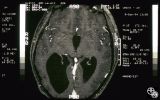
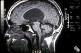
Orbital Tumors | Mitchell J. Wolin, MD | Cavernous hemangiomas of the orbit usually result in painless orbital signs such as proptosis or visual loss. Orbital imaging of the lesion, which usually is a well-defined orbital mass, is demonstrated in this study. The lesion is benign and usually occurs in young to middle-aged adults. Surgical e... |
| 206 |
 |
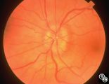
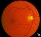
Optic Neuropathies | Rosa A. Tang, MD | Optic atrophy might be a false-localizing sign of optic nerve disease, as retinal disease may secondarily produce optic atrophy. In retinitis pigmentosa, for example, patients may exhibit a waxy disc palor. Fundus imaging. Anatomy: Retina. Disease/Diagnosis: Retinitis pigmentosa; Optic atrophy. |
| 207 |
 |
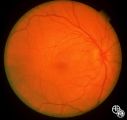
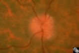
Isolated Optic Neuritis/Neuropathy | Ralph A. Sawyer, MD | Papilledema usually results in bilateral optic disc edema without visual loss. The blind spot may enlarge initially, but progressive visual field loss may occur with chronic optic disc edema. Asymmetric or frankly unilateral optic disc edema may occur due to structural disc fractures that prevent th... |
| 208 |
 |
Isolated Optic Neuritis/Neuropathy | Ralph A. Sawyer, MD | Papilledema usually results in bilateral optic disc edema without visual loss. The blind spot may enlarge initially, but progressive visual field loss may occur with chronic optic disc edema. Asymmetric or frankly unilateral optic disc edema may occur due to structural disc fractures that prevent th... |
| 209 |
 |
Neuro-Ophthalmic Vascular Disease | Robert F. Saul, MD | In image 93_29, taken during the episode, note the change in caliber of the blood vessels. |
| 210 |
 |
Neuro-Ophthalmic Vascular Disease | Robert F. Saul, MD | Image 93_30 is immediately after the attack, note the slight redness to the macula. |
| 211 |
 |
Neuro-Ophthalmic Vascular Disease | Robert F. Saul, MD | Image 93_28 shows the fundus before the attack. |
| 212 |
 |
Neuro-Ophthalmic Vascular Disease | Larry P. Frohman, MD | This 23-year-old woman has had insulin-dependent diabetes mellitus since age 3. She was diagnosed with Sydenham's chorea in early childhood and had grand mal seizures from age 13 to 15. She has been hypertensive since age 18. Her vision was 20/25 OD and 20/40 OS, with dyschromatopsia OS, and a 1.8 l... |
| 213 |
 |
Neuro-Ophthalmic Vascular Disease | Larry P. Frohman, MD | This 27-year-old woman had no past ocular history and presented with 3 weeks of redness OS that has been treated by the referring doctor as allergic conjunctivitis. She was referred for evaluation when she developed binocular diplopia. Her past medical history included phlebitis and one miscarriage ... |
| 214 |
 |
Isolated Optic Neuritis/Neuropathy | Daniel M. Jacobson MD | This 35-year-old otherwise-healthy woman developed typical optic neuritis OD with excellent recovery. She had no clinical evidence of multiple sclerosis at that time. She presented in August of 1991, at which time perivenous sheathing was seen in the retinal periphery OU. A limited workup was negati... |
| 215 |
 |
Isolated Optic Neuritis/Neuropathy | Daniel M. Jacobson MD | This 35-year-old otherwise-healthy woman developed typical optic neuritis OD with excellent recovery. She had no clinical evidence of multiple sclerosis at that time. She presented in August of 1991, at which time perivenous sheathing was seen in the retinal periphery OU. A limited workup was negati... |
| 216 |
 |
Chiasmal Syndromes | Shlomo A. Dotan, MD | A 52-year-old, morbidly obese man with a past medical history that included ischemic cardiac disease with a history of angioplasty, COPD, hypertension, and NIDDM, presented with a severe headache. The next day he had a frozen OD, complete right ptosis, and an unreactive middilated right pupil with V... |
| 217 |
 |
Chiasmal Syndromes | Shlomo A. Dotan, MD | A 52-year-old, morbidly obese man with a past medical history that included ischemic cardiac disease with a history of angioplasty, COPD, hypertension, and NIDDM, presented with a severe headache. The next day he had a frozen OD, complete right ptosis, and an unreactive middilated right pupil with V... |
| 218 |
 |
Chiasmal Syndromes | Shlomo A. Dotan, MD | A 52-year-old, morbidly obese man with a past medical history that included ischemic cardiac disease with a history of angioplasty, COPD, hypertension, and NIDDM, presented with a severe headache. The next day he had a frozen OD, complete right ptosis, and an unreactive middilated right pupil with V... |
| 219 |
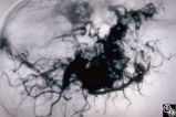 |
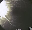
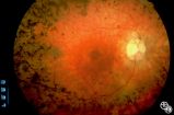
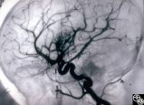
Ocular Manifestations of Congenital/Inherited Diseases | Mark J. Kupersmith, MD | This 9-year-old girl, who had complained of recurrent spontaneous bleeding from the palate and slight swelling and increased warmth over the left cheek, was found to have Wyburn-Mason syndrome. Image 1993_16 shows a small area of arteriovenous shunt on the left optic disc in this patient, who has no... |
| 220 |
 |
Ocular Manifestations of Congenital/Inherited Diseases | Mark J. Kupersmith, MD | This 9-year-old girl, who had complained of recurrent spontaneous bleeding from the palate and slight swelling and increased warmth over the left cheek, was found to have Wyburn-Mason syndrome. Image 1993_16 shows a small area of arteriovenous shunt on the left optic disc in this patient, who has no... |
| 221 |
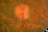 |
Isolated Optic Neuritis/Neuropathy | Anthony C. Arnold, MD | This 42-year-old male with pseudotumor cerebri and chronic papilledema demonstrated refractile bodies, which can be seen with chronic optic disc edema. This image exhibits decreased disc edema and resolution of the refractile bodies OD after therapy. Pair with 96_01, 96_02, 96_03, 96_05, and 96_06. |
| 222 |
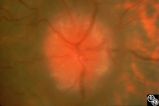 |
Isolated Optic Neuritis/Neuropathy | Anthony C. Arnold, MD | This 42-year-old male with pseudotumor cerebri and chronic papilledema demonstrated refractile bodies, which can been seen with chronic optic disc edema. This image demonstrates later recurrence of the refractile bodies with worsening papilledema OD. Pair with 96_01, 96_02, 96_03, 96_04, and 96_06. |
| 223 |
 |
Isolated Optic Neuritis/Neuropathy | Anthony C. Arnold, MD | This 42-year-old male with pseudotumor cerebri and chronic papilledema demonstrated refractile bodies, which can be seen with chronic optic disc edema. This image demonstrates later recurrence of the refractile bodies with worsening papilledema OD. Pair with 96_01, 96_02, 96_03, 96_04, and 96_05. |
| 224 |
 |
Ocular Manifestations of Congenital/Inherited Diseases | Larry P. Frohman, MD | This 14-year-old boy presented with sudden visual loss of the right eye that occurred 3 weeks before and due to a central retinal vein occlusion. His ocular history was quite complicated. He had had a resection of a lymphangioma of the left upper lid at age 7 months and underwent left orbitotomy for... |
| 225 |
 |
Ocular Manifestations of Congenital/Inherited Diseases | Larry P. Frohman, MD | This 14-year-old boy presented with sudden visual loss of the right eye that occurred 3 weeks before and due to a central retinal vein occlusion. His ocular history was quite complicated. He had had a resection of a lymphangioma of the left upper lid at age 7 months and underwent left orbitotomy for... |