AAO-NANOS Neuro-Ophthalmology Clinical Collection: Derived from the AAO-NANOS Clinical Neuro-Ophthalmology collection produced on CD. The images are of selected cases from the NANOS teaching slide exchange, and the CD was produced under the direction of Larry Frohman, MD and Andrew Lee, MD.
The American Academy of Ophthalmology (AAO); The North American Neuro-Ophthalmology Association (NANOS).
NOVEL: https://novel.utah.edu/
TO
| Title | Creator | Description | ||
|---|---|---|---|---|
| 201 |
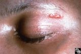 |
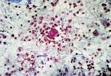
Systemic Disorders With Optic Nerve and Retinal Findings | Larry P. Frohman, MD | Skin rashes occur in about 30 percent of patients with sarcoid. When seen, the rashes offer an accessible site for obtaining histologic material for confirmation of the clinical diagnosis. Pair with 91_69. |
| 202 |
 |
Systemic Disorders With Optic Nerve and Retinal Findings | Larry P. Frohman, MD | A 42-year old woman presented with a history of severe brow pain and 4 days of progressive visual loss OD. There was no increased pain on ocular rotation. Aside from heavy menses, she denied any significant past medical history. Her examination revealed acuity NLP OD, 20/25 OS; color vision 9/10 OS;... |
| 203 |
 |
Systemic Disorders With Optic Nerve and Retinal Findings | Larry P. Frohman, MD | A 42-year old woman presented with a history of severe brow pain and 4 days of progressive visual loss OD. There was no increased pain on ocular rotation. Aside from heavy menses, she denied any significant past medical history. Her examination revealed acuity NLP OD, 20/25 OS; color vision 9/10 OS;... |
| 204 |
 |
Systemic Disorders With Optic Nerve and Retinal Findings | Larry P. Frohman, MD | A 42-year old woman presented with a history of severe brow pain and 4 days of progressive visual loss OD. There was no increased pain on ocular rotation. Aside from heavy menses, she denied any significant past medical history. Her examination revealed acuity NLP OD, 20/25 OS; color vision 9/10 OS;... |
| 205 |
 |
Systemic Disorders With Optic Nerve and Retinal Findings | Larry P. Frohman, MD | A 42-year old woman presented with a history of severe brow pain and 4 days of progressive visual loss OD. There was no increased pain on ocular rotation. Aside from heavy menses, she denied any significant past medical history. Her examination revealed acuity NLP OD, 20/25 OS; color vision 9/10 OS;... |
| 206 |
 |
Systemic Disorders With Optic Nerve and Retinal Findings | Larry P. Frohman, MD | A 42-year old woman presented with a history of severe brow pain and 4 days of progressive visual loss OD. There was no increased pain on ocular rotation. Aside from heavy menses, she denied any significant past medical history. Disease/Diagnosis: Syphilitic optic neuritis/perineuritis, fundus, OS -... |
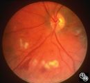
| 207 |
 |
Systemic Disorders With Optic Nerve and Retinal Findings | Larry P. Frohman, MD | This is a 32-year-old HIV-positive man with anterior uveitis, vitritis, and bilateral papillitis from syphilis. With intravenous penicillin treatment, the optic discs and vision returned to normal. |
| 208 |
 |
Systemic Disorders With Optic Nerve and Retinal Findings | Larry P. Frohman, MD | This is a 32-year-old HIV-positive man with anterior uveitis, vitritis, and bilateral papillitis from syphilis. With intravenous penicillin treatment, the optic discs and vision returned to normal. |
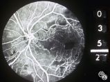
| 209 |
 |
Chiasmal Syndromes | Larry P. Frohman, MD | This 39-year-old HIV-positive man presented in 1982 with 1 month of bilateral vision loss. His prior evaluation had included 2 CT scans, which suggested a fullness to the chiasm, and a spinal tap that showed 6 monocytes, 14 red cells, a protein of 81 mg/dl, and a glucose of 56 mg/dl, with a negative... |
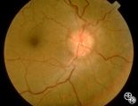
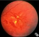
| 210 |
 |
Systemic Disorders With Optic Nerve and Retinal Findings | Larry P. Frohman, MD | This 25-year-old man presented to the eye service with a history of 3 days of decreased vision OD. His past medical history was unremarkable. His examination showed acuities of 20/25 OU, with intact color plates, a 0.3 log unit of RAPD OD, and an inferior arcuate scotoma. The photos (Images 95_42, 9... |
| 211 |
 |
Magnetic Resonance Imaging in Detection of Extracranial Internal Carotid Artery Dissection | Marilyn C. Kay, MD | This 28-year-old woman presented with a 4-week history of bilateral visual loss. She had a known history of multiple sclerosis. Her vision was 20/60 OD and 20/40 OS, with an RAPD OS and optic pallor OU. Her fields and MRI are shown. Optic tract lesions usually result in an incongruous homonymous hem... |
| 212 |
 |
Neuro-Ophthalmic Case With Notable Field Changes | Marilyn C. Kay, MD | This 28-year-old woman presented with a 4-week history of bilateral visual loss. She had a known history of multiple sclerosis. Her vision was 20/60 OD and 20/40 OS, with an RAPD OS and optic pallor OU. Her fields and MRI are shown. Optic tract lesions usually result in an incongruous homonymous hem... |
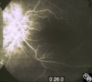
| 213 |
 |
Neuro-Ophthalmic Imaging-Cerebral Angiography | Mark J. Kupersmith, MD | Ehlers-Danlos syndrome is a connective tissue disorder that may affect blood vessels and predispose some affected patients to development of carotid cavernous fistula. Most patients with high-flow direct carotid cavernous sinus fistulas have suffered acute traumatic tears in the internal carotid art... |
| 214 |
 |
Neuro-Ophthalmic Consequences of Therapy | Mark J. Kupersmith, MD | radiation retinopathy may mimic diabetic or hypertensive optic neuropathy. A history of irradiation to the eye, orbit, or head is mandatory. Radiation retinopathy usually occurs many months after radiation therapy. |
| 215 |
 |
Neuro-Ophthalmic Consequences of Therapy | Mark J. Kupersmith, MD | radiation retinopathy may mimic diabetic or hypertensive optic neuropathy. A history of irradiation to the eye, orbit, or head is mandatory. Radiation retinopathy usually occurs many months after radiation therapy. |
| 216 |
 |
Neuro-Ophthalmic Consequences of Therapy | Mark J. Kupersmith, MD | radiation retinopathy may mimic diabetic or hypertensive optic neuropathy. A history of irradiation to the eye, orbit, or head is mandatory. Radiation retinopathy usually occurs many months after radiation therapy. |
| 217 |
 |
Neuro-Ophthalmic Consequences of Therapy | Mark J. Kupersmith, MD | Radiation causes a vascular retinopathy that may mimic diabetic or hypertensive retinopathy. It does not develop until many months or several years after radiation therapy to the eye, orbit or head. |
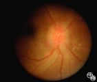
| 218 |
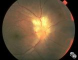
 |
Isolated Optic Neuritis/Neuropathy | Mark J. Kupersmith, MD | Papilledema may produce visual loss due to chronic atrophic papilledema, secondary macular hemorrhage, exudate or edema, secondary ischemic optic neuropathy, or secondary subretinal neovascular membrane formation. Patients with papilledema and visual loss should be suspected of harboring one of thes... |
| 219 |
 |
Isolated Optic Neuritis/Neuropathy | Mark J. Kupersmith, MD | Papilledema may produce visual loss due to chronic atrophic papilledema, secondary macular hemorrhage, exudate or edema, secondary ischemic optic neuropathy, or secondary subretinal neovascular membrane formation. Patients with papilledema and visual loss should be suspected of harboring one of thes... |
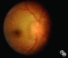
| 220 |
 |
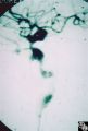
Systemic Disorders With Optic Nerve and Retinal Findings | Mark J. Kupersmith, MD | Sarcoidosis is an inflammatory granulomatous disease that may result in inflammatory or infiltrative optic neuropathology or retinal vasculitis. Pair with 91_34. |
| 221 |
 |
Systemic Disorders With Optic Nerve and Retinal Findings | Mark J. Kupersmith, MD | Sarcoidosis is an inflammatory granulomatous disease that may result in inflammatory or infiltrative optic neuropathology or retinal vasculitis. Pair with 91_35. |
| 222 |
 |
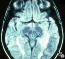
Neuro-Ophthalmic Vascular Disease | Mark J. Kupersmith, MD | A 9-year-old boy had recurrent ischemic episodes that had begun 2 years prior to evaluation. A significant right hemiparesis and a significant speech, learning, and memory disorder were present. His noncontrast axial view CT scan demonstrated multiple cerebral infarcts. Cerebral angiography revealed... |
| 223 |
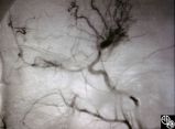 |
Neuro-Ophthalmic Vascular Disease | Mark J. Kupersmith, MD | A 9-year-old boy had recurrent ischemic episodes that had begun 2 years prior to evaluation. A significant right hemiparesis and a significant speech, learning, and memory disorder were present. His noncontrast axial view CT scan demonstrated multiple cerebral infarcts. Cerebral angiography revealed... |
| 224 |
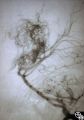 |
Neuro-Ophthalmic Vascular Disease | Mark J. Kupersmith, MD | A 9-year-old boy had recurrent ischemic episodes that had begun 2 years prior to evaluation. A significant right hemiparesis and a significant speech, learning, and memory disorder were present. His noncontrast axial view CT scan demonstrated multiple cerebral infarcts. Cerebral angiography revealed... |
| 225 |
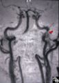 |
Neuro-Ophthalmic Vascular Disease | Mark J. Kupersmith, MD | MR angiography was performed on this 33-year-old woman, who complained of the onset of a bad taste in her mouth followed by pain along the left forehead and development of the left third-order Horner's syndrome during pregnancy. Except for the Horner's syndrome, the patient was neurologically intact... |