AAO-NANOS Neuro-Ophthalmology Clinical Collection: Derived from the AAO-NANOS Clinical Neuro-Ophthalmology collection produced on CD. The images are of selected cases from the NANOS teaching slide exchange, and the CD was produced under the direction of Larry Frohman, MD and Andrew Lee, MD.
The American Academy of Ophthalmology (AAO); The North American Neuro-Ophthalmology Association (NANOS).
NOVEL: https://novel.utah.edu/
TO
Filters: Collection: ehsl_novel_aao_nanos
| Title | Description | Subject | ||
|---|---|---|---|---|
| 176 |
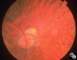 |
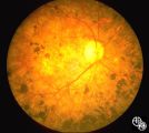
Systemic Disorders With Optic Nerve and Retinal Findings | Intraocular lymphoma may present with an unexplained vitritis, optic disc infiltration, or choroidal infiltration. One unusual manifestation of large-cell lymphoma is this leopard-spot appearance. Pair with 94_33, 94_34, and 94_35. This is a fundus photo. | Lymphoma; Leopard Spot Fundus; Lymphoma; Optic Nerve |
| 177 |
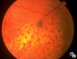 |
Systemic Disorders With Optic Nerve and Retinal Findings | Intraocular lymphoma may present with an unexplained vitritis, optic disc infiltration, or choroidal infiltration. One unusual manifestation of large-cell lymphoma is this leopard-spot appearance. Pair with 94_32, 94_34, and 94_35. This is a fundus photo. | Lymphoma; Leopard Spot Fundus; Lymphoma; Optic Nerve |
| 178 |
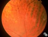 |
Systemic Disorders With Optic Nerve and Retinal Findings | Intraocular lymphoma may present with an unexplained vitritis, optic disc infiltration, or choroidal infiltration. One unusual manifestation of large-cell lymphoma is this leopard-spot appearance. Pair with 94_32, 94_33, and 94_35. This is a fundus photo. | Lymphoma; Leopard Spot Fundus; Lymphoma; Optic Nerve |
| 179 |
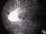 |
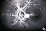
Systemic Disorders With Optic Nerve and Retinal Findings | Intraocular lymphoma may present with an unexplained vitritis, optic disc infiltration, or choroidal infiltration. One unusual manifestation of large-cell lymphoma is this leopard-spot appearance. Pair with 94_32, 94_33, and 94_34. This is a fluorescein angiogram. | Lymphoma; Leopard Spot Fluorescein Angiogram; Lymphoma; Optic Nerve |
| 180 |
 |
Systemic Disorders With Optic Nerve and Retinal Findings | This patent has known pseudoxanthoma elasticum (an uncommon elastic tissue disorder characterized by plaque-like skin folds [plucked chicken skin], and degeneration of collagen fibers involving multiple systems, including the GI tract and heart), angioid streaks, and optic disc drusen. | Pseudoxanthoma Elasticum |
| 181 |
 |
Systemic Disorders With Optic Nerve and Retinal Findings | This patent has known pseudoxanthoma elasticum (an uncommon elastic tissue disorder characterized by plaque-like skin folds [plucked chicken skin], and degeneration of collagen fibers involving multiple systems, including the GI tract and heart), angioid streaks, and optic disc drusen. | Pseudoxanthoma Elasticum |
| 182 |
 |
Systemic Disorders With Optic Nerve and Retinal Findings | This patent has known pseudoxanthoma elasticum (an uncommon elastic tissue disorder characterized by plaque-like skin folds [plucked chicken skin], and degeneration of collagen fibers involving multiple systems, including the GI tract and heart), angioid streaks, and optic disc drusen. | Pseudoxanthoma Elasticum |
| 183 |
 |
Systemic Disorders With Optic Nerve and Retinal Findings | This patent has known pseudoxanthoma elasticum (an uncommon elastic tissue disorder characterized by plaque-like skin folds [plucked chicken skin], and degeneration of collagen fibers involving multiple systems, including the GI tract and heart), angioid streaks, and optic disc drusen. Imaging of a... | Pseudoxanthoma Elasticum |
| 184 |
 |
Systemic Disorders With Optic Nerve and Retinal Findings | This patent has known pseudoxanthoma elasticum (an uncommon elastic tissue disorder characterized by plaque-like skin folds [plucked chicken skin], and degeneration of collagen fibers involving multiple systems, including the GI tract and heart), angioid streaks, and optic disc drusen. Imaging of a... | Pseudoxanthoma Elasticum |
| 185 |
 |
Ocular Manifestations of Systemic Disorders | Thyroid eye disease may cause proptosis and extraocular muscle enlargement that may be seen on orbital imaging studies. In general, coronal images allow the best visualization of the extraocular muscle enlargement. Pair with 94_45 and 94_46. | Thyroid Disease; Thyroid Orbitopathy; Thyroid Eye Disease; Myopathy; Proptosis; Chemosis; Periorbital Edema; Lid Retraction |
| 186 |
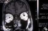 |
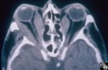
Ocular Manifestations of Systemic Disorders | Thyroid eye disease may cause proptosis and extraocular muscle enlargement that may be seen on orbital imaging studies. In general, coronal images allow the best visualization of the extraocular muscle enlargement. Pair with 94_44 and 94_46. | Thyroid Disease; Thyroid Eye Disease; Myopathy; Proptosis; Chemosis; Periorbital Edema; Lid Retraction; MRI |
| 187 |
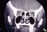 |
Ocular Manifestations of Systemic Disorders | Thyroid eye disease may cause proptosis and extraocular muscle enlargement that may be seen on orbital imaging studies. In general, coronal images allow the best visualization of the extraocular muscle enlargement. Pair with 94_44 and 94_45. | Thyroid Disease; Thyroid Eye Disease; Myopathy; Proptosis; Chemosis; Periorbital Edema; Lid Retraction; CT |
| 188 |
 |
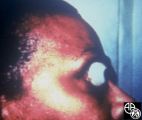
Ocular Manifestations of Systemic Disorders | Systemic lymphoma may occur in the orbit and may involve the lacrimal gland. Patients usually present with a painless, progressive proptosis or a mass. CT scan usually demonstrates an irregularly shaped lesion conforming to the globe or lacrimal fossa, and bone erosion is not usually found. Pair wit... | Lymphoma; Systemic Lymphoma; Proptosis; Lacrimal Gland |
| 189 |
 |
Ocular Manifestations of Systemic Disorders | Systemic lymphoma may occur in the orbit and may involve the lacrimal gland. Patients usually present with a painless, progressive proptosis or a mass. CT scan usually demonstrates an irregularly shaped lesion conforming to the globe or lacrimal fossa, and bone erosion is not usually found. Pair wit... | Lymphoma; Systemic Lymphoma; MRI Orbit; Lacrimal Gland |
| 190 |
 |
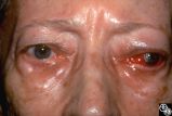
Ocular Manifestations of Systemic Disorders | Thyroid eye disease may result in proptosis and restrictive external ophthalmoplegia. The extracoular muscles are often diffusely enlarged with sparing of the tendons. | Thyroid Disease; Restriction Syndromes; Thyroid Eye Disease; Thyroid-Associated Ophthalmopathy; Blow-Out Fracture; Thyroid Orbitopathy; Thyroid Eye Disease; Myopathy; Proptosis; Chemosis; Periorbital Edema; Lid Retraction; CT |
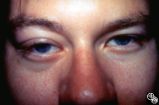
| 191 |
 |
Ocular Manifestations of Systemic Disorders | Thyroid eye disease can result in significant upper eyelid retraction and axial proptosis resulting in exposure keratopathy. | Thyroid Disease; Thyroid Orbitopathy; Thyroid Eye Disease; Myopathy; Proptosis; Chemosis; Periorbital Edema; Lid Retraction |
| 192 |
 |
Ocular Manifestations of Congenital/Inherited Diseases | Patients with olivopontocerebellar atrophy may exhibit signs of ocular motor deficits, such as ocular motor apraxia or cerebellar eye signs, and peripheral pigmentary retinopathy and optic atrophy. Pair with 94_55. | Olivopontocerebellar Degeneration |
| 193 |
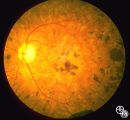 |
Ocular Manifestations of Congenital/Inherited Diseases | Patients with olivopontocerebellar atrophy may exhibit signs of ocular motor deficits, such as ocular motor apraxia or cerebellar eye signs, and peripheral pigmentary retinopathy and optic atrophy. Pair with 94_54. | Olivopontocerebellar Degeneration |
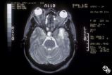
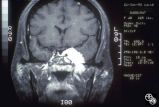
| 194 |
 |
Orbital Tumors | Cavernous hemangiomas of the orbit usually result in painless orbital signs such as proptosis or visual loss. Orbital imaging of the lesion, which usually is a well-defined orbital mass, is demonstrated in this study. The lesion is benign and usually occurs in young to middle-aged adults. Surgical e... | Cavernous Hemangioma |
| 195 |
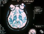 |
Orbital Tumors | Cavernous hemangiomas of the orbit usually result in painless orbital signs such as proptosis or visual loss. Orbital imaging of the lesion, which usually is a well-defined orbital mass, is demonstrated in this study. The lesion is benign and usually occurs in young to middle-aged adults. Surgical e... | Cavernous Hemangioma |
| 196 |
 |
Orbital Tumors | Cavernous hemangiomas of the orbit usually result in painless orbital signs such as proptosis or visual loss. Orbital imaging of the lesion, which usually is a well-defined orbital mass, is demonstrated in this study. The lesion is benign and usually occurs in young to middle-aged adults. Surgical e... | Cavernous Hemangioma |
| 197 |
 |
Chiasmal Syndromes | This 36-year-old woman presented in 1988 with 3 weeks of vertical binocular diplopia. She was a known amblyope OD. Her examination was notable for a right hyperdeviation of 1 PD present in right gaze and a subtle left noncongruous homonymous field defect. She was lost to follow-up, but 5 years later... | Sarcoidosis; MRI |
| 198 |
 |
Chiasmal Syndromes | This 36-year-old woman presented in 1988 with 3 weeks of vertical binocular diplopia. She was a known amblyope OD. Her examination was notable for a right hyperdeviation of 1 PD present in right gaze and a subtle left noncongruous homonymous field defect. She was lost to follow-up, but 5 years later... | Sarcoidosis; MRI |
| 199 |
 |
Chiasmal Syndromes | This 36-year-old woman presented in 1988 with 3 weeks of vertical binocular diplopia. She was a known amblyope OD. Her examination was notable for a right hyperdeviation of 1 PD present in right gaze and a subtle left noncongruous homonymous field defect. She was lost to follow-up, but 5 years later... | Sarcoidosis; MRI |
| 200 |
 |
Motility Disturbances | This man had a posttraumatic right sixth nerve paresis. He is shown in primary gaze before Botox (botulinum toxin; image 94_64) | Trochlear Palsy |