Best known for his world-renowned neuro-ophthalmology unit based at the University of California, San Francisco, William Hoyt, MD collected here more than 850 of his best images covering a wide range of disorders.
William F. Hoyt, MD, Professor Emeritus of Ophthalmology, Neurology and Neurosurgery, Department of Ophthalmology, University of California, San Francisco.
NOVEL: https://novel.utah.edu/
TO
| Title | Description | Type | ||
|---|---|---|---|---|
| 176 |
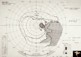 |
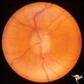
H51 Segmental Hypoplasia, Retinal-Nasal Hypoplasia | Visual field showing flag-like temporal field defect in patient shown in H_50. Anatomy: Optic disc. Pathology: Nasal segmental disc hypoplasia. Disease/ Diagnosis: Cogenital anomaly. | Image |
| 177 |
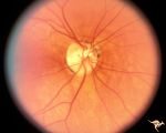 |
H50 Segmental Hypoplasia, Retinal-Nasal Hypoplasia | Right eye. Nasal hypoplasia with nasal pit. Same patient as H_51. Anatomy: Optic disc. Pathology: Nasal segmental disc hypoplasia. Disease/ Diagnosis: Congential anomaly. | Image |
| 178 |
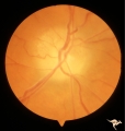 |
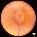
P55a Chronic Papilledema, Late Stage | Right eye. Optic atrophy secondary to chronic papilledema. Glioblastoma. Chemotherapy patient. Anatomy: Optic disc. Pathology: Papilledema. Disease/ Diagnosis: Chronic papilledema with atrophy. | Image |
| 179 |
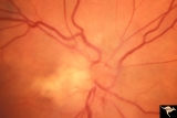 |
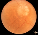
F101 Optic Disc Lymphosarcoma | Optic disc lymphosarcoma. This disc has been infiltrated by neoplastic cells. Anatomy: Optic disc. Pathology: Lymphosarcoma. Disease/ Diagnosis: Lymphosarcoma. | Image |
| 180 |
 |
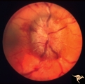
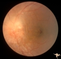
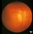
Bilateral Chronic Papilledema | Left eye. Frisen's stage 5. Patient with long standing aqueductal stenosis. Bilateral Chronic Papilledema. Man. Anatomy: Optic disc. Pathology: Papilledema. Disease/Diagnosis: Papilledema from aqueductal stenosis. | Image |
| 181 |
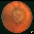 |
Bilateral Chronic Papilledema | Right eye. Frisen's stage 5. Patient with long standing aqueductal stenosis. Bilateral Chronic Papilledema. Man. Anatomy: Optic disc. Pathology: Papilledema. Disease/Diagnosis: Papilledema from aqueductal stenosis. | Image |
| 182 |
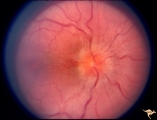 |
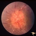
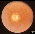
Bilateral Papilledema | Right eye. Bilateral Papilledema in patient with cardiopulmonary insufficiency. Woman. Anatomy: Optic disc. Pathology: Papilledema. Disease/Diagnosis: cardiopulmonary insufficiency causing intracranial hypertension. Clinical notes: headache. | Image |
| 183 |
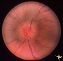 |
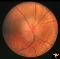
Bilateral Papilledema | Left eye. Bilateral Papilledema with hypoparathyroidism. Woman. Anatomy: Optic disc. Pathology: Papilledema. Papilledema with hypopararthyroidism. | Image |
| 184 |
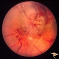 |
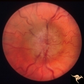
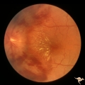
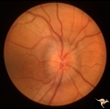
Bilateral Papilledema from Occipital Tumor | Left eye. Bilateral hemorrhagic papilledema. Occipital glioma. Woman. Anatomy: Optic disc. Pathology: Papilledema. Disease/Diagnosis: Hemorrhagic papilledema from occipital glioma. | Image |
| 185 |
 |
Bilateral Papilledema from Occipital Tumor | Right eye. Bilateral hemorrhagic papilledema. Occipital glioma. Right hemianopia. Woman. Anatomy: Optic disc. Pathology: Papilledema. Disease/Diagnosis: Hemorrhagic papilledema from occipital glioma. | Image |
| 186 |
 |
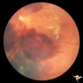
Chronic Atrophic Papilledema | Left eye. Left eye blind. Chronic Atrophic Papilledema. Obese woman (300 lbs) with large tentorial meningioma. "Pseudo Pseudotumor". Anatomy: Optic disc. Pathology: Papilledema. Disease/Diagnosis: Papilledema from large tentorial meningioma. | Image |
| 187 |
 |
Chronic Atrophic Papilledema | Right eye. Chronic Atrophic Papilledema. Obese woman (300 lbs) with large tentorial meningioma. "Pseudo Pseudotumor" Anatomy: Optic disc. Pathology: Papilledema. Disease/Diagnosis: Papilledema from large tentorial meningioma. | Image |
| 188 |
 |
Chronic Papilledema in Resolution. Sequence | Left eye 2 weeks after presentation. Chronic papilledema in resolution. Note first evidence of a vertical choroidal fold. Anatomy: Optic disc. Pathology: Papilledema. Disease/Diagnosis: Papilledema. | Image |
| 189 |
 |
Chronic Papilledema in Resolution. Sequence | Left eye at presentation. Chronic papilledema. Anatomy: Optic disc Pathology: Papilledema Disease/Diagnosis: Papilledema | Image |
| 190 |
 |
Chronic Papilledema with Hemorrhagic and Exudative Complications | Left eye one month after presentation. Resolving hemorrhage. Chronic papilledema with hemorrhagic and exudative complications due to Pseudotumor cerebri. Anatomy: Optic disc. Pathology: Papilledema. Disease/Diagnosis: Chronic papilledema with hemorrhagic and exudative complications | Image |
| 191 |
 |
Chronic Papilledema with Hemorrhagic and Exudative Complications | Left eye at presesntation. Chronic papilledema with hemorrhagic and exudative complications due to Pseudotumor cerebri. Anatomy: Optic disc Pathology: Papilledema Disease/Diagnosis: Chronic papilledema with hemorrhagic and exudative complications. | Image |
| 192 |
 |
Chronic Papilledema with Pseudo Drusen | Left eye. Chronic papilledema with pseudo drusen due to cerebral pontine angle tumor. Anatomy: Optic disc. Pathology: Papilledema Disease/Diagnosis: Chronic papilledema with pseudo drusen. | Image |
| 193 |
 |
Chronic Papilledema with Pseudo Drusen | Right eye. Chronic papilledema with pseudo drusen due to cerebral pontine angle tumor. Anatomy: Optic disc Pathology: Papilledema Disease/Diagnosis: Chronic papilledema with pseudo drusen | Image |
| 194 |
 |
Chronic Papilledema with Pseudo Drusen | Right eye. Meningioma. Pseudo drusen from chronic papilledema. Woman. Anatomy: Optic disc Pathology: Papilledema Disease/Diagnosis: Chronic papilledema with pseudo drusen | Image |
| 195 |
 |
Chronic Papilledema with Pseudo Drusen | Left eye of 51 year old, 220 pound black woman. Pseudotumor cerebri, pseudo drusen, exudates. Anatomy: Optic disc. Pathology: Papilledema Disease/Diagnosis: Chronic papilledema with pseudo drusen | Image |
| 196 |
 |
Chronic Papilledema with Pseudo Drusen | Chronic papilledema with pseudo drusen. Residual choroidal folds. Pseudo drusen. Anatomy: Optic disc. Pathology: Papilledema. Disease/Diagnosis: Chronic papilledema with pseudo drusen. | Image |
| 197 |
 |
Chronic Papilledema with Pseudo Drusen | Left eye. Meningioma. Pseudo drusen from chronic papilledema. The patient's meningioma had blinded her left eye and caused chronic elevated intracranial pressure. Woman. Anatomy: Optic disc Pathology: Papilledema Disease/Diagnosis: Chronic papilledema with pseudo drusen | Image |
| 198 |
 |
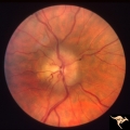
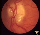
D101 Disc Edema with Systemic Lupus | Unilateral disc swelling with narrowed arterioles. No decrease in visual acuity or field. 19 year old woman. Patient died of cerebral lupus within two months. Optociliary veins dumping into disc edge at 4:00, 9:00, and 11:00. Anatomy: Optic disc. Pathology: Axoplasmic stasis due to vasculitis (Lupu... | Image |
| 199 |
 |
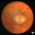
D102 Disc Edema with Systemic Lupus | 28 year old woman. Vision 20/20 but blind spot enlarged. Same patient as D1_03. Right eye. Anatomy: Optic disc. Pathology: Axoplasmic stasis due to vasculitis (Lupus). Disease/ Diagnosis: Lupus papillopathy. Clinical: Normal vision with enlarged blind spot on visual field. | Image |
| 200 |
 |
D103 Disc Edema with Systemic Lupus | 28 year old woman with systemic Lupus erythematosus. Vision 20/20 but blind spot enlarged. Same patient as D1_02. Magnified. Anatomy: Optic disc. Pathology: Axoplasmic stasis due to vasculitis (Lupus). Disease/ Diagnosis: Lupus papillopathy. Clinical: Normal vision with enlarged blind spot on visual... | Image |