AAO-NANOS Neuro-Ophthalmology Clinical Collection: Derived from the AAO-NANOS Clinical Neuro-Ophthalmology collection produced on CD. The images are of selected cases from the NANOS teaching slide exchange, and the CD was produced under the direction of Larry Frohman, MD and Andrew Lee, MD.
The American Academy of Ophthalmology (AAO); The North American Neuro-Ophthalmology Association (NANOS).
NOVEL: https://novel.utah.edu/
TO
| Title | Creator | Description | ||
|---|---|---|---|---|
| 176 |
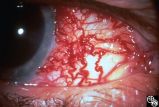 |
Neuro-Ophthalmic Vascular Disease | Anthony C. Arnold, MD | This 76-year-old woman has a 7-month history of redness and pressure sensation in both eyes that is worse in the morning. She has noted intermittent horizontal diplopia during this time. Angiography demonstrated a right dural cavernous sinus fistula, which was successfully occluded with direct injec... |
| 177 |
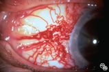 |
Neuro-Ophthalmic Vascular Disease | Anthony C. Arnold, MD | This 76-year-old woman has a 7-month history of redness and pressure sensation in both eyes that is worse in the morning. She has noted intermittent horizontal diplopia during this time. Angiography demonstrated a right dural cavernous sinus fistula, which was successfully occluded with direct injec... |
| 178 |
 |
Congenitally Tilted Optic Disc | Anthony C. Arnold, MD | Colobomas or defects of the optic nerve may exhibit spontaneous pulsations. Disease/Diagnosis: Coloboma. |
| 179 |
 |
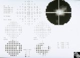
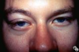
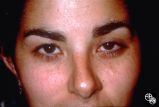
Neuro-Ophthalmic Imaging-CT Scan | Larry P. Frohman, MD | This 39-year-old woman's initial sign was painless, progressive, symmetric ptosis OU, without diurnal variation, that manifested when she was age 17 living in the Dominican Republic. At that time, she had no diplopia or systemic signs. She had no family history of ocular or muscle disease, and no ot... |
| 180 |
 |
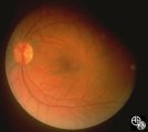
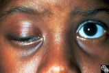
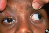
Ocular Manifestations of Congenital/Inherited Diseases | Larry P. Frohman, MD | This 14-year-old boy presented with sudden visual loss of the right eye that occurred 3 weeks before and due to a central retinal vein occlusion. His ocular history was quite complicated. He had had a resection of a lymphangioma of the left upper lid at age 7 months and underwent left orbitotomy for... |
| 181 |
 |
Isolated Optic Neuritis/Neuropathy | Larry P. Frohman, MD | The patient is a 62-year-old female who presented in August 1996 with visual loss OD that she first noted as loss of her superior field in May 1996. She felt that it had been static since, and perhaps was even a little better in the week before she was seen. There was no pain, even with ocular rotat... |
| 182 |
 |
Neuro-Ophthalmic Vascular Disease | Larry P. Frohman, MD | This 32-year-old woman was referred with a history of 4 days of loss of vision OD. She had a history of manic depressive illness and IV drug abuse; she had been HIV tested 4 weeks before and was negative. She said she last injected cocaine 5 days before being seen, the night before she awoke with th... |
| 183 |
 |
Neuro-Ophthalmic Vascular Disease | Larry P. Frohman, MD | This 32-year-old woman was referred with a history of 4 days of loss of vision OD. She had a history of manic depressive illness and IV drug abuse; she had been HIV tested 4 weeks before and was negative. She said she last injected cocaine 5 days before being seen, the night before she awoke with th... |
| 184 |
 |
Neuro-Ophthalmic Vascular Disease | Larry P. Frohman, MD | This 32-year-old woman was referred with a history of 4 days of loss of vision OD. She had a history of manic depressive illness and IV drug abuse; she had been HIV tested 4 weeks before and was negative. She said she last injected cocaine 5 days before being seen, the night before she awoke with th... |
| 185 |
 |
Isolated Optic Neuritis/Neuropathy | Daniel M. Jacobson MD | This 35-year-old otherwise-healthy woman developed typical optic neuritis OD with excellent recovery. She had no clinical evidence of multiple sclerosis at that time. She presented in August of 1991, at which time perivenous sheathing was seen in the retinal periphery OU. A limited workup was negati... |
| 186 |
 |
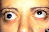
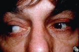
Motility Disturbances | Rosa A. Tang, MD | Cyclical oculomotor paresis may occur in patients as an intermittent phenomenon, with a paretic phase and diplopia and intervals that are nonparetic. The history and examination are classic for the disorder. Pair with Images 95_19 and 95_20. |
| 187 |
 |
Motility Disturbances | Rosa A. Tang, MD | Cyclical oculomotor paresis may occur in patients as an intermittent phenomenon, with a paretic phase and diplopia and intervals that are nonparetic. The history and examination are classic for the disorder. Pair with Images 95_18 and 95_19. |
| 188 |
 |
Ocular Manifestations of Systemic Disorders | Rosa A. Tang, MD | Systemic lymphoma may occur in the orbit and may involve the lacrimal gland. Patients usually present with a painless, progressive proptosis or a mass. CT scan usually demonstrates an irregularly shaped lesion conforming to the globe or lacrimal fossa, and bone erosion is not usually found. Pair wit... |
| 189 |
 |
Ocular Manifestations of Systemic Disorders | Rosa A. Tang, MD | Systemic lymphoma may occur in the orbit and may involve the lacrimal gland. Patients usually present with a painless, progressive proptosis or a mass. CT scan usually demonstrates an irregularly shaped lesion conforming to the globe or lacrimal fossa, and bone erosion is not usually found. Pair wit... |
| 190 |
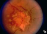 |
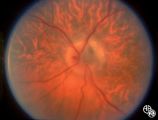
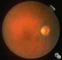
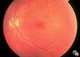
Systemic Disorders With Optic Nerve and Retinal Findings | Rosa A. Tang, MD | Neoplasms may result in an optic neuropathy by direct metastatic involvement. In this patient, a lung adenocarcinoma was metastatic to the optic nerve.This is a fundus photo. |
| 191 |
 |
Motility Disturbances | Larry P. Frohman, MD | This young woman had bilateral sixth nerve paresis from a motor vehicle accident. The images show the results of a successful Jensen procedure. |
| 192 |
 |
Motility Disturbances | Larry P. Frohman, MD | This young woman had bilateral sixth nerve paresis from a motor vehicle accident. The images show the results of a successful Jensen procedure. |
| 193 |
 |
Motility Disturbances | Larry P. Frohman, MD | This patient sustained a traumatic avulsion of the left medial rectus. Image 94_75 shows the successful postoperative result. |
| 194 |
 |
Motility Disturbances | Rosa A. Tang, MD | Cyclical oculomotor paresis may occur in patients as an intermittent phenomenon, with a paretic phase and diplopia and intervals that are nonparetic. The history and examination are classic for the disorder. Pair with Images 95_18 and 95_20. |
| 195 |
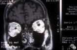 |
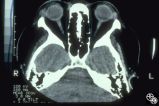
Ocular Manifestations of Systemic Disorders | Rosa A. Tang, MD | Thyroid eye disease may cause proptosis and extraocular muscle enlargement that may be seen on orbital imaging studies. In general, coronal images allow the best visualization of the extraocular muscle enlargement. Pair with 94_44 and 94_46. |
| 196 |
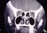 |
Ocular Manifestations of Systemic Disorders | Rosa A. Tang, MD | Thyroid eye disease may cause proptosis and extraocular muscle enlargement that may be seen on orbital imaging studies. In general, coronal images allow the best visualization of the extraocular muscle enlargement. Pair with 94_44 and 94_45. |
| 197 |
 |
Neuro-Ophthalmic Imaging-MRI | Rosa A. Tang, MD | Aneurisms may result in neuro-ophthalmologic sign and symptoms by direct compression of the afferent or efferent systems or by the secondary effects of hemorrhage. Basilar aneurisms may result in ocular motor deficits such as a unilateral or bilateral third nerve palsy. |
| 198 |
 |
Motility Disturbances | Larry P. Frohman, MD | This man had a posttraumatic right sixth nerve paresis. Image 94_66 demonstrates the adduction deficit that the Botox induced. |
| 199 |
 |
Motility Disturbances | Larry P. Frohman, MD | This man had a posttraumatic right sixth nerve paresis. He is shown in primary gaze before Botox (botulinum toxin; image 94_64) |
| 200 |
 |
Motility Disturbances | Larry P. Frohman, MD | This man had a posttraumatic right sixth nerve paresis. He is shown in primary gaze after Botox (image 94_65). |