AAO-NANOS Neuro-Ophthalmology Clinical Collection: Derived from the AAO-NANOS Clinical Neuro-Ophthalmology collection produced on CD. The images are of selected cases from the NANOS teaching slide exchange, and the CD was produced under the direction of Larry Frohman, MD and Andrew Lee, MD.
The American Academy of Ophthalmology (AAO); The North American Neuro-Ophthalmology Association (NANOS).
NOVEL: https://novel.utah.edu/
TO
Filters: Collection: ehsl_novel_aao_nanos
| Title | Description | Subject | ||
|---|---|---|---|---|
| 151 |
 |
Optic Tract Syndrome Due to Carotid Artery Dolichoectasia | This 43-year-old man was referred for evaluation of 6 months of visual loss OU. In retrospect, he had noticed increasing difficulty with his tennis game dating back over 3 years, as balls would pass him unexpectedly when hit to his backhand (left) side. The patient did not think this was progressive... | Dolichoectasia |
| 152 |
 |
Optic Tract Syndrome Due to Carotid Artery Dolichoectasia | This 43-year-old man was referred for evaluation of 6 months of visual loss OU. In retrospect, he had noticed increasing difficulty with his tennis game dating back over 3 years, as balls would pass him unexpectedly when hit to his backhand (left) side. The patient did not think this was progressive... | Dolichoectasia |
| 153 |
 |
Optic Tract Syndrome Due to Carotid Artery Dolichoectasia | This 43-year-old man was referred for evaluation of 6 months of visual loss OU. In retrospect, he had noticed increasing difficulty with his tennis game dating back over 3 years, as balls would pass him unexpectedly when hit to his backhand (left) side. The patient did not think this was progressive... | Dolichoectasia |
| 154 |
 |
Optic Tract Syndrome Due to Carotid Artery Dolichoectasia | This 43-year-old man was referred for evaluation of 6 months of visual loss OU. In retrospect, he had noticed increasing difficulty with his tennis game dating back over 3 years, as balls would pass him unexpectedly when hit to his backhand (left) side. The patient did not think this was progressive... | Dolichoectasia |
| 155 |
 |
Optic Tract Syndrome Due to Carotid Artery Dolichoectasia | This 43-year-old man was referred for evaluation of 6 months of visual loss OU. In retrospect, he had noticed increasing difficulty with his tennis game dating back over 3 years, as balls would pass him unexpectedly when hit to his backhand (left) side. The patient did not think this was progressive... | Dolichoectasia |
| 156 |
 |
Neuro-Ophthalmic Vascular Disease | Occlusion of a branch or central retinal artery may result in acute visual loss. The ophthalmoscopic findings are retinal whitening due to ischemic retina in the distribution of the occluded artery. Sparing or selective involvement of cilioretinal artery branches may occur. Patients with a central r... | Central/Branch Retinal Artery Occlusion |
| 157 |
 |
Neuro-Ophthalmic Vascular Disease | Occlusion of a branch or central retinal artery may result in acute visual loss. The ophthalmoscopic findings are retinal whitening due to ischemic retina in the distribution of the occluded artery. Sparing or selective involvement of cilioretinal artery branches may occur. Patients with a central r... | Central/Branch Retinal Artery Occlusion |
| 158 |
 |
Neuro-Ophthalmic Vascular Disease | Occlusion of a branch or central retinal artery may result in acute visual loss. The ophthalmoscopic findings are retinal whitening due to ischemic retina in the distribution of the occluded artery. Sparing or selective involvement of cilioretinal artery branches may occur. Patients with a central r... | Central/Branch Retinal Artery Occlusion |
| 159 |
 |
Neuro-Ophthalmic Vascular Disease | Occlusion of a branch or central retinal artery may result in acute visual loss. The ophthalmoscopic findings are retinal whitening due to ischemic retina in the distribution of the occluded artery. Sparing or selective involvement of cilioretinal artery branches may occur. Patients with a central r... | Central/Branch Retinal Artery Occlusion |
| 160 |
 |
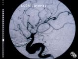
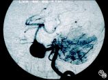
Neuro-Ophthalmic Vascular Disease | This 76-year-old woman has a 7-month history of redness and pressure sensation in both eyes that is worse in the morning. She has noted intermittent horizontal diplopia during this time. Angiography demonstrated a right dural cavernous sinus fistula, which was successfully occluded with direct injec... | Dural Arteriovenous Malformation |
| 161 |
 |
Neuro-Ophthalmic Vascular Disease | This 76-year-old woman has a 7-month history of redness and pressure sensation in both eyes that is worse in the morning. She has noted intermittent horizontal diplopia during this time. Angiography demonstrated a right dural cavernous sinus fistula, which was successfully occluded with direct injec... | Dural Arteriovenous Malformation |
| 162 |
 |
Neuro-Ophthalmic Vascular Disease | This 76-year-old woman has a 7-month history of redness and pressure sensation in both eyes that is worse in the morning. She has noted intermittent horizontal diplopia during this time. Angiography demonstrated a right dural cavernous sinus fistula, which was successfully occluded with direct injec... | Dural Arteriovenous Malformation |
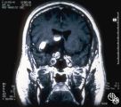
| 163 |
 |
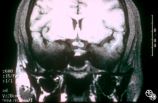
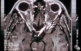
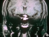
Neuro-Ophthalmic Vascular Disease | This coronal CT scan shows the enlarged superior ophthalmic vein in the left orbit. This 76-year-old woman has a 7-month history of redness and pressure sensation in both eyes that is worse in the morning. She has noted intermittent horizontal diplopia during this time. Angiography demonstrated a ri... | Dural Arteriovenous Malformation |
| 164 |
 |
Neuro-Ophthalmic Vascular Disease | This 76-year-old woman has a 7-month history of redness and pressure sensation in both eyes that is worse in the morning. She has noted intermittent horizontal diplopia during this time. Angiography demonstrated a right dural cavernous sinus fistula, which was successfully occluded with direct injec... | Dural Arteriovenous Malformation |
| 165 |
 |
Neuro-Ophthalmic Vascular Disease | This 76-year-old woman has a 7-month history of redness and pressure sensation in both eyes that is worse in the morning. She has noted intermittent horizontal diplopia during this time. Angiography demonstrated a right dural cavernous sinus fistula, which was successfully occluded with direct injec... | Dural Arteriovenous Malformation |
| 166 |
 |
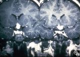
Neuro-Ophthalmic Imaging-MRI | A 59-year-old untreated hypertensive man had a sudden onset of vomiting, gait ataxia, dysarthria, and left-sided weakness. The eyes were deviated downward and to the left. (Note position of gaze in 94_11). Extraocular motility was full. There was rotary nystagmus with the fast phase to the left; the... | Wallenberg Syndrome; Lateral Medullary Syndrome; Posterior Inferior Cerebellar Artery |
| 167 |
 |
Neuro-Ophthalmic Imaging-MRI | This 23-year-old right-handed man had a history of idiopathic recurrent optic neuritis. The patient presented with acuity of 20/400 OD and 20/100 OS, with a central scotoma OD and a complete temporal defect OS. MRI with fat suppression and gadolinium revealed enhancement of the intracranial nerve an... | Chiasmal Optic Neuritis |
| 168 |
 |
Neuro-Ophthalmic Imaging-MRI | This 23-year-old right-handed man had a history of idiopathic recurrent optic neuritis. The patient presented with acuity of 20/400 OD and 20/100 OS, with a central scotoma OD and a complete temporal defect OS. MRI with fat suppression and gadolinium revealed enhancement of the intracranial nerve an... | Chiasmal Optic Neuritis |
| 169 |
 |
Neuro-Ophthalmic Imaging-MRI | This 23-year-old right-handed man had a history of idiopathic recurrent optic neuritis. The patient presented with acuity of 20/400 OD and 20/100 OS, with a central scotoma OD and a complete temporal defect OS. MRI with fat suppression and gadolinium revealed enhancement of the intracranial nerve an... | Chiasmal Optic Neuritis |
| 170 |
 |
Ocular Manifestations of Congenital/Inherited Diseases | This 21-year-old woman had a 2-year history of blurred vision. A computerized visual field demonstrated a temporal defect OS. MRI confirmed a chiasmal mass lesion. The pathology was consistent with hemangioblastoma. Further workup revealed retinal angiomas and multiple other hemangioblastomas of the... | von Hippel-Lindau Disease |
| 171 |
 |
Ocular Manifestations of Congenital/Inherited Diseases | This 21-year-old woman had a 2-year history of blurred vision. A computerized visual field demonstrated a temporal defect OS. MRI confirmed a chiasmal mass lesion. The pathology was consistent with hemangioblastoma. Further workup revealed retinal angiomas and multiple other hemangioblastomas of the... | von Hippel-Lindau Disease |
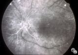
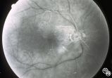
| 172 |
 |
Ocular Manifestations of Congenital/Inherited Diseases | This 21-year-old woman had a 2-year history of blurred vision. A computerized visual field demonstrated a temporal defect OS. MRI confirmed a chiasmal mass lesion. The pathology was consistent with hemangioblastoma. Further workup revealed retinal angiomas and multiple other hemangioblastomas of the... | von Hippel-Lindau Disease |
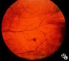
| 173 |
 |
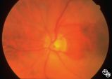
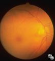
Systemic Disorders With Optic Nerve and Retinal Findings | This fondus image shows a white-centered hemorrhage in a leukemia patient with orbital aspergillosis. | Leukemia; Orbital Aspergilosis; Retinal Hemorrhage |
| 174 |
 |
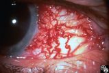
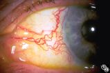
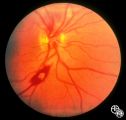
Ocular Manifestations of Congenital/Inherited Diseases | This 38-year-old man had unexplained poor vision in his left eye all his life and was told that he had some type of congenital vascular anomaly. He had no neurocutaneous problems. The photographs demonstrate a complex arteriovenous malformation with dilated loops of veins extending out from the nerv... | Wyburn-Mason Syndrome |
| 175 |
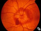 |
Isolated Optic Neuritis/Neuropathy | Papilledema is a term reserved for optic disc edema related to increased intracranial pressure (eg. Papilledema, sixth nerve palsy, headache), a normal neuroimaging study, and an elevated opening pressure with normal cerebrospinal fluid contents. | Pseudotumor Cerebri/Papilledema; Edema |