Best known for his world-renowned neuro-ophthalmology unit based at the University of California, San Francisco, William Hoyt, MD collected here more than 850 of his best images covering a wide range of disorders.
William F. Hoyt, MD, Professor Emeritus of Ophthalmology, Neurology and Neurosurgery, Department of Ophthalmology, University of California, San Francisco.
NOVEL: https://novel.utah.edu/
TO
| Title | Description | Type | ||
|---|---|---|---|---|
| 151 |
 |
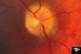
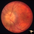
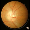
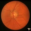
Medullated Nerve Fibers with Papilledema | Right eye. Papilledema superimposed upon medullated nerve fibers. Man with metastatic gastric carcinoma. Anatomy: Optic disc. Pathology: Papilledema. Disease/Diagnosis: Papilledema plus medullated nerve fibers. | Image |
| 152 |
 |
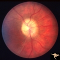
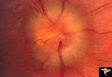
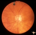
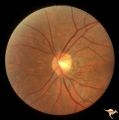
Papilledema due to Brain Tumor - Natural History | Right eye 33 months after presentation. Atrophy appears about the same. Notice especially the narrowing of retinal arterioles. Visual loss is severe in both eyes. Papilledema due to brain tumor - 3 year natural history. Patient refused treatment. Man. Anatomy: Optic disc. Pathology: Papilledema. Dis... | Image |
| 153 |
 |
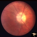
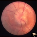
Papilledema due to Brain Tumor - Natural History | Right eye at 27 months after presentation. Atrophy is more profound in both eyes. Papilledema due to brain tumor - 3 year natural history. Patient refused treatment. Man. Anatomy: Optic disc. Pathology: Papilledema. Disease/Diagnosis: Papilledema from acoustic neuronoma. | Image |
| 154 |
 |
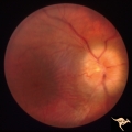
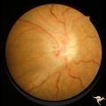
Papilledema due to Brain Tumor - Natural History | Left eye at 25 months after presentation. Atrophy is more profound in both eyes. Papilledema due to brain tumor - 3 year natural history. Patient refused treatment. Man. Anatomy: Optic disc. Pathology: Papilledema. Disease/Diagnosis: Papilledema from acoustic neuronoma. | Image |
| 155 |
 |
Papilledema due to Brain Tumor - Natural History | Right eye 23 months after presentation. Atrophy is replacing papilledema in both eyes. Papilledema due to brain tumor - 3 year natural history. Patient refused treatment. Man. Anatomy: Optic disc. Pathology: Papilledema. Disease/Diagnosis: Papilledema from acoustic neuronoma. | Image |
| 156 |
 |
Papilledema due to Brain Tumor - Natural History | Left eye 23 months after presentation. Atrophy is replacing papilledema in both eyes. Papilledema due to brain tumor - 3 year natural history. Patient refused treatment. Man. Anatomy: Optic disc. Pathology: Papilledema. Disease/Diagnosis: Papilledema from acoustic neuronoma. | Image |
| 157 |
 |
Papilledema due to Brain Tumor - Natural History | Right eye at presentation. Papilledema due to acoustic neuronoma(tumor)- 3 year natural history. Patient refused treatment. Man. Anatomy: Optic disc. Pathology: Papilledema. Disease/Diagnosis: Papilledema from acoustic neuronoma. | Image |
| 158 |
 |
Papilledema due to Brain Tumor - Natural History | Left eye at presentation. Papilledema due to acoustic neuronoma(tumor) - 3 year natural history. Patient refused treatment. Man. Anatomy: Optic disc. Pathology: Papilledema. Disease/Diagnosis: Papilledema from acoustic neuronoma. | Image |
| 159 |
 |
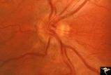
Post Papilledema Retinal Choroidal Bypass (Optociliary) | Right eye. Post papilledema retinal choroidal bypass (optociliary). Arterial venous malformation draining into saggital sinus causing papilledema and retinal choroidal collaterals. Anatomy: Optic disc. Pathology: Post papilledema. Disease/Diagnosis: Post papilledema with retinal choroidal bypass ves... | Image |
| 160 |
 |
Post Papilledema Retinal Choroidal Bypass (Optociliary) | Left eye. Post papilledema retinal choroidal bypass (optociliary). Arterial venous malformation draining into saggital sinus causing papilledema and retinal choroidal collaterals. Anatomy: Optic disc. Pathology: Post papilledema. Disease/Diagnosis: Post papilledema with retinal choroidal bypass vess... | Image |
| 161 |
 |
IC102a Central Retinal Artery Occlusion with Cilioretinal Collaterals | Left eye, 1988, Central retinal artery with cilioretinal collaterals due to calcific embolic behind the lamina cribrosa due to calcific valvular heart disease. Collaterals have been called "Nettleship Collaterals", recognizing the British physician who first described them in 1892. Anatomy: Optic di... | Image |
| 162 |
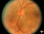 |
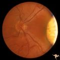
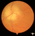
C105 Disc Edema with Systemic Lupus | Mild disc edema blurs the inferior disc margin. Flourescein angiogram in D1_06. Same patient as D1_06 an D1_07. Man. Anatomy: Optic disc. Pathology: Axoplasmic stasis due to vasculitis (Lupus). Disease/ Diagnosis: Lupus papillopathy. | Image |
| 163 |
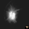 |
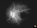
D107 Disc Edema with Systemic Lupus | Late stage Flourescein angiogram showing flourescein leakage on the disc and around the neighboring vessels. Note this amount of edema could not be appreciated in the colored fundus image D1_05. Same patient as D1_06 an D1_05. Anatomy: Optic disc. Pathology: Axoplasmic stasis due to vasculitis (Lupu... | Image |
| 164 |
 |
D106 Disc Edema with Systemic Lupus | Flourescein angiogram shows evidence of vascular papillopathy. (Lupus) Same patient as D1_05 an D1_07. Anatomy: Optic disc. Pathology: Axoplasmic stasis due to vasculitis (Lupus). Disease/ Diagnosis: Lupus papillopathy. | Image |
| 165 |
 |
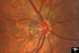
H35 Segmental Hypoplasia, Retinal-Congenital Toxo | Left eye. Moving out temporally to see large chorioretinal scar. Temporal sector hypoplasia from congenital retinal toxoplasmosis. Same patient as H_34. Anatomy: Optic disc; Retina. Pathology: Hypoplasia secondary to retinal lesion. Disease/ Diagnosis: Segmental optic disc hypoplasia | Image |
| 166 |
 |
H48 Segmental Hypoplasia, Retinal-Nasal Hypoplasia | Bilateral nasal hypoplasia with absence of nasal nerve fiber layer and corresponding flag-like temporal field defect. Right eye. Same patient as H_49. Anatomy: Optic disc. Pathology: Nasal segmental disc hypoplasia. Disease/ Diagnosis: Congential anomaly. | Image |
| 167 |
 |
H49 Segmental Hypoplasia, Retinal-Nasal Hypoplasia | Bilateral nasal hypoplasia with absence of nasal nerve fiber layer and corresponding flag-like temporal field defect. Left eye. Same patient as H_48. Anatomy: Optic disc. Pathology: Nasal segmental disc hypoplasia. Disease/ Diagnosis: Congential anomaly. | Image |
| 168 |
 |
H46 Segmental Hypoplasia, Retinal-Nasal Hypoplasia | Nasal hypoplasia with suspected nasal pit about 3:00. Right eye. Man with temporal sector field defect. Same patient as H_47. Anatomy: Optic disc. Pathology: Nasal segmental disc hypoplasia. Disease/ Diagnosis: Congenital anomaly | Image |
| 169 |
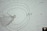 |
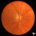
H42 Segmental Hypoplasia, Retinal, Nasal Hypoplasia | Visual field of patient in H_43. Flag-like temporal field defect from patient with nasal segmental disc hypoplasia. Anatomy: Optic disc. Pathology: Nasal segmental disc hypoplasia. Disease/ Diagnosis: Congenital anomaly. | Image |
| 170 |
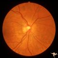 |
H44 Segmental Hypoplasia, Retinal, Nasal Hypoplasia | Bilateral nasal hypoplasia with bilateral flag-like temporal field defect. Right eye. Same patient as H_45. Anatomy: Optic disc. Pathology: Nasal segmental disc hypoplasia. Disease/ Diagnosis: Congenital anomaly. | Image |
| 171 |
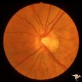 |
H45 Segmental Hypoplasia, Retinal-Nasal Hypoplasia | Bilateral nasal hypoplasia with bilateral flag-like temporal field defect. Left eye. Same patient as H_44. Anatomy: Optic disc. Pathology: Nasal segmental disc hypoplasia. Disease/ Diagnosis: Congenital anomaly. | Image |
| 172 |
 |
H47 Segmental Hypoplasia, Retinal-Nasal Hypoplasia | Normal left eye. Same patient as H_46. Anatomy: Optic disc. Pathology: Nasal segmental disc hypoplasia. Disease/ Diagnosis: Congenital anomaly. | Image |
| 173 |
 |
H43 Segmental Hypoplasia, Retinal, Nasal Hypoplasia | Nasal hypoplasia barely perceptible on disc. Nasal retinal nerve fibers are completely absent from 7:00 - 12:00. Anaotmy: Optic disc. Pathology: Nasal segmental disc hypoplasia. Disease/ Diagnosis: Congenital anomaly. | Image |
| 174 |
 |
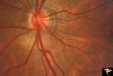
H34 Segmental Hypoplasia, Retinal-Congenital Toxo | Left eye. Temporal sector hypoplasia from congenital retinal toxoplasmosis. Note the sector shaped nerve fiber loss between 2:00 and 4:00. Same patient as H_35. Anatomy: Optic disc; retina. Pathology: Hypoplasia secondary to retinal lesion. Disease/ Diagnosis: Segmental optic disc hypoplasia. | Image |
| 175 |
 |
P55b Chronic Papilledema, Late Stage | Left eye. Optic atrophy secondary to chronic papilledema, late stage with retinal choroidal bypass vein. Opticocilliary shunt. Glioblastoma, chemotherapy patient. Anatomy: Optic disc. Pathology: Papilledema. Disease/ Diagnosis: Chronic papilledema with atrophy. | Image |