Best known for his world-renowned neuro-ophthalmology unit based at the University of California, San Francisco, William Hoyt, MD collected here more than 850 of his best images covering a wide range of disorders.
William F. Hoyt, MD, Professor Emeritus of Ophthalmology, Neurology and Neurosurgery, Department of Ophthalmology, University of California, San Francisco.
NOVEL: https://novel.utah.edu/
TO
| Title | Description | Type | ||
|---|---|---|---|---|
| 151 |
 |
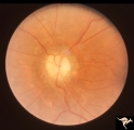
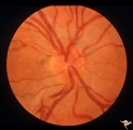
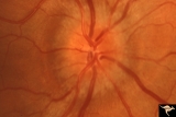
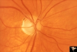
Chronic Papilledema with Pseudo Drusen | Right eye. Chronic papilledema with pseudo drusen due to cerebral pontine angle tumor. Anatomy: Optic disc Pathology: Papilledema Disease/Diagnosis: Chronic papilledema with pseudo drusen | Image |
| 152 |
 |
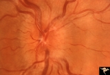
Chronic Papilledema with Pseudo Drusen | Right eye. Meningioma. Pseudo drusen from chronic papilledema. Woman. Anatomy: Optic disc Pathology: Papilledema Disease/Diagnosis: Chronic papilledema with pseudo drusen | Image |
| 153 |
 |
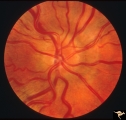
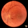
Chronic Papilledema with Pseudo Drusen | Left eye of 51 year old, 220 pound black woman. Pseudotumor cerebri, pseudo drusen, exudates. Anatomy: Optic disc. Pathology: Papilledema Disease/Diagnosis: Chronic papilledema with pseudo drusen | Image |
| 154 |
 |
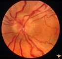
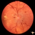
Chronic Papilledema with Pseudo Drusen | Chronic papilledema with pseudo drusen. Residual choroidal folds. Pseudo drusen. Anatomy: Optic disc. Pathology: Papilledema. Disease/Diagnosis: Chronic papilledema with pseudo drusen. | Image |
| 155 |
 |
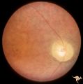
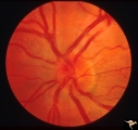
Chronic Papilledema with Pseudo Drusen | Left eye. Meningioma. Pseudo drusen from chronic papilledema. The patient's meningioma had blinded her left eye and caused chronic elevated intracranial pressure. Woman. Anatomy: Optic disc Pathology: Papilledema Disease/Diagnosis: Chronic papilledema with pseudo drusen | Image |
| 156 |
 |
Congenital Retinal Cerebellar Degeneration | Congenital retinal blindness due to cerebellar degeneration syndrome. Granular retinal pigmentary degeneration. Pair with R2_B1_1a. Anatomy: Retina. Pathology: Optic atrophy. Disease/Diagnosis: Congenital retinal cerebellar degeneration. Clinical: Severe mental retardation and blindness. | Image |
| 157 |
 |
Congenital Retinal Cerebellar Degeneration | Congenital retinal blindness due to cerebellar degeneration syndrome. Optic disc pallor with arteriolar attenuation. Pair with R2_B1_1b. Anatomy: Retina. Pathology: Optic atrophy. Disease/ Diagnosis: Congenital retinal cerebellar degeneration. Clinical: Severe mental retardation and blindness. | Image |
| 158 |
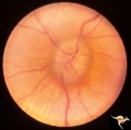
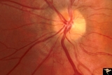 |
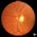
Crowded Disc | PP7a: right eye crowded disc with blurred margin. Note anomalous vascular pattern; PP7b- left disc is cupless disc and normal. 10 year old girl with gonadal dysgenesis and growth retardation. Anatomy: Optic disc Pathology: Normal variation of the optic disc Disease/Diagnosis: Normal variation of the... | Image |
| 159 |
 |
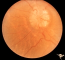
Crowded Disc | PP5a: left eye; PP5b: left eye X 2 magnification; congenital disc blurring. Boy. Anatomy: Optic disc. Pathology: Normal variation of the optic disc. Disease/Diagnosis: Normal variation of the optic disc. Congenital blurred disc. Clinical: Blurred disc margin. Beautiful example of difficult different... | Image |
| 160 |
 |
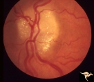
Crowded Disc (Family) | Right eye. PP3 a & b: sister; PP4 a & b brother; Congenital disc margin blurring with crowded discs. Excellent example of pseudo papilledema. Anatomy: Optic disc. Pathology: Normal variant of the optic disc. Disease/Diagnosis: Normal variant of the optic disc. Crowded disc. Clinical: Appearance is ... | Image |
| 161 |
 |
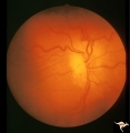
Crowded Disc (Family) | Right eye. PP3 a & b: sister; PP4 a & b brother; Congenital disc margin blurring with crowded discs. Excellent example of pseudo papilledema that caused serious diagnostic confusion which led to a pneumoencephalogram (PEG) and arteriogram. Anatomy: Optic disc. Pathology: Normal variation of the opt... | Image |
| 162 |
 |
Crowded Disc (Family) | Left eye. PP3 a & a: sister; PP4 a & b brother; Congenital disc margin blurring with crowded discs. Excellent example of pseudo papilledema that caused serious diagnostic confusion which led to a pneumoencephalogram (PEG) and arteriogram. Anatomy: Optic disc. Pathology: Normal variation of the opti... | Image |
| 163 |
 |
Crowded Disc (Family) | Left eye. PP3 a & b: sister; PP4 a & b brother; Congenital disc margin blurring with crowded discs. Excellent example of pseudo papilledema. Anatomy: Optic disc. Pathology: Normal variation of the optic disc. Disease/Diagnosis: Normal variation of the optic disc. Crowded disc. Clinical: Appearance ... | Image |
| 164 |
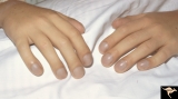 |
Cyanotic Heart Disease with Clubbing of Fingernails | Note the cyanotic nail beds and clubbing. Anatomy: Optic disc. Pathology: Papilledema. Disease/Diagnosis: Pseudotumor due to cyanotic heart disease. Clinical: Young boy with clubbing. | Image |
| 165 |
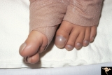 |
Cyanotic Heart Disease with Clubbing of Toes | Bilateral Papilledema with cyanotic heart disease. Anatomy: Optic disc. Pathology: Papilledema. Disease/Diagnosis: Pseudotumor due to cyanotic heart disease. Clinical: Young boy with clubbing. | Image |
| 166 |
 |
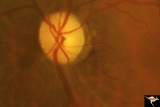
D101 Disc Edema with Systemic Lupus | Unilateral disc swelling with narrowed arterioles. No decrease in visual acuity or field. 19 year old woman. Patient died of cerebral lupus within two months. Optociliary veins dumping into disc edge at 4:00, 9:00, and 11:00. Anatomy: Optic disc. Pathology: Axoplasmic stasis due to vasculitis (Lupu... | Image |
| 167 |
 |
D102 Disc Edema with Systemic Lupus | 28 year old woman. Vision 20/20 but blind spot enlarged. Same patient as D1_03. Right eye. Anatomy: Optic disc. Pathology: Axoplasmic stasis due to vasculitis (Lupus). Disease/ Diagnosis: Lupus papillopathy. Clinical: Normal vision with enlarged blind spot on visual field. | Image |
| 168 |
 |
D103 Disc Edema with Systemic Lupus | 28 year old woman with systemic Lupus erythematosus. Vision 20/20 but blind spot enlarged. Same patient as D1_02. Magnified. Anatomy: Optic disc. Pathology: Axoplasmic stasis due to vasculitis (Lupus). Disease/ Diagnosis: Lupus papillopathy. Clinical: Normal vision with enlarged blind spot on visual... | Image |
| 169 |
 |
D104 Disc Edema with Systemic Lupus | Unilateral disc swelling and enlarged blind spot. Patient had episcleritis 4 weeks before this image was taken. 14 year old girl. Anatomy: Optic disc. Pathology: Axoplasmic stasis due to vasculitis (Lupus). Disease/ Diagnosis: Lupus papillitis. Clinical: No visual loss. History of episcleritis. Big ... | Image |
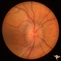
| 170 |
 |
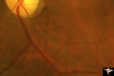
D201 Disc Edema with Systemic Hypertension | Left eye. Note generalized arterial narrowing. Low grade disc edema and multiple splinter hemorrhages. The patient had severe hypertension from kidney failure. Additional yellow intraretinal exudate at the macula. 20 year old male patient. Right eye. Pair with D2_02. Anatomy: Optic disc; Retina; Ret... | Image |
| 171 |
 |
D202 Disc Edema with Systemic Hypertension | Right eye. Note generalized arteriole narrowing. Low grade disc edema and multiple splinter hemorrhages. The patient had severe hypertension from kidney failure. Additional yellow intraretinal exudate at the macula. 20 year old male patient. Pair with D2_01. Anatomy: Optic disc; Retina; Retinal art... | Image |
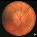
| 172 |
 |
Diffuse Atrophy | Primary optic atrophy following optic neuritis. 1960. Note absence of all retinal nerve fiber layer reflex in the peripapillary retina. The retinal vessels appear to lie on the retina without any tissue surrounding them. Normal looking arterioles. Anatomy: Optic disc. Pathology: Optic atrophy. Disea... | Image |
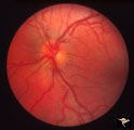
| 173 |
 |
Diffuse Atrophy | Primary optic atrophy from optic nerve compression by aneurysm. Note narrowing of retinal arterioles. Close up showing arcuate streaks of nerve fibers entering inferior optic disc. Pair with IIA1_7. Anatomy: Optic disc. Pathology: Optic atrophy. Disease/Diagnosis: Optic atrophy due to giant aneurysm... | Image |
| 174 |
 |
Diffuse Atrophy | Primary optic atrophy following head injury. 1982. Normal looking arterioles. Anatomy: Optic disc. Pathology: Optic atrophy. Disease/Diagnosis: Optic atrophy after trauma. Clinical: Blindness. | Image |
| 175 |
 |
Diffuse Atrophy | Primary optic atrophy from optic nerve compression by aneurysm. Note narrowing of retinal arterioles. Pair with IIA1_8. Anatomy: Optic disc. Pathology: Optic atrophy. Disease/Diagnosis: Optic atrophy due to giant aneurysm. Clinical: Blindness. | Image |