Collection of materials relating to neuro-ophthalmology as part of the Neuro-Ophthalmology Virtual Education Library.
NOVEL: https://novel.utah.edu/
TO
- NOVEL728
| Title | Creator | Description | Subject | ||
|---|---|---|---|---|---|
| 151 |
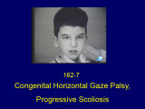 |
Congenital Horizontal Gaze Palsy, Progressive Scoliosis | Shirley H. Wray, MD, PhD, FRCP | The patient is an 8 year old boy with a rare autosomal recessive disorder characterized by congenital absence of conjugate horizontal eye movements preservation of vertical gaze and convergence and progressive scoliosis (HGPPS) developing in childhood. The child was referred to Dr. Cogan with a diag... | Congenital Horizontal Gaze Palsy; Progressive Scoliosis; Mutation of the ROBO 3 Gene on Chromosome 11q23-q25; Congenital Cranial Disinnervation Syndrome; Mobius Syndrome |
| 152 |
 |
Congenital Hydrocephalus | Mays El-Dairi, MD | Presentation covering an overview of congenital hydrocephalus. | Congenital Hydrocephalus |
| 153 |
 |
Congenital and Secondary Syphilis | Gregory P. Van Stavern, MD | Images showing evideince of Congenital and Secondary Syphilis | Syphilis |
| 154 |
 |
Conjunctival Examination | Sovik De Sirkar, MSIII; Ore-ofe Adesina, MD, | Description of the conjunctival examination. | Conjunctival Examination |
| 155 |
 |
Constructional Apraxia | Shirley H. Wray, MD, PhD, FRCP | Slideshow describing condition. | Apraxia of the Left Hand; Constructional Apraxia; Degenerative CNS Disease; Dressing Apraxia; Progressive Lobar Atrophy; Right Parietal Lobe |
| 156 |
 |
Constructional Apraxia | Shirley H. Wray, MD, PhD, FRCP | The patient is a 72 year old right handed woman who presented in November 1995 with the sudden onset of impaired coordination of visual and motor skills following an inner right ear infection. One of her problems was difficulty sitting on a chair as she tended to place her body incorrectly. By late ... | Dressing Apraxia; Apraxia of the Left Hand; Constructional Apraxia; Right Parietal Lobe; Progressive Lobar Atrophy; Degenerative CNS Disease; Apraxia |
| 157 |
 |
Contrast Sensitivity | Sean Gratton, MD | Explanation of contrast sensitivity. | Contrast Sensitivity |
| 158 |
 |
Convergence Insufficiency | Shirley H. Wray, MD, PhD, FRCP | A PowerPoint slideshow describing the condition. | Basal Ganglia; Blepharoclonus; Convergence Insufficiency; Parkinson's Disease- Dopamine deficiency; Saccadic Breakdown of Smooth Pursuit; Slow Hypometric Horizontal Saccades; Slow Hypometric Saccades |
| 159 |
 |
Convergence Insufficiency | Shirley H. Wray, MD, PhD, FRCP | The patient is a 73 year old man with a ten year history of idiopathic Parkinson's disease characterized by difficulty in walking, generalized rigidity and a mild tremor of his hands at rest with deterioration in his handwriting. He denied any memory impairment or loss of cognitive function. He was ... | Basal Ganglia; Blepharoclonus; Convergence Insufficiency; Slow Hypometric Saccades; Saccadic Breakdown of Smooth Pursuit; Parkinson's Disease- Dopamine deficiency; Slow Hypometric Horizontal Saccades; Convergence |
| 160 |
 |
Corectopia | Meagan Seay, DO | These are photos of a patient with unilateral corectopia. This patient's corectopia is of unclear etiology and possibly related to birth trauma. | Corectopia; Unilateral; Photos |
| 161 |
 |
Corneal Staining | Sovik De Sirkar, MSIII; Ore-ofe Adesina, MD | Description of the corneal staining technique. | Corneal Staining |
| 162 |
 |
Cotton Wool Spots: The Basics | Arnav Gupta, BHSc; Rahul Sharma, MD, MPH | A presentation describing cotton wool spots, an abnormal finding on funduscopic exam of the retina of the eye. | Cotton Wool Spots; Retina |
| 163 |
 |
Craniopharyngioma and Optic Atrophy | Kathleen B. Digre, MD; James J. Corbett, MD | Slideshow describing condition. | Craniopharayngioma; Otpic Atrophy |
| 164 |
 |
Crowded Disc - Family | William F. Hoyt, PhD | Left eye. PP3 a & b: sister; PP4 a&b: brother; Congenital disc margin blurring with crowded discs. Excellent example of pseudo papilledema. Pathology: Normal variation of the optic disc. Disease/Diagnosis: Normal variation of the optic disc. Crowded disc. Clinical notes: Appearance due to too many f... | Pseudopapilledema; Congenital Blurred Disc |
| 165 |
 |
Decompensated Phoria | Alex Christoff, MD | An overview of decompensated phoria and its treatment. | Decompensated Phoria |
| 166 |
 |
Dementia with Lewy Bodies: Overview and Neuro-ophthalmologic features | Pavan Vaswani, MD, PhD; Ali G. Hamedani, MD, MHS | Objectives: Recognize the difference between Dementia with Lewy Bodies and Parkinson disease dementia; Recognize the clinical presentation of DLB and differentiating features from Alzheimer disease dementia; Understand the symptomatic therapies and prognosis | Dementia; Lewy Bodies |
| 167 |
 |
Dementia: Overview and Classification | Molly Cincotta, MD; Whitley Aamodt, MD; Ali G. Hamedani, MD, MHS | PowerPoint providing a broad overview of dementia, including definition, clinical findings, work up, diagnosis, classification, and management. | Dementia |
| 168 |
 |
Diagnosis and Evaluation of Stroke in the Pons | Padmaja Sudhakar; Fatai Momodu | This short power point describes the anatomy of the pons, followed by description of stroke in the pons along with clinical presentation and work up. | Crossed Signs; One and Half Syndrome; Pontine Stroke |
| 169 |
 |
Diagnostic Error of Neuro-ophthalmologic Conditions: State of the Science | Leanne Stunkel, MD; David E. Newman-Toker, MD, PhD; Nancy J. Newman, MD; Valérie Biousse, MD | Diagnostic error is prevalent and costly, occurring in up to 15% of US medical encounters and affecting up to 5% of the US population. One-third of malpractice payments are related to diagnostic error. A complex and specialized diagnostic process makes neuro-ophthalmologic conditions particularly vu... | Diagnostic Errors |
| 170 |
 |
Diffusion Tensor Imaging (DTI) | Devin D. Mackay, MD | Explanation of using diffusion tensor imaging (DTI) in examinations. | Diffusion Tensor Imaging (DTI) |
| 171 |
 |
Diffusion Tensor Imaging (DTI) | John Pula, MD | Diffusion tensor (DT) MRI applies the direction of water diffusion through tissues to map out neural pathways in the brain, such as white matter tracts. | Diffusion Tensor Imaging; DTI |
| 172 |
 |
Diffusion Weighted Imaging (DWI) | John Pula, MD | Diffusion weighted imaging sequences are often included as part of a routine brain MRI protocol. Imaging provides examples of DWI. | Diffusion Weighted Imaging; DWI |
| 173 |
 |
Digital Subtraction Angiography | Devin D. Mackay, MD | Explanation of using digital subtraction angiography in examinations. | Digital Subtraction Angiography |
| 174 |
 |
Direct Carotid Cavernous Fistula | Emory Eye Center | Slideshow describing condition. | Fistula |
| 175 |
 |
Disability Evaluation Under Social Security | John Pula, MD | A. How do we evaluate visual disorders? 1. What are visual disorders? Visual disorders are abnormalities of the eye, the optic nerve, the optic tracts, or the brain that may cause a loss of visual acuity or visual fields. A loss of visual acuity limits your ability to distinguish detail, read, or do... | Visual Impairment; Visual Disorders; Legal Blindness |
